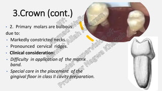
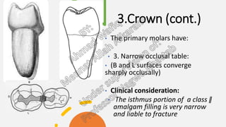
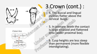
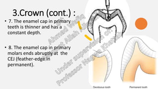
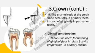
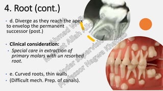
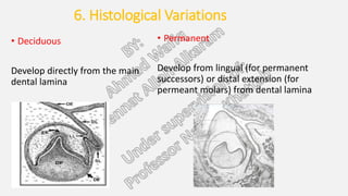
morphological difference
betweendeciduous and
permanent teeth.
BY:
Ahmad Wafiq
Mennat Allah Alkaram
Under supervision of:
Professor Nagwa Khattab
1.Size
1.Size
• Smaller in all dimensions
• MD width of primary incisors
and canines is less than their
permanent successors
• The MD width of the primary
molars is wider than their
successors(premolars) but
smaller than their
corresponding permanents.
Thickness of E and D in
primary teeth is half the
thickness in permanent
teeth.
 Clinical consideration:
 The occlusal cavity in
primary teeth must be
shallower.
2. Color:
2. Color:
• Primary teeth color is
bluish white
• The color of permanent
teeth ranges from grayish
white to yellowish white
3.crown
3.crown
• Wider in MD diameter
than in OG height
• Ant. teeth cup -
shaped
• Post. Teeth square -
shaped
3.Crown (cont.)
• 2. Primary molars are bulbous
due to:
• Markedly constricted necks.
• Pronounced cervical ridges.
• Clinical consideration:
• Difficulty in application of the matrix
band.
• Special care in the placement of the
gingival floor in class II cavity preparation.
3.Crown (cont.)
• The primary molars have:
• 3. Narrow occlusal table:
• (B and L surfaces converge
sharply occlusally)
• Clinical consideration:
• The isthmus portion of a class ǁ
amalgam filling is very narrow
and liable to fracture
3.Crown (cont.) :
• 4. The buccal and lingual
inclines flatter above the
cervical bulge.
• 5. In primary teeth the contact
is large ellipsoid and flattened
area (wider proximal box).
• 6. Cusp heights are less steep
than permanent (more flexible
interdigitating).
3.Crown (cont.) :
• 7. The enamel cap in primary
teeth is thinner and has a
constant depth.
• 8. The enamel cap in primary
molars ends abruptly at the
CEJ (feather-edge in
permanent).
3.Crown (cont.) :
• 9. The enamel rods at the cervix
slope occlusally in primary teeth
instead of gingivally in permanent
teeth.
• Clinical consideration
• There is no need for beveling
of gingival floor in class ǁ cavity
preparation in primary molars.
4. Roots
4. Roots
• The roots of primary teeth are:
• Narrower mesiodistally.
• Longer and more slender in comparison to
the crown (1:2).
• Flare out near the cervix leaving no root
trunk.
4. Root (cont.)
• d. Diverge as they reach the apex
to envelop the permanent
successor (post.)
• Clinical consideration:
• Special care in extraction of
primary molars with un resorbed
root.
• e. Curved roots, thin walls
• (Difficult mech. Prep. of canals).
5. Pulp
5. Pulp
• The pulp outline follows the DEJ more
closely in primary than in permanent
teeth.
• The pulp chambers are proportionally
larger than permanent teeth .
• The pulp horns are higher in primary
molars especially the mesial horn.
• Clinical consideration:
• Special attention should be taken when
establishing the depth of cavities in primary
teeth.
5. Pulp (cont.)
• The root canals of the primary molars show:
• D. more lateral branching and apical
ramifications.
• Clinical consideration:
• This makes it impossible to remove all pulp
tissue in the root canals during root canal
therapy.
•
• E. The apical foramina in primary teeth are
relatively wider than in permanent teeth.
6. Histological
Variations
6. Histological Variations
• Deciduous
Develop directly from the main
dental lamina
• Permanent
Develop from lingual (for permanent
successors) or distal extension (for
permeant molars) from dental lamina
• Deciduous
Enamel and Dentin are less
mineralized
More prone to acidic attack
Easier in cavity preparation
• Permanent
More Mineralized
Less prone
More difficult
Histological Variations
POC
Deciduous Permanent
Cellularity and
Vascularity
High degree Low degree
Density of
Innervation
Less More
Localization of
Infection
Poorer Better
THANK YOU

Morphological differences between permanent and deciduous teeth.pptx

  • 1.
    morphological difference betweendeciduous and permanentteeth. BY: Ahmad Wafiq Mennat Allah Alkaram Under supervision of: Professor Nagwa Khattab
  • 3.
  • 4.
    1.Size • Smaller inall dimensions • MD width of primary incisors and canines is less than their permanent successors • The MD width of the primary molars is wider than their successors(premolars) but smaller than their corresponding permanents.
  • 5.
    Thickness of Eand D in primary teeth is half the thickness in permanent teeth.  Clinical consideration:  The occlusal cavity in primary teeth must be shallower.
  • 6.
  • 7.
    2. Color: • Primaryteeth color is bluish white • The color of permanent teeth ranges from grayish white to yellowish white
  • 8.
  • 9.
    3.crown • Wider inMD diameter than in OG height • Ant. teeth cup - shaped • Post. Teeth square - shaped
  • 10.
    3.Crown (cont.) • 2.Primary molars are bulbous due to: • Markedly constricted necks. • Pronounced cervical ridges. • Clinical consideration: • Difficulty in application of the matrix band. • Special care in the placement of the gingival floor in class II cavity preparation.
  • 11.
    3.Crown (cont.) • Theprimary molars have: • 3. Narrow occlusal table: • (B and L surfaces converge sharply occlusally) • Clinical consideration: • The isthmus portion of a class ǁ amalgam filling is very narrow and liable to fracture
  • 12.
    3.Crown (cont.) : •4. The buccal and lingual inclines flatter above the cervical bulge. • 5. In primary teeth the contact is large ellipsoid and flattened area (wider proximal box). • 6. Cusp heights are less steep than permanent (more flexible interdigitating).
  • 13.
    3.Crown (cont.) : •7. The enamel cap in primary teeth is thinner and has a constant depth. • 8. The enamel cap in primary molars ends abruptly at the CEJ (feather-edge in permanent).
  • 14.
    3.Crown (cont.) : •9. The enamel rods at the cervix slope occlusally in primary teeth instead of gingivally in permanent teeth. • Clinical consideration • There is no need for beveling of gingival floor in class ǁ cavity preparation in primary molars.
  • 15.
  • 16.
    4. Roots • Theroots of primary teeth are: • Narrower mesiodistally. • Longer and more slender in comparison to the crown (1:2). • Flare out near the cervix leaving no root trunk.
  • 17.
    4. Root (cont.) •d. Diverge as they reach the apex to envelop the permanent successor (post.) • Clinical consideration: • Special care in extraction of primary molars with un resorbed root. • e. Curved roots, thin walls • (Difficult mech. Prep. of canals).
  • 18.
  • 19.
    5. Pulp • Thepulp outline follows the DEJ more closely in primary than in permanent teeth. • The pulp chambers are proportionally larger than permanent teeth . • The pulp horns are higher in primary molars especially the mesial horn. • Clinical consideration: • Special attention should be taken when establishing the depth of cavities in primary teeth.
  • 20.
    5. Pulp (cont.) •The root canals of the primary molars show: • D. more lateral branching and apical ramifications. • Clinical consideration: • This makes it impossible to remove all pulp tissue in the root canals during root canal therapy. • • E. The apical foramina in primary teeth are relatively wider than in permanent teeth.
  • 21.
  • 22.
    6. Histological Variations •Deciduous Develop directly from the main dental lamina • Permanent Develop from lingual (for permanent successors) or distal extension (for permeant molars) from dental lamina
  • 23.
    • Deciduous Enamel andDentin are less mineralized More prone to acidic attack Easier in cavity preparation • Permanent More Mineralized Less prone More difficult Histological Variations
  • 24.
    POC Deciduous Permanent Cellularity and Vascularity Highdegree Low degree Density of Innervation Less More Localization of Infection Poorer Better
  • 25.