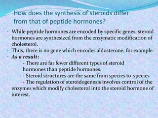
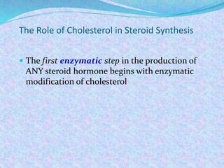
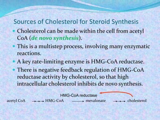
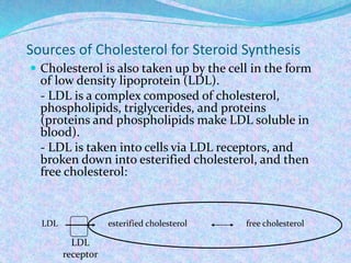
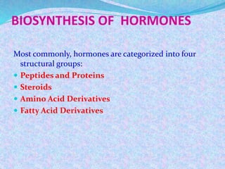
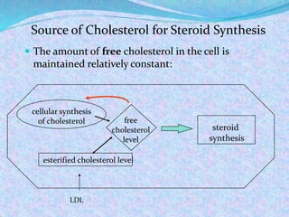
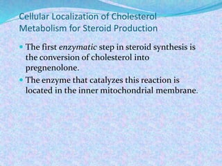
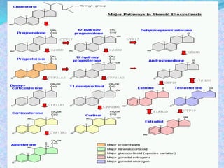
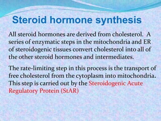
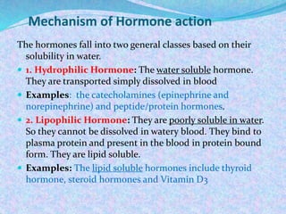
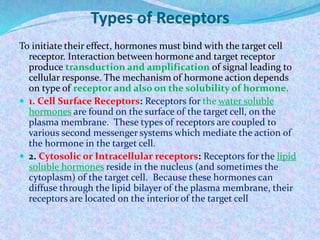
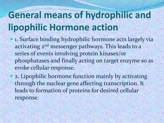
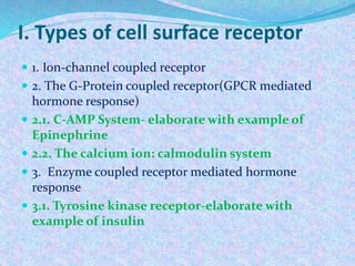
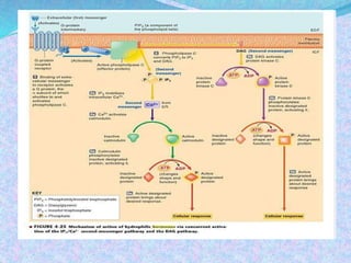
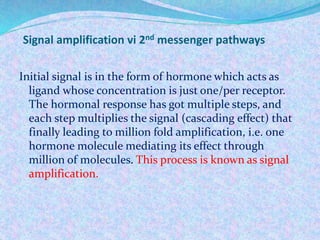
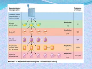
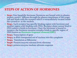
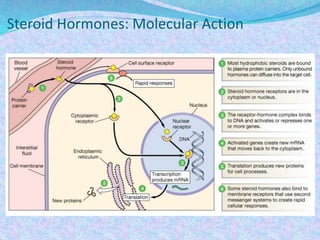
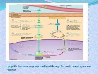
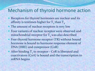
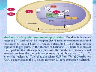
By
Dr Khaled S. Algariri
MOLECULAR
ENDOCRINOLOGY
-2-
BIOSYNTHESIS OF HORMONES
Most commonly, hormones are categorized into four
structural groups:
 Peptides and Proteins
 Steroids
 Amino Acid Derivatives
 Fatty Acid Derivatives
1. Peptide and Protein 1. Steroids 1. Amino acid
derivatives
1. Fatty acid
derivatives
Insulin Testosterone Epinephrine Eicosanoids-
prostaglandins,
prostacyclins,
leukotrienes and
thromboxanes.
Glucagon Estrogen Nor-epinephrine
ACTH Progesteron Thyroxine
TSH Cortisol
Thyrotropin releasing
hormone
Aldosteron
1- Peptides and Proteins Hormones
Biosynthesis
Protein and peptide hormones are encoded in genes,
with each hormone usually represented only once
in the genome. The genes are transcribed in the
nucleus and undergo transcriptional processing to
yield mature mRNA, which is then transported
from nucleus to the cytoplasm. On export from the
nucleus, the mRNA transcripts attach to
ribosomes, where they are translated into protein.
 Several important peptide hormones are secreted from
the pituitary gland. The anterior pituitary secretes
three: prolactin, which acts on the mammary gland;
adrenocorticotropic hormone (ACTH), which acts on
the adrenal cortex to regulate the secretion of
glucocorticoids; and growth hormone, which acts on
bone, muscle, and the liver. The posterior pituitary
gland secretes antidiuretic hormone, also called
vasopressin, and oxytocin
Protein/Peptide hormones are synthesized in
endoplasmic reticulum, transferred to the Golgi
and packaged into secretory vesicles for export.
They can be secreted by one of two pathways:
 Regulated secretion: The cell stores hormone in
secretory granules and releases them in "bursts" when
stimulated. This is the most commonly used pathway
and allows cells to secrete a large amount of hormone
over a short period of time.
 Constitutive secretion: The cell does not store
hormone, but secretes it from secretory vesicles as it is
synthesized
2-Steroid hormones
 Steroid hormones are not water soluble so have to
be carried in the blood complexed to specific
binding globulins.
 Corticosteroid binding globulin carries cortisol
 Sex steroid binding globulin carries testosterone
and estradiol
 In some cases a steroid is secreted by one cell and
is converted to the active steroid by the target cell:
an example is androgen which secreted by the
gonad and converted into estrogen in the brain
Steroid Hormones
 Steroid hormones are nonpolar (no net charge), and
can thus diffuse across lipid membranes (such as the
plasma membrane). They leave cells shortly after
synthesis.
phospholipid
Polar substances are water soluble (dissolve in water),
nonpolar substances are lipid soluble.
Functions of Steroid Hormones
 Steroid hormones play important roles in:
- carbohydrate regulation (glucocorticoids)
- mineral balance (mineralocorticoids)
- reproductive functions (gonadal steroids)
 Steroids also play roles in inflammatory
responses, stress responses, bone metabolism,
cardiovascular fitness, behavior, cognition, and
mood.
How does the synthesis of steroids differ
from that of peptide hormones?
• While peptide hormones are encoded by specific genes, steroid
hormones are synthesized from the enzymatic modification of
cholesterol.
• Thus, there is no gene which encodes aldosterone, for example.
• As a result:
- There are far fewer different types of steroid
hormones than peptide hormones.
- Steroid structures are the same from species to species
- The regulation of steroidogenesis involves control of the
enzymes which modify cholesterol into the steroid hormone of
interest.
The Role of Cholesterol in Steroid Synthesis
 The first enzymatic step in the production of
ANY steroid hormone begins with enzymatic
modification of cholesterol
Sources of Cholesterol for Steroid Synthesis
 Cholesterol can be made within the cell from acetyl
CoA (de novo synthesis).
 This is a multistep process, involving many enzymatic
reactions.
 A key rate-limiting enzyme is HMG-CoA reductase.
 There is negative feedback regulation of HMG-CoA
reductase activity by cholesterol, so that high
intracellular cholesterol inhibits de novo synthesis.
acetyl CoA HMG-CoA mevalonate cholesterol
HMG-CoA reductase
Sources of Cholesterol for Steroid Synthesis
 Cholesterol is also taken up by the cell in the form
of low density lipoprotein (LDL).
- LDL is a complex composed of cholesterol,
phospholipids, triglycerides, and proteins
(proteins and phospholipids make LDL soluble in
blood).
- LDL is taken into cells via LDL receptors, and
broken down into esterified cholesterol, and then
free cholesterol:
LDL
receptor
LDL esterified cholesterol free cholesterol
 The amount of free cholesterol in the cell is
maintained relatively constant:
Source of Cholesterol for Steroid Synthesis
steroid
synthesis
free
cholesterol
level
esterified cholesterol level
cellular synthesis
of cholesterol
LDL
Cellular Localization of Cholesterol
Metabolism for Steroid Production
 The first enzymatic step in steroid synthesis is
the conversion of cholesterol into
pregnenolone.
 The enzyme that catalyzes this reaction is
located in the inner mitochondrial membrane.
Steroid hormone synthesis
All steroid hormones are derived from cholesterol. A
series of enzymatic steps in the mitochondria and ER
of steroidogenic tissues convert cholesterol into all of
the other steroid hormones and intermediates.
The rate-limiting step in this process is the transport of
free cholesterol from the cytoplasm into mitochondria.
This step is carried out by the Steroidogenic Acute
Regulatory Protein (StAR)
Transport of Cholesterol
 Cholesterol is lipid soluble, and mostly located
associated with the external mitochondrial
membrane.
 The conversion of cholesterol to steroids occurs in
the internal mitochondrial membrane.
 Now, to see if you have been paying attention…
 How does cholesterol get from the external
membrane to the internal membrane?
 Answer: Steroidogenic acute regulatory protein
(StAR), which transports cholesterol into the
mitochondria, moving it from the outer membrane
to the inner membrane.
3. Amino Acid Derivatives
 There are two groups of hormones derived from the amino
acid tyrosine:
 Thyroid hormones are basically a "double" tyrosine with
the critical incorporation of 3 or 4 iodine atoms.
 Catecholamines include epinephrine and norepinephrine,
which are used as both hormones and neurotransmitters.
 The pathways to synthesis of these hormones is provided in
the sections on the thyroid gland and the adrenal medulla.
The circulating halflife of thyroid hormones is on the
order of a few days. They are inactivated primarily by
intracellular deiodinases. Catecholamines, on the
other hand, are rapidly degraded, with circulating
halflives of only a few minutes.
 Two other amino acids are used for synthesis of
hormones:
 Tryptophan is the precursor to serotonin and the
pineal hormone melatonin
 Glutamic acid is converted to histamine
4. Fatty Acid Derivatives - Eicosanoids
Eicosanoids are a large group of molecules derived from
polyunsaturated fatty acids. The principal groups of
hormones of this class are prostaglandins, rostacyclins,
leukotrienes and thromboxanes.
 Arachadonic acid is the most abundant precursor for these
hormones. Stores of arachadonic acid are present in
membrane lipids and released through the action of
various lipases. The specific eicosanoids synthesized by a
cell are dictated by the battery of processing enzymes
expressed in that cell.
 These hormones are rapidly inactivated by being
metabolized, and are typically active for only a few seconds
Mechanism of Hormone action
The hormones fall into two general classes based on their
solubility in water.
 1. Hydrophilic Hormone: The water soluble hormone.
They are transported simply dissolved in blood
 Examples: the catecholamines (epinephrine and
norepinephrine) and peptide/protein hormones.
 2. Lipophilic Hormone: They are poorly soluble in water.
So they cannot be dissolved in watery blood. They bind to
plasma protein and present in the blood in protein bound
form. They are lipid soluble.
 Examples: The lipid soluble hormones include thyroid
hormone, steroid hormones and Vitamin D3
Types of Receptors
To initiate their effect, hormones must bind with the target cell
receptor. Interaction between hormone and target receptor
produce transduction and amplification of signal leading to
cellular response. The mechanism of hormone action depends
on type of receptor and also on the solubility of hormone.
 1. Cell Surface Receptors: Receptors for the water soluble
hormones are found on the surface of the target cell, on the
plasma membrane. These types of receptors are coupled to
various second messenger systems which mediate the action of
the hormone in the target cell.
 2. Cytosolic or Intracellular receptors: Receptors for the lipid
soluble hormones reside in the nucleus (and sometimes the
cytoplasm) of the target cell. Because these hormones can
diffuse through the lipid bilayer of the plasma membrane, their
receptors are located on the interior of the target cell
General means of hydrophilic and
lipophilic Hormone action
 1. Surface binding hydrophilic hormone acts largely via
activating 2nd messenger pathways. This leads to a
series of events involving protein kinases/or
phosphatases and finally acting on target enzyme so as
evoke cellular response.
 2. Lipophilic hormone function mainly by activating
through the nuclear gene affecting transcription. It
leads to formation of proteins for desired cellular
response.
I. Types of cell surface receptor
 1. Ion-channel coupled receptor
 2. The G-Protein coupled receptor(GPCR mediated
hormone response)
 2.1. C-AMP System- elaborate with example of
Epinephrine
 2.2. The calcium ion: calmodulin system
 3. Enzyme coupled receptor mediated hormone
response
 3.1. Tyrosine kinase receptor-elaborate with
example of insulin
Signal amplification vi 2nd messenger pathways
Initial signal is in the form of hormone which acts as
ligand whose concentration is just one/per receptor.
The hormonal response has got multiple steps, and
each step multiplies the signal (cascading effect) that
finally leading to million fold amplification, i.e. one
hormone molecule mediating its effect through
million of molecules. This process is known as signal
amplification.
STEPS OF ACTION OF HORMONES
 Step1: Free lipophilic hormone (hormone not bound with its plasma
protein carrier) diffuses through the plasma membrane of the target
cell and binds with the receptor which is intracellularly located inside
the cytosol/or in the nucleus.
 Step2. Each receptor has specific binding region with hormone and
another region with binding with DNA. Receptor alone cannot bind to
DNA unless it binds to hormone. Once the hormone is bound to
receptor, the hormone receptor complex binds to specific region of
DNA known as Hormone response element(HRE).
 Step3: Transcription of gene
 Step4: m RNA transported out of nucleus into the cytoplasm
 Step5: Translation at Ribosome
 Step6: Protein/enzyme released from ribosome
 Step7: protein/enzyme mediate ultimate response
Steroid Hormones: Molecular Action
Lipophilic hormone response mediated through Cytosolic receptor/nuclear
receptor
Mechanism of thyroid hormone action
 Receptors for thyroid hormones are nuclear and its
affinity is tentimes higher for T3 than T4
 The amount of nuclear receptors is very low
 Four variants of nuclear receptor were observed and
mitochondrial receptor for T3 was also described
 Free thyroid hormone receptor (TR) without bound
hormone is bound to hormone response element of
DNA (HRE) and corepressor (CoR)
 After binding T3 to receptor - CoR is liberated and
coactivators (CoA) is bound and the transcription to
mRNA begins
Mechanism of thyroid hormone receptor action. The thyroid hormone
receptor (TR) and retinoid X receptor (RXR) form heterodimers that bind
specifically to thyroid hormone response elements (TRE) in the promoter
regions of target genes. In the absence of hormone, TR binds co-repressor
(CoR) proteins that silence gene expression. The numbers refer to a series of
ordered reactions that occur in response to thyroid hormone: (1) T4 or T3
enters the nucleus; (2) T3 binding dissociates CoR from TR; (3) Coactivators
(CoA) are recruited to the T3-bound receptor; (4) gene expression is altered.
THANK YOU

Molecular endocrine2

  • 1.
    By Dr Khaled S.Algariri MOLECULAR ENDOCRINOLOGY -2-
  • 2.
    BIOSYNTHESIS OF HORMONES Mostcommonly, hormones are categorized into four structural groups:  Peptides and Proteins  Steroids  Amino Acid Derivatives  Fatty Acid Derivatives
  • 3.
    1. Peptide andProtein 1. Steroids 1. Amino acid derivatives 1. Fatty acid derivatives Insulin Testosterone Epinephrine Eicosanoids- prostaglandins, prostacyclins, leukotrienes and thromboxanes. Glucagon Estrogen Nor-epinephrine ACTH Progesteron Thyroxine TSH Cortisol Thyrotropin releasing hormone Aldosteron
  • 4.
    1- Peptides andProteins Hormones Biosynthesis Protein and peptide hormones are encoded in genes, with each hormone usually represented only once in the genome. The genes are transcribed in the nucleus and undergo transcriptional processing to yield mature mRNA, which is then transported from nucleus to the cytoplasm. On export from the nucleus, the mRNA transcripts attach to ribosomes, where they are translated into protein.
  • 6.
     Several importantpeptide hormones are secreted from the pituitary gland. The anterior pituitary secretes three: prolactin, which acts on the mammary gland; adrenocorticotropic hormone (ACTH), which acts on the adrenal cortex to regulate the secretion of glucocorticoids; and growth hormone, which acts on bone, muscle, and the liver. The posterior pituitary gland secretes antidiuretic hormone, also called vasopressin, and oxytocin
  • 7.
    Protein/Peptide hormones aresynthesized in endoplasmic reticulum, transferred to the Golgi and packaged into secretory vesicles for export. They can be secreted by one of two pathways:  Regulated secretion: The cell stores hormone in secretory granules and releases them in "bursts" when stimulated. This is the most commonly used pathway and allows cells to secrete a large amount of hormone over a short period of time.  Constitutive secretion: The cell does not store hormone, but secretes it from secretory vesicles as it is synthesized
  • 8.
    2-Steroid hormones  Steroidhormones are not water soluble so have to be carried in the blood complexed to specific binding globulins.  Corticosteroid binding globulin carries cortisol  Sex steroid binding globulin carries testosterone and estradiol  In some cases a steroid is secreted by one cell and is converted to the active steroid by the target cell: an example is androgen which secreted by the gonad and converted into estrogen in the brain
  • 9.
    Steroid Hormones  Steroidhormones are nonpolar (no net charge), and can thus diffuse across lipid membranes (such as the plasma membrane). They leave cells shortly after synthesis. phospholipid Polar substances are water soluble (dissolve in water), nonpolar substances are lipid soluble.
  • 10.
    Functions of SteroidHormones  Steroid hormones play important roles in: - carbohydrate regulation (glucocorticoids) - mineral balance (mineralocorticoids) - reproductive functions (gonadal steroids)  Steroids also play roles in inflammatory responses, stress responses, bone metabolism, cardiovascular fitness, behavior, cognition, and mood.
  • 11.
    How does thesynthesis of steroids differ from that of peptide hormones? • While peptide hormones are encoded by specific genes, steroid hormones are synthesized from the enzymatic modification of cholesterol. • Thus, there is no gene which encodes aldosterone, for example. • As a result: - There are far fewer different types of steroid hormones than peptide hormones. - Steroid structures are the same from species to species - The regulation of steroidogenesis involves control of the enzymes which modify cholesterol into the steroid hormone of interest.
  • 12.
    The Role ofCholesterol in Steroid Synthesis  The first enzymatic step in the production of ANY steroid hormone begins with enzymatic modification of cholesterol
  • 13.
    Sources of Cholesterolfor Steroid Synthesis  Cholesterol can be made within the cell from acetyl CoA (de novo synthesis).  This is a multistep process, involving many enzymatic reactions.  A key rate-limiting enzyme is HMG-CoA reductase.  There is negative feedback regulation of HMG-CoA reductase activity by cholesterol, so that high intracellular cholesterol inhibits de novo synthesis. acetyl CoA HMG-CoA mevalonate cholesterol HMG-CoA reductase
  • 14.
    Sources of Cholesterolfor Steroid Synthesis  Cholesterol is also taken up by the cell in the form of low density lipoprotein (LDL). - LDL is a complex composed of cholesterol, phospholipids, triglycerides, and proteins (proteins and phospholipids make LDL soluble in blood). - LDL is taken into cells via LDL receptors, and broken down into esterified cholesterol, and then free cholesterol: LDL receptor LDL esterified cholesterol free cholesterol
  • 15.
     The amountof free cholesterol in the cell is maintained relatively constant: Source of Cholesterol for Steroid Synthesis steroid synthesis free cholesterol level esterified cholesterol level cellular synthesis of cholesterol LDL
  • 16.
    Cellular Localization ofCholesterol Metabolism for Steroid Production  The first enzymatic step in steroid synthesis is the conversion of cholesterol into pregnenolone.  The enzyme that catalyzes this reaction is located in the inner mitochondrial membrane.
  • 18.
    Steroid hormone synthesis Allsteroid hormones are derived from cholesterol. A series of enzymatic steps in the mitochondria and ER of steroidogenic tissues convert cholesterol into all of the other steroid hormones and intermediates. The rate-limiting step in this process is the transport of free cholesterol from the cytoplasm into mitochondria. This step is carried out by the Steroidogenic Acute Regulatory Protein (StAR)
  • 19.
    Transport of Cholesterol Cholesterol is lipid soluble, and mostly located associated with the external mitochondrial membrane.  The conversion of cholesterol to steroids occurs in the internal mitochondrial membrane.  Now, to see if you have been paying attention…  How does cholesterol get from the external membrane to the internal membrane?  Answer: Steroidogenic acute regulatory protein (StAR), which transports cholesterol into the mitochondria, moving it from the outer membrane to the inner membrane.
  • 20.
    3. Amino AcidDerivatives  There are two groups of hormones derived from the amino acid tyrosine:  Thyroid hormones are basically a "double" tyrosine with the critical incorporation of 3 or 4 iodine atoms.  Catecholamines include epinephrine and norepinephrine, which are used as both hormones and neurotransmitters.  The pathways to synthesis of these hormones is provided in the sections on the thyroid gland and the adrenal medulla.
  • 21.
    The circulating halflifeof thyroid hormones is on the order of a few days. They are inactivated primarily by intracellular deiodinases. Catecholamines, on the other hand, are rapidly degraded, with circulating halflives of only a few minutes.  Two other amino acids are used for synthesis of hormones:  Tryptophan is the precursor to serotonin and the pineal hormone melatonin  Glutamic acid is converted to histamine
  • 22.
    4. Fatty AcidDerivatives - Eicosanoids Eicosanoids are a large group of molecules derived from polyunsaturated fatty acids. The principal groups of hormones of this class are prostaglandins, rostacyclins, leukotrienes and thromboxanes.  Arachadonic acid is the most abundant precursor for these hormones. Stores of arachadonic acid are present in membrane lipids and released through the action of various lipases. The specific eicosanoids synthesized by a cell are dictated by the battery of processing enzymes expressed in that cell.  These hormones are rapidly inactivated by being metabolized, and are typically active for only a few seconds
  • 23.
    Mechanism of Hormoneaction The hormones fall into two general classes based on their solubility in water.  1. Hydrophilic Hormone: The water soluble hormone. They are transported simply dissolved in blood  Examples: the catecholamines (epinephrine and norepinephrine) and peptide/protein hormones.  2. Lipophilic Hormone: They are poorly soluble in water. So they cannot be dissolved in watery blood. They bind to plasma protein and present in the blood in protein bound form. They are lipid soluble.  Examples: The lipid soluble hormones include thyroid hormone, steroid hormones and Vitamin D3
  • 24.
    Types of Receptors Toinitiate their effect, hormones must bind with the target cell receptor. Interaction between hormone and target receptor produce transduction and amplification of signal leading to cellular response. The mechanism of hormone action depends on type of receptor and also on the solubility of hormone.  1. Cell Surface Receptors: Receptors for the water soluble hormones are found on the surface of the target cell, on the plasma membrane. These types of receptors are coupled to various second messenger systems which mediate the action of the hormone in the target cell.  2. Cytosolic or Intracellular receptors: Receptors for the lipid soluble hormones reside in the nucleus (and sometimes the cytoplasm) of the target cell. Because these hormones can diffuse through the lipid bilayer of the plasma membrane, their receptors are located on the interior of the target cell
  • 25.
    General means ofhydrophilic and lipophilic Hormone action  1. Surface binding hydrophilic hormone acts largely via activating 2nd messenger pathways. This leads to a series of events involving protein kinases/or phosphatases and finally acting on target enzyme so as evoke cellular response.  2. Lipophilic hormone function mainly by activating through the nuclear gene affecting transcription. It leads to formation of proteins for desired cellular response.
  • 26.
    I. Types ofcell surface receptor  1. Ion-channel coupled receptor  2. The G-Protein coupled receptor(GPCR mediated hormone response)  2.1. C-AMP System- elaborate with example of Epinephrine  2.2. The calcium ion: calmodulin system  3. Enzyme coupled receptor mediated hormone response  3.1. Tyrosine kinase receptor-elaborate with example of insulin
  • 28.
    Signal amplification vi2nd messenger pathways Initial signal is in the form of hormone which acts as ligand whose concentration is just one/per receptor. The hormonal response has got multiple steps, and each step multiplies the signal (cascading effect) that finally leading to million fold amplification, i.e. one hormone molecule mediating its effect through million of molecules. This process is known as signal amplification.
  • 30.
    STEPS OF ACTIONOF HORMONES  Step1: Free lipophilic hormone (hormone not bound with its plasma protein carrier) diffuses through the plasma membrane of the target cell and binds with the receptor which is intracellularly located inside the cytosol/or in the nucleus.  Step2. Each receptor has specific binding region with hormone and another region with binding with DNA. Receptor alone cannot bind to DNA unless it binds to hormone. Once the hormone is bound to receptor, the hormone receptor complex binds to specific region of DNA known as Hormone response element(HRE).  Step3: Transcription of gene  Step4: m RNA transported out of nucleus into the cytoplasm  Step5: Translation at Ribosome  Step6: Protein/enzyme released from ribosome  Step7: protein/enzyme mediate ultimate response
  • 31.
  • 32.
    Lipophilic hormone responsemediated through Cytosolic receptor/nuclear receptor
  • 33.
    Mechanism of thyroidhormone action  Receptors for thyroid hormones are nuclear and its affinity is tentimes higher for T3 than T4  The amount of nuclear receptors is very low  Four variants of nuclear receptor were observed and mitochondrial receptor for T3 was also described  Free thyroid hormone receptor (TR) without bound hormone is bound to hormone response element of DNA (HRE) and corepressor (CoR)  After binding T3 to receptor - CoR is liberated and coactivators (CoA) is bound and the transcription to mRNA begins
  • 34.
    Mechanism of thyroidhormone receptor action. The thyroid hormone receptor (TR) and retinoid X receptor (RXR) form heterodimers that bind specifically to thyroid hormone response elements (TRE) in the promoter regions of target genes. In the absence of hormone, TR binds co-repressor (CoR) proteins that silence gene expression. The numbers refer to a series of ordered reactions that occur in response to thyroid hormone: (1) T4 or T3 enters the nucleus; (2) T3 binding dissociates CoR from TR; (3) Coactivators (CoA) are recruited to the T3-bound receptor; (4) gene expression is altered.
  • 35.