4. • Master Gland
– Controls growth & activity of the Thyroid Gland,
Adrenal Gland, Gonads, & Liver
• “Middleman” between the brain (CNS) and
the peripheral endocrine organs
• Pituitary hormones act on endocrine and
non-endocrine tissue
• Located outside the blood-brain barrier
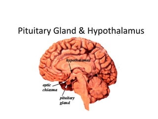
Pituitary Gland
6. • Secretes releasing and inhibiting hormones
that control the release of hormones by the
pituitary gland.
• They reach the pituitary gland via the
hypophyseal portal system.
Hypothalamus
10. a.k.a. Somatotropin
• Most abundant AP hormone
• Produced by somatotrophs
• Account for majority of cells present in AP
• 1° - acts on liver (IGF)
• GHRH stimulates; SST/GHIH inhibits
Human Growth Hormone (hGH/GH)
13. a.k.a. Thyrotopin
• Produced by thyrotrophs
• 1°- acts on thyroid (T3/T4).
In mammals thermogenesis
• Stimulated by TRH
Thyroid-Stimulating Hormone (TSH)
14. a.k.a Follitropin
• Produced by gonadotrophs
• Specifically, FSH-gonadotroph
• 1°- in men: promotes spematogenesis;
in women: follicular growth
(estrogen/progesterone)
• Stimulated by GnRH
Follicle-Stimulating Hormone (FSH)
15. a.k.a. Lutropin
• Produced by gonadotrophs
• Specifically, LH-gonadotroph
• 1° - in men: acts on testes (testosterone);
in women: acts on ovaries (ovulation/CL)
Stimulated by GnRH
Luteinizing Hormone (LH)
16. • Produced by lactotrophs
• A.k.a. mammotrophs
• PRL increases during pregnancy and reaches
maximal values at parturition
• Milk production in post-partum women
• Stimulated by nursing infant
Prolactin (PRL)
17. a.k.a. Corticotropin
• Produced by corticotrophs
• 1° action- stimulates steroid biosynthesis
within the adrenal cortex; cortisol
• Stimulated by CRH
• High cortisol = Cushings Disease
• Low cortisol = Addisons Disease
Adrenocorticotropic Hormone (ACTH)
18. a.k.a. Melanotropin
• Produced by corticotrophs
• Disperse melanin pigment in melanocytes in
the skin
• Not secreted in large amounts by AP
Melanocyte-Stimulating Hormone (MSH)
The pituitary is composed of the:
Anterior pituitary (glandular)
Posterior pituitary (neural)
Alternative names:
Anterior pituitary: adenohypophysis, pars distalis, pars anterior
Posterior pituitary: neurohypophysis, pars nervosa
Pars intermedia – separates anterior from posterior
PVN – paraventricular hypothalamic nucleus
SON – supraoptic hypothalamic nucleus
Hormone secreting cells of the neurohypophysis (posterior pituitary), the adrenal medulla and the pineal gland are regulated by direct neural innervation.
Stimulation of hormone secretion by nerves is referred to as neuroendocrine transduction.
Hypophyseal arteries come off of the carotid artery
The arteries of the pituitary gland arise from the internal carotid arteries as the inferior and superior hypophyseal arteries. The inferior hypophyseal arteries mainly supply the pars nervosa before forming short portal vessels to supply the pars distalis. The superior hypophyseal arteries supply the floor of the hypothalamus, the median eminence. The capillary plexus formed by the superior hypophyseal artery in the median eminence form into long portal vessels which run down in the pituitary stalk to form a capillary plexus in the pars distalis. In the median eminence the capillary plexus receives secretions from releasing factor cells.
The blood is drained from the pituitary by inferior hypophyseal veins into the dural venous sinuses.
Coming from carotid
Superior – anterior pituitary
Inferior – posterior pituitary
Trophic- “nourishing”
Somatostatin (SST)
Growth Hormone Releasing Hormone (GHRH)
SST: decreases GH secretion at AP and by decreasing GHRH
If you have high IGF-I it will feedback to increase SST and then decrease GH and GHRH
Long Loop Feedback – target tissue secretes hormone that affect the pituitary and the hypothalamus
Short Loop Feedback– pituitary (hypophyseal) hormone feeds back to the hypothalamus to shut it down
Ultrashort Feedback – Hormone released from the pituitary shuts itself off.
growth hormone-releasing hormone (GHRH) and growth hormone-inhibiting hormone (GHIH).
Increases LH receptors in Leydig cells (sperm production)
Corpus lutem
Prolactin-inhibiting factor- a.k.a. dopamine via tonic inhibition
Primary Cushings- high cortisol/ Secondary Cushings – high ACTH
Primary Addisons- low cortisol / Secondary Addisons- low ACTH
Cushings Disease
Excessive secretion of ACTH
Characterized by excess cortisol and alterations of glucose metabolism
Hyperpigmentation, wasting of muscle, puffiness (water retention)
Pituitary or hypothalamus tumor
Addison’s Disease
Absence of ACTH secretion
Characterized by decreased cortisol secretion