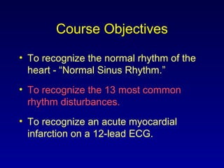
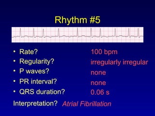
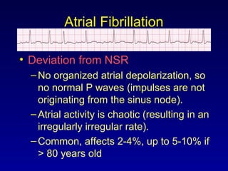
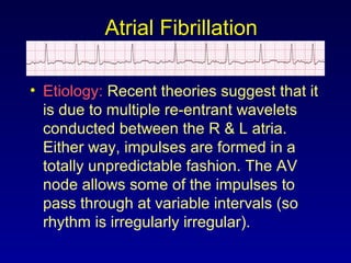
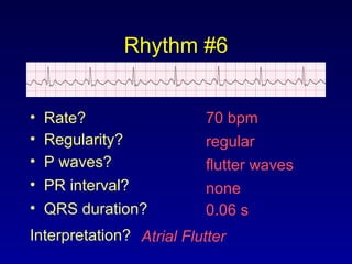
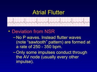
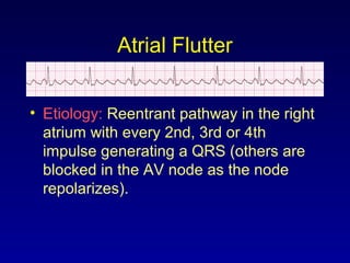
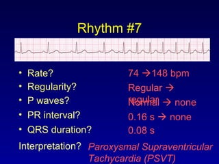
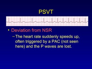
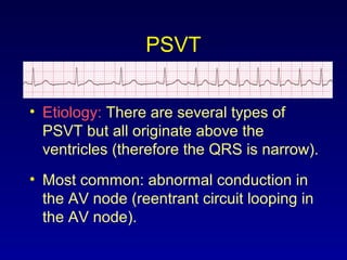
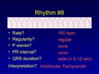
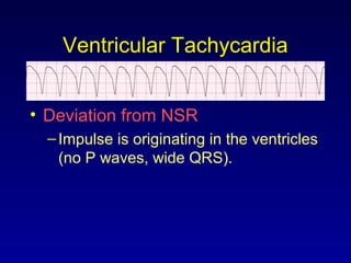
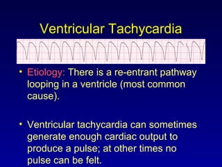
ECG Rhythm Interpretation
Supraventricular and
Ventricular Arrhythmias
Course Objectives
• To recognize the normal rhythm of the
heart - “Normal Sinus Rhythm.”
• To recognize the 13 most common
rhythm disturbances.
• To recognize an acute myocardial
infarction on a 12-lead ECG.
Learning Modules
• ECG Basics
• How to Analyze a Rhythm
• Normal Sinus Rhythm
• Heart Arrhythmias
• Diagnosing a Myocardial Infarction
• Advanced 12-Lead Interpretation
Arrhythmias
• Sinus Rhythms
• Premature Beats
• Supraventricular Arrhythmias
• Ventricular Arrhythmias
• AV Junctional Blocks
Supraventricular Arrhythmias
• Atrial Fibrillation
• Atrial Flutter
• Paroxysmal Supraventricular
Tachycardia
Rhythm #5
100 bpm• Rate?
• Regularity? irregularly irregular
none
0.06 s
• P waves?
• PR interval? none
• QRS duration?
Interpretation? Atrial Fibrillation
Atrial Fibrillation
• Deviation from NSR
–No organized atrial depolarization, so
no normal P waves (impulses are not
originating from the sinus node).
–Atrial activity is chaotic (resulting in an
irregularly irregular rate).
–Common, affects 2-4%, up to 5-10% if
> 80 years old
Atrial Fibrillation
• Etiology: Recent theories suggest that it
is due to multiple re-entrant wavelets
conducted between the R & L atria.
Either way, impulses are formed in a
totally unpredictable fashion. The AV
node allows some of the impulses to
pass through at variable intervals (so
rhythm is irregularly irregular).
Rhythm #6
70 bpm• Rate?
• Regularity? regular
flutter waves
0.06 s
• P waves?
• PR interval? none
• QRS duration?
Interpretation? Atrial Flutter
Atrial Flutter
• Deviation from NSR
–No P waves. Instead flutter waves
(note “sawtooth” pattern) are formed at
a rate of 250 - 350 bpm.
–Only some impulses conduct through
the AV node (usually every other
impulse).
Atrial Flutter
• Etiology: Reentrant pathway in the right
atrium with every 2nd, 3rd or 4th
impulse generating a QRS (others are
blocked in the AV node as the node
repolarizes).
Rhythm #7
74 148 bpm• Rate?
• Regularity? Regular 
regularNormal  none
0.08 s
• P waves?
• PR interval? 0.16 s  none
• QRS duration?
Interpretation? Paroxysmal Supraventricular
Tachycardia (PSVT)
PSVT
• Deviation from NSR
–The heart rate suddenly speeds up,
often triggered by a PAC (not seen
here) and the P waves are lost.
PSVT
• Etiology: There are several types of
PSVT but all originate above the
ventricles (therefore the QRS is narrow).
• Most common: abnormal conduction in
the AV node (reentrant circuit looping in
the AV node).
Ventricular Arrhythmias
• Ventricular Tachycardia
• Ventricular Fibrillation
Rhythm #8
160 bpm• Rate?
• Regularity? regular
none
wide (> 0.12 sec)
• P waves?
• PR interval? none
• QRS duration?
Interpretation? Ventricular Tachycardia
Ventricular Tachycardia
• Deviation from NSR
–Impulse is originating in the ventricles
(no P waves, wide QRS).
Ventricular Tachycardia
• Etiology: There is a re-entrant pathway
looping in a ventricle (most common
cause).
• Ventricular tachycardia can sometimes
generate enough cardiac output to
produce a pulse; at other times no
pulse can be felt.
Rhythm #9
none• Rate?
• Regularity? irregularly irreg.
none
wide, if recognizable
• P waves?
• PR interval? none
• QRS duration?
Interpretation? Ventricular Fibrillation
Ventricular Fibrillation
• Deviation from NSR
–Completely abnormal.
Ventricular Fibrillation
• Etiology: The ventricular cells are
excitable and depolarizing randomly.
• Rapid drop in cardiac output and death
occurs if not quickly reversed

ECG PART 5

  • 1.
    ECG Rhythm Interpretation Supraventricularand Ventricular Arrhythmias
  • 2.
    Course Objectives • Torecognize the normal rhythm of the heart - “Normal Sinus Rhythm.” • To recognize the 13 most common rhythm disturbances. • To recognize an acute myocardial infarction on a 12-lead ECG.
  • 3.
    Learning Modules • ECGBasics • How to Analyze a Rhythm • Normal Sinus Rhythm • Heart Arrhythmias • Diagnosing a Myocardial Infarction • Advanced 12-Lead Interpretation
  • 4.
    Arrhythmias • Sinus Rhythms •Premature Beats • Supraventricular Arrhythmias • Ventricular Arrhythmias • AV Junctional Blocks
  • 5.
    Supraventricular Arrhythmias • AtrialFibrillation • Atrial Flutter • Paroxysmal Supraventricular Tachycardia
  • 6.
    Rhythm #5 100 bpm•Rate? • Regularity? irregularly irregular none 0.06 s • P waves? • PR interval? none • QRS duration? Interpretation? Atrial Fibrillation
  • 7.
    Atrial Fibrillation • Deviationfrom NSR –No organized atrial depolarization, so no normal P waves (impulses are not originating from the sinus node). –Atrial activity is chaotic (resulting in an irregularly irregular rate). –Common, affects 2-4%, up to 5-10% if > 80 years old
  • 8.
    Atrial Fibrillation • Etiology:Recent theories suggest that it is due to multiple re-entrant wavelets conducted between the R & L atria. Either way, impulses are formed in a totally unpredictable fashion. The AV node allows some of the impulses to pass through at variable intervals (so rhythm is irregularly irregular).
  • 9.
    Rhythm #6 70 bpm•Rate? • Regularity? regular flutter waves 0.06 s • P waves? • PR interval? none • QRS duration? Interpretation? Atrial Flutter
  • 10.
    Atrial Flutter • Deviationfrom NSR –No P waves. Instead flutter waves (note “sawtooth” pattern) are formed at a rate of 250 - 350 bpm. –Only some impulses conduct through the AV node (usually every other impulse).
  • 11.
    Atrial Flutter • Etiology:Reentrant pathway in the right atrium with every 2nd, 3rd or 4th impulse generating a QRS (others are blocked in the AV node as the node repolarizes).
  • 12.
    Rhythm #7 74 148bpm• Rate? • Regularity? Regular  regularNormal  none 0.08 s • P waves? • PR interval? 0.16 s  none • QRS duration? Interpretation? Paroxysmal Supraventricular Tachycardia (PSVT)
  • 13.
    PSVT • Deviation fromNSR –The heart rate suddenly speeds up, often triggered by a PAC (not seen here) and the P waves are lost.
  • 14.
    PSVT • Etiology: Thereare several types of PSVT but all originate above the ventricles (therefore the QRS is narrow). • Most common: abnormal conduction in the AV node (reentrant circuit looping in the AV node).
  • 15.
    Ventricular Arrhythmias • VentricularTachycardia • Ventricular Fibrillation
  • 16.
    Rhythm #8 160 bpm•Rate? • Regularity? regular none wide (> 0.12 sec) • P waves? • PR interval? none • QRS duration? Interpretation? Ventricular Tachycardia
  • 17.
    Ventricular Tachycardia • Deviationfrom NSR –Impulse is originating in the ventricles (no P waves, wide QRS).
  • 18.
    Ventricular Tachycardia • Etiology:There is a re-entrant pathway looping in a ventricle (most common cause). • Ventricular tachycardia can sometimes generate enough cardiac output to produce a pulse; at other times no pulse can be felt.
  • 19.
    Rhythm #9 none• Rate? •Regularity? irregularly irreg. none wide, if recognizable • P waves? • PR interval? none • QRS duration? Interpretation? Ventricular Fibrillation
  • 20.
    Ventricular Fibrillation • Deviationfrom NSR –Completely abnormal.
  • 21.
    Ventricular Fibrillation • Etiology:The ventricular cells are excitable and depolarizing randomly. • Rapid drop in cardiac output and death occurs if not quickly reversed