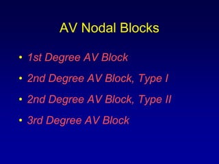
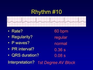
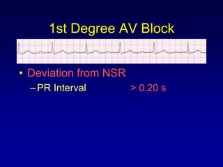
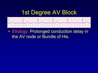
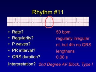
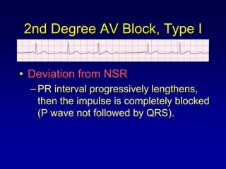
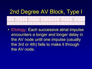
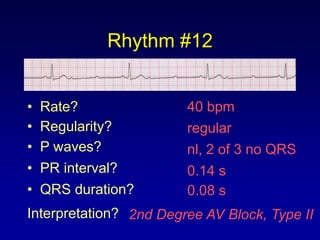
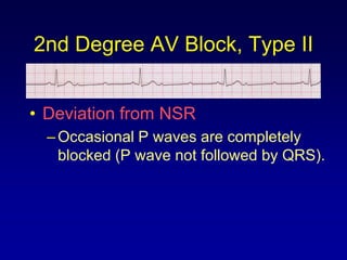
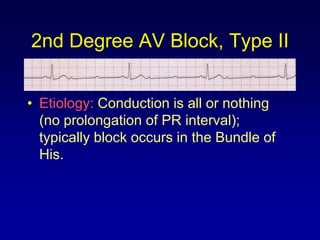
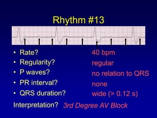
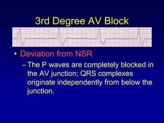
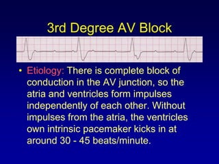
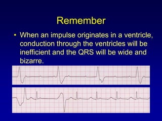
ECG Rhythm Interpretation


        Module IV c

    AV Junctional Blocks
Course Objectives

• To recognize the normal rhythm of the
  heart - “Normal Sinus Rhythm.”
• To recognize the 13 most common
  rhythm disturbances.
• To recognize an acute myocardial
  infarction on a 12-lead ECG.
Learning Modules

•   ECG Basics
•   How to Analyze a Rhythm
•   Normal Sinus Rhythm
•   Heart Arrhythmias
•   Diagnosing a Myocardial Infarction
•   Advanced 12-Lead Interpretation
Arrhythmias

•   Sinus Rhythms
•   Premature Beats
•   Supraventricular Arrhythmias
•   Ventricular Arrhythmias
•   AV Junctional Blocks
AV Nodal Blocks

• 1st Degree AV Block
• 2nd Degree AV Block, Type I
• 2nd Degree AV Block, Type II
• 3rd Degree AV Block
Rhythm #10


•   Rate?               60 bpm
•   Regularity?         regular
•   P waves?            normal
•   PR interval?        0.36 s
•   QRS duration?       0.08 s
Interpretation? 1st Degree AV Block
1st Degree AV Block


• Deviation from NSR
  – PR Interval        > 0.20 s
1st Degree AV Block


• Etiology: Prolonged conduction delay in
  the AV node or Bundle of His.
Rhythm #11


•   Rate?               50 bpm
•   Regularity?         regularly irregular
•   P waves?            nl, but 4th no QRS
•   PR interval?        lengthens
•   QRS duration?       0.08 s
Interpretation? 2nd Degree AV Block, Type I
2nd Degree AV Block, Type I


• Deviation from NSR
  – PR interval progressively lengthens,
    then the impulse is completely blocked
    (P wave not followed by QRS).
2nd Degree AV Block, Type I


• Etiology: Each successive atrial impulse
  encounters a longer and longer delay in
  the AV node until one impulse (usually
  the 3rd or 4th) fails to make it through
  the AV node.
Rhythm #12


•   Rate?               40 bpm
•   Regularity?         regular
•   P waves?            nl, 2 of 3 no QRS
•   PR interval?        0.14 s
•   QRS duration?       0.08 s
Interpretation? 2nd Degree AV Block, Type II
2nd Degree AV Block, Type II


• Deviation from NSR
  – Occasional P waves are completely
    blocked (P wave not followed by QRS).
2nd Degree AV Block, Type II


• Etiology: Conduction is all or nothing
  (no prolongation of PR interval);
  typically block occurs in the Bundle of
  His.
Rhythm #13


•   Rate?               40 bpm
•   Regularity?         regular
•   P waves?            no relation to QRS
•   PR interval?        none
•   QRS duration?       wide (> 0.12 s)
Interpretation? 3rd Degree AV Block
3rd Degree AV Block


• Deviation from NSR
  – The P waves are completely blocked in
    the AV junction; QRS complexes
    originate independently from below the
    junction.
3rd Degree AV Block


• Etiology: There is complete block of
  conduction in the AV junction, so the
  atria and ventricles form impulses
  independently of each other. Without
  impulses from the atria, the ventricles
  own intrinsic pacemaker kicks in at
  around 30 - 45 beats/minute.
Remember
• When an impulse originates in a ventricle,
  conduction through the ventricles will be
  inefficient and the QRS will be wide and
  bizarre.

Ekg module 4c

  • 1.
    ECG Rhythm Interpretation Module IV c AV Junctional Blocks
  • 2.
    Course Objectives • Torecognize the normal rhythm of the heart - “Normal Sinus Rhythm.” • To recognize the 13 most common rhythm disturbances. • To recognize an acute myocardial infarction on a 12-lead ECG.
  • 3.
    Learning Modules • ECG Basics • How to Analyze a Rhythm • Normal Sinus Rhythm • Heart Arrhythmias • Diagnosing a Myocardial Infarction • Advanced 12-Lead Interpretation
  • 4.
    Arrhythmias • Sinus Rhythms • Premature Beats • Supraventricular Arrhythmias • Ventricular Arrhythmias • AV Junctional Blocks
  • 5.
    AV Nodal Blocks •1st Degree AV Block • 2nd Degree AV Block, Type I • 2nd Degree AV Block, Type II • 3rd Degree AV Block
  • 6.
    Rhythm #10 • Rate? 60 bpm • Regularity? regular • P waves? normal • PR interval? 0.36 s • QRS duration? 0.08 s Interpretation? 1st Degree AV Block
  • 7.
    1st Degree AVBlock • Deviation from NSR – PR Interval > 0.20 s
  • 8.
    1st Degree AVBlock • Etiology: Prolonged conduction delay in the AV node or Bundle of His.
  • 9.
    Rhythm #11 • Rate? 50 bpm • Regularity? regularly irregular • P waves? nl, but 4th no QRS • PR interval? lengthens • QRS duration? 0.08 s Interpretation? 2nd Degree AV Block, Type I
  • 10.
    2nd Degree AVBlock, Type I • Deviation from NSR – PR interval progressively lengthens, then the impulse is completely blocked (P wave not followed by QRS).
  • 11.
    2nd Degree AVBlock, Type I • Etiology: Each successive atrial impulse encounters a longer and longer delay in the AV node until one impulse (usually the 3rd or 4th) fails to make it through the AV node.
  • 12.
    Rhythm #12 • Rate? 40 bpm • Regularity? regular • P waves? nl, 2 of 3 no QRS • PR interval? 0.14 s • QRS duration? 0.08 s Interpretation? 2nd Degree AV Block, Type II
  • 13.
    2nd Degree AVBlock, Type II • Deviation from NSR – Occasional P waves are completely blocked (P wave not followed by QRS).
  • 14.
    2nd Degree AVBlock, Type II • Etiology: Conduction is all or nothing (no prolongation of PR interval); typically block occurs in the Bundle of His.
  • 15.
    Rhythm #13 • Rate? 40 bpm • Regularity? regular • P waves? no relation to QRS • PR interval? none • QRS duration? wide (> 0.12 s) Interpretation? 3rd Degree AV Block
  • 16.
    3rd Degree AVBlock • Deviation from NSR – The P waves are completely blocked in the AV junction; QRS complexes originate independently from below the junction.
  • 17.
    3rd Degree AVBlock • Etiology: There is complete block of conduction in the AV junction, so the atria and ventricles form impulses independently of each other. Without impulses from the atria, the ventricles own intrinsic pacemaker kicks in at around 30 - 45 beats/minute.
  • 18.
    Remember • When animpulse originates in a ventricle, conduction through the ventricles will be inefficient and the QRS will be wide and bizarre.