Micturition
•Download as PPTX, PDF•
98 likes•51,381 views

Micturition is the process of urinating that involves two main steps - the bladder filling with urine until tension triggers the micturition reflex, causing the bladder to empty. This reflex is controlled by the spinal cord but can be inhibited or facilitated by the brain. The urinary bladder stores urine and empties through contraction of the detrusor muscle. Urine enters the bladder via the ureters and exits through the urethra. The micturition reflex maintains control of urination but damage to nerves can cause abnormalities like an atonic bladder with no control or an automatic bladder that empties without brain input.

Recommended
More Related Content
What's hot
What's hot (20)
Similar to Micturition
Similar to Micturition (20)
More from DrChintansinh Parmar
More from DrChintansinh Parmar (20)
Recently uploaded
Recently uploaded (20)
Micturition
- 2. Micturition • Micturition is the process by which the urinary bladder empties when it becomes filled. • This involves two main steps: • First, the bladder fills progressively until the tension in its walls rises above a threshold level; • this elicits the second step, which is a nervous reflex called the micturition reflex that empties the bladder or, if this fails, at least causes a conscious desire to urinate. • Although the micturition reflex is an autonomic spinal cord reflex, it can also be inhibited or facilitated by centers in the cerebral cortex or brain stem.
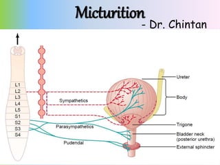
- 3. Physiologic Anatomy • The urinary bladder is a smooth muscle chamber composed of two main parts: • (1) the body, which is the major part of the bladder in which urine collects, and • (2) the neck, which is a funnel-shaped extension of the body, passing inferiorly and anteriorly into the urogenital triangle and connecting with the urethra. • The smooth muscle of the bladder is called the detrusor muscle - contraction of the detrusor muscle is a major step in emptying the bladder
- 5. Innervation of the Bladder • The principal nerve supply of the bladder is by way of the pelvic nerves, which connect with the spinal cord through the sacral plexus, mainly connecting with cord segments S-2 and S-3. • Coursing through the pelvic nerves are both sensory nerve fibers and motor nerve fibers. The sensory fibers detect the degree of stretch in the bladder wall. • The motor nerves transmitted in the pelvic nerves are parasympathetic fibers. These terminate on ganglion cells located in the wall of the bladder. Short postganglionic nerves then innervate the detrusor muscle.
- 6. Innervation of the Bladder • skeletal motor fibers transmitted through the pudendal nerve to the external bladder sphincter - somatic nerve fibers that innervate and control the voluntary skeletal muscle of the sphincter. • the bladder receives sympathetic innervation from the sympathetic chain through the hypogastric nerves, connecting mainly with the L-2 segment of the spinal cord. • These sympathetic fibers stimulate mainly the blood vessels - sensory nerve fibers also pass by way of the sympathetic nerves and important in the sensation of fullness and pain
- 8. Transport of Urine • Urine flowing from the collecting ducts into the renal calyces stretches the calyces and increases their intrinsic pacemaker activity, • which in turn initiates peristaltic contractions that spread to the renal pelvis and then downward along the length of the ureter • peristaltic contractions in the ureter are enhanced by parasympathetic stimulation and • inhibited by sympathetic stimulation
- 9. Transport of Urine • The normal tone of the detrusor muscle in the bladder wall have a tendency to compress the ureter, • thereby preventing backflow of urine from the bladder when pressure builds up in the bladder during micturition or bladder compression • Vesicoureteral reflux – enlargement of the ureters - can increase the pressure in the renal calyces and structures of the renal medulla, causing damage - hydronephrosis
- 10. The Cystometrogram • When there is no urine in the bladder, the intravesicular pressure is about 0, • but by the time 30 to 50 milliliters of urine has collected, the pressure rises to 5 to 10 centimeters of water. • Additional urine — 200 to 300 milliliters — can collect with only a small additional rise in pressure; this constant level of pressure is caused by intrinsic tone of the bladder wall. • Beyond 300 to 400 milliliters, collection of more urine in the bladder causes the pressure to rise rapidly.
- 12. The Cystometrogram • Superimposed on the tonic pressure changes during filling of the bladder are periodic acute increases in pressure that last from a few seconds to more than a minute. • The pressure peaks may rise only a few centimeters of water or may rise to more than 100 centimeters of water. • These pressure peaks are called micturition waves in the Cystometrogram and are caused by the micturition reflex.
- 13. Micturition Reflex • micturition contractions are the result of a stretch reflex initiated by sensory stretch receptors in the bladder wall, • Especially by the receptors in the posterior urethra when this area begins to fill with urine at the higher bladder pressures. • Sensory signals from the bladder stretch receptors are conducted to the sacral segments of the cord through the pelvic nerves • and then reflexively back again to the bladder through the parasympathetic nerve fibers by way of these same nerves.
- 14. Micturition Reflex • When the bladder is only partially filled, these micturition contractions usually relax spontaneously after a fraction of a minute, the detrusor muscles stop contracting, and pressure falls back to the baseline. • As the bladder continues to fill, the micturition reflexes become more frequent and cause greater contractions of the detrusor muscle. • Once a micturition reflex begins, it is “self-regenerative.” • initial contraction of the bladder activates the stretch receptors to cause a greater increase in sensory impulses to the bladder and posterior urethra, which causes a further increase in reflex contraction of the bladder
- 15. Micturition Reflex • cycle is repeated again and again until the bladder has reached a strong degree of contraction. • Then, after a few seconds to more than a minute, the self- regenerative reflex begins to fatigue and the regenerative cycle of the micturition reflex stops, permitting the bladder to relax. • the micturition reflex is a single complete cycle of • (1) progressive and rapid increase of pressure, • (2) a period of sustained pressure, and • (3) return of the pressure to the basal tone of the bladder
- 16. Micturition Reflex • Once a micturition reflex has occurred but has not succeeded in emptying the bladder, • the nervous elements of this reflex usually remain in an inhibited state for a few minutes to 1 hour or more before another micturition reflex occurs. • As the bladder becomes more and more filled, micturition reflexes occur more and more often and more and more powerfully.
- 17. Micturition Reflex • Once the micturition reflex becomes powerful enough, it causes another reflex, which passes through the pudendal nerves to the external sphincter to inhibit it. • If this inhibition is more potent in the brain than the voluntary constrictor signals to the external sphincter, urination will occur. • If not, urination will not occur until the bladder fills still further and the micturition reflex becomes more powerful.
- 18. Role of the Brain • The micturition reflex is a completely autonomic spinal cord reflex, but it can be inhibited or facilitated by centers in the brain. • These centers include • (1) strong facilitative and inhibitory centers in the brain stem, located mainly in the pons, and • (2) several centers located in the cerebral cortex that are mainly inhibitory but can become excitatory. • The micturition reflex is the basic cause of micturition, but the higher centers normally exert final control of micturition
- 19. Role of the Brain • 1. The higher centers keep the micturition reflex partially inhibited, except when micturition is desired. • 2. The higher centers can prevent micturition, even if the micturition reflex occurs, by continual tonic contraction of the external bladder sphincter until a convenient time presents itself. • 3. When it is time to urinate, the cortical centers can facilitate the sacral micturition centers to help initiate a micturition reflex • and at the same time inhibit the external urinary sphincter so that urination can occur.
- 20. Voluntary urination • First, a person voluntarily contracts his or her abdominal muscles, which increases the pressure in the bladder • and allows extra urine to enter the bladder neck and posterior urethra under pressure, thus stretching their walls. • This stimulates the stretch receptors, which excites the micturition reflex and simultaneously inhibits the external urethral sphincter. • Ordinarily, all the urine will be emptied, with rarely more than 5 to 10 milliliters left in the bladder.
- 21. Abnormalities of Micturition • Atonic Bladder Caused by Destruction of Sensory Nerve Fibers - preventing transmission of stretch signals from the bladder. • person loses bladder control, despite intact efferent fibers from the cord to the bladder and despite intact neurogenic connections within the brain. • Instead of emptying periodically, the bladder fills to capacity and overflows a few drops at a time through the urethra - overflow incontinence. • crush injury to the sacral region of the spinal cord - syphilis can cause constrictive fibrosis around the dorsal root nerve fibers (tabes dorsalis)
- 22. Abnormalities of Micturition • Automatic Bladder Caused by Spinal Cord Damage Above the Sacral Region. • If the sacral cord segments are still intact, typical micturition reflexes can still occur but they are no longer controlled by the brain. • first micturition reflexes are suppressed because of the state of “spinal shock” • if the bladder is emptied periodically by catheterization, the excitability of the micturition reflex gradually increases - then, periodic automatic bladder emptying occurs. • Some patients can still control urination in this condition by stimulating the skin (scratching or tickling) in the genital region, which sometimes elicits a micturition reflex.
- 23. Abnormalities of Micturition • Uninhibited Neurogenic Bladder Caused by Lack of Inhibitory Signals from the Brain. • frequent and relatively uncontrolled micturition. • partial damage in the spinal cord or the brain stem that interrupts most of the inhibitory signals. • facilitative impulses passing continually down the cord keep the sacral centers so excitable that even a small quantity of urine elicits an uncontrollable micturition reflex, thereby promoting frequent urination.