SlideShare a Scribd company logo
1 of 100
Dr. ChintanDr. Chintan
Two Mechanisms which control respirationTwo Mechanisms which control respiration
1) Neural Mechanism
2) Chemical Mechanism
Neural control of respiration
1) Voluntary control of respiration
2) Automatic control of respiration
3) Reflex control of respiration
Voluntary control of respiration
Respiration is under automatic or involuntary control
basically.
But to some extent it can be controlled voluntary as
most of the respiratory muscles are voluntary
muscles.
Respiration can be modified both in rate and depth
at will for a specific period only.
Voluntary hyperventilation or hypoventilation,
breath holding, forceful inspiration or expiration
efforts etc, are examples of this.
Centre for voluntary control isCentre for voluntary control is motor cortexmotor cortex
Centre is in motor cortex which sends impulses via
corticospinal tract to the respiratory muscles
For inspiration, Phrenic nerve motor neurons in
C3,4,5 and external intercostal muscles motor
neurons in ventral horn cells of T1,2 stimulate the resp
muscles
Corticobulbar tract which modifies activity of
medullary neurons
Ondine’s curseOndine’s curse
In conditions like Bulbar Poliomyelitis, Tumors
of brain stem etc automatic control is lost and
voluntary control is present.
This is because impulses arising from brain stem
control automatic respiration.
This is known as Ondine’s curse.
Death is due to exhaustion.
Automatic control of respirationAutomatic control of respiration
The center is located in brainstem in pons and medulla
bilaterally.
 It is divided into three major collections of neurons:
(1) a dorsal respiratory group, located in the dorsal portion of
the medulla, which mainly causes inspiration;
(2) a ventral respiratory group, located in the ventrolateral
part of the medulla, which mainly causes expiration;
(3) the pneumotaxic center, located dorsally in the superior
portion of the pons, which mainly controls rate and depth of
breathing.
4) Apneustic center in lower
pons.
It is thought to have an
excitatory effect on Inspiratory
neurons .
Respiratory centersRespiratory centers
Experimental studies done on cat toExperimental studies done on cat to
show the centersshow the centers
1) Section A – made above Pons- normal respiration-
eupnoea.
2) Section B - made below medulla- stops respiration
– apnoea.
3) Section C - in pons between Pneumotaxic center
and Apneustic center - Apneustic breathing,
4) Section D - below Apneustic but above medulla-
more or less respiration continues rhythmically
showing evidence of reciprocal innervation between
inspiratory and expiratory neurons.
DRG- in NTS - controls inspiration and rhythmDRG- in NTS - controls inspiration and rhythm
The dorsal respiratory group of neurons extends most of the
length of the medulla.
 Most of its neurons are located within the nucleus of the
tractus solitarius, although additional neurons in the
adjacent reticular substance of the medulla also play
important roles in respiratory control.
The NTS is the sensory termination of both the vagal and
the glossopharyngeal nerves, which transmit sensory signals
into the respiratory center from
(1) peripheral chemoreceptors, (2) baroreceptors, and
(3) several types of receptors in the lungs.
Rhythmical inspiratory discharges from DRGRhythmical inspiratory discharges from DRG
The basic rhythm of respiration is generated
mainly in the dorsal respiratory group of neurons.
Even when all the peripheral nerves entering
the medulla have been sectioned and the brain
stem transected both above and below the
medulla,
this group of neurons still emits repetitive
bursts of inspiratory neuronal action
potentials.
Inspiratory Ramp SignalInspiratory Ramp Signal
The nervous signal that is transmitted to the
inspiratory muscles, mainly the diaphragm, is not an
instantaneous burst of action potentials.
Instead, in normal respiration, it begins weakly and
increases steadily in a ramp manner for about 2
seconds. Then it ceases abruptly for approximately
the next 3 seconds,
which turns off the excitation of the diaphragm
and allows elastic recoil of the lungs and the chest wall
to cause expiration.
Ramp signal advantageousRamp signal advantageous
The inspiratory signal begins again for another
cycle; this cycle repeats again and again, with
expiration occurring in between - ramp signal.
It causes a steady increase in the volume of
the lungs during inspiration, rather than
inspiratory gasps.
Two qualities of inspiratory rampTwo qualities of inspiratory ramp
controlled as followscontrolled as follows
1. Control of the rate of increase of the ramp
signal, so that during heavy respiration, the ramp
increases rapidly and therefore fills the lungs rapidly.
2. Control of the limiting point at which the ramp
suddenly stops.
This is the usual method for controlling the rate of
respiration;
the earlier the ramp ceases → the shorter the
duration of inspiration → shortens the duration of
expiration → the frequency of respiration is
increased
Newer conceptNewer concept
Rhythmic respiration is initiated by a small group of
synaptically coupled pacemaker cells in the
pre-Botzinger complex on either side of the medulla
between the nucleus ambiguous and the lateral
reticular nucleus.
These neurons discharge rhythmically, and they
produce rhythmic discharges in phrenic motor
neurons that are abolished by sections between the
pre-Botzinger complex and these motor neurons.
They also contact the hypoglossal nuclei, and the
tongue is involved in the regulation of airway
resistance.
Pre Botzinger areaPre Botzinger area
Rhythmic discharge (tracing
below) of neurons in the
pre-Botzinger complex (pre-
BOTC) in a brain slice
from a neonatal rat.
IO, inferior olive;
LRN, lateral reticular
nucleus;
NA, nucleus ambiguous;
XII, nucleus of 12th cranial
nerve;
5SP, spinal nucleus of
trigeminal nerve
VRG- functions in both inspiration andVRG- functions in both inspiration and
expirationexpiration
Located in each side of the medulla,
about 5 millimeters anterior and
lateral to the dorsal respiratory group
of neurons, is the ventral respiratory
group of neurons,
found in the nucleus ambiguous
rostrally and the nucleus
retroambiguous caudally.
VRG differs functionally from DRG as followsVRG differs functionally from DRG as follows
1) The neurons of the ventral respiratory group
remain almost totally inactive during normal
quiet respiration.
normal quiet breathing is caused only by
repetitive inspiratory signals from the dorsal
respiratory group transmitted mainly to the
diaphragm,
and expiration results from elastic recoil of the
lungs and thoracic cage.
2) When the respiratory drive for increased
pulmonary ventilation becomes greater than
normal,
respiratory signals spill over into the
ventral respiratory neurons from the basic
oscillating mechanism of the dorsal respiratory
area.
 As a consequence, the ventral respiratory area
contributes extra respiratory drive
3) Electrical stimulation of a few of the neurons
in the ventral group causes inspiration,
whereas stimulation of others causes
expiration.
These neurons contribute to both inspiration
and expiration.
They are especially important in providing the
powerful expiratory signals to the
abdominal muscles during very heavy
expiration.
Newer conceptNewer concept
Both DRG and VRG are interconnected
and equally important for rhythmic
respiration along with several other
neurons in the vicinity.
There is reciprocal innervation
between I and E neurons.
Pneumotaxic centerPneumotaxic center
It Limits the Duration of Inspiration and Increases
the Respiratory Rate.
It is located dorsally in the nucleus Para brachialis
of the upper pons, transmits signals to the
inspiratory area.
The primary effect of this center is to control the
“switch-off” point of the inspiratory ramp, thus
controlling the duration of the filling phase of the lung
cycle.
Limits duration of inspirationLimits duration of inspiration
When the pneumotaxic signal is
strong, inspiration might last for as
little as 0.5 second, thus filling the
lungs only slightly;
 when the Pneumotaxic signal is
weak, inspiration might continue for
5 or more seconds, thus filling the
lungs with a great excess of air.
Limitation of inspiration affects rate ofLimitation of inspiration affects rate of
respirationrespiration
A secondary effect of increasing the rate of
breathing, because limitation of inspiration also
shortens expiration and the entire period of each
respiration.
A strong pneumotaxic signal can increase the
rate of breathing to 30 to 40 breaths per
minute,
 Whereas a weak pneumotaxic signal may
reduce the rate to only 3 to 5 breaths per
minute.
Pneumotaxic center alsoPneumotaxic center also
inhibits the Apneustic centerinhibits the Apneustic center
Thus it prevents Apneusis
which is a type of arrested
breathing.
Apneustic centerApneustic center
It is located in in lower part of Pons.
It stimulates the inspiratory center and increases the
inspiration.
It gets feedback from vagal afferents and also from other
respiratory centers .
Pneumotaxic center inhibits it.
It works in association with Pneumotaxic center to
control depth of inspiration.
Its function is demonstrated by sectioning of vagus
nerve to medulla and by blocking the connection
from Pneumotaxic center by transecting PONS in
mid region.
This causes loss of inhibitory control over
Apneustic center which sends signals to DRG and
prevents switch off of ramp signals
so lungs become completely filled with air and
only occasional expiratory gasps occur.
Thus if vagus is intact regular
rhythm of respiration continues.
If vagus is cut arrest of
respiration occurs called
Apneusis.
Reflex control of respirationReflex control of respiration
1) By receptors inside respiratory system
a) Hering Breuer Reflexes due to stretch receptors in
muscular wall of bronchi and bronchioles,
b) pulmonary irritant receptors,
c) J receptors
2) By receptors outside respiratory system
A) Those which stimulate respiration -
proprioceptors, nociceptors, thermoceptors, emotional
stimuli from hypothalamus and limbic system.
B)Those which inhibit respiration- Baroreceptors
and visceroreceptors
Hering Breuer ReflexesHering Breuer Reflexes
Located in the muscular portions of
the walls of the bronchi and
bronchioles throughout the lungs are
stretch receptors
transmit signals through the vagi
into the dorsal respiratory group of
neurons when the lungs become
overstretched.
These signals act in same way as signalsThese signals act in same way as signals
from Pneumotaxic centerfrom Pneumotaxic center
 when the lungs become overly inflated,
the stretch receptors activate an
appropriate feedback response that
“switches off” the inspiratory ramp and thus
stops further inspiration.
This is called the Hering - Breuer
inflation reflex. This reflex also increases
the rate of respiration, as is true for
signals from the pneumotaxic center
Importance of H-B inflation reflexImportance of H-B inflation reflex
1) This reflex operates only when Tidal
volume exceeds 1.5 liters as in exercise
and not in normal ventilation and so
prevents over inflation of lungs and is thus
protective
2) This effect is not seen in man if vagi
are cut and also so in transplanted lungs
which are denervated.
Hering Breuer Deflation reflexHering Breuer Deflation reflex
It is seen when there is extreme deflation of
lungs as seen in collapse of lungs, pneumothorax,
hydrothorax, etc.
The receptors are stimulated by change in shape
of small airways in collapsed area so also called as
compression receptors.
So forceful deflation of lungs causes stimulation
of receptors → stimulation of inspiration via
vagus
Effect of irritant receptors in theEffect of irritant receptors in the
airwaysairways
The epithelium of the trachea, bronchi and
bronchioles is supplied with sensory nerve
endings called pulmonary irritant receptors and are
rapidly adapting and innervated by myelinated vagal
nerve fibers.
They are stimulated by many incidents like lung
hyperinflation, exogenous subs like SO2, smoke
and endogenous subs like histamine, PGs.
These cause hyperpnoea, coughing and sneezing,
bronchial constriction and mucus secretion in
diseases as asthma and emphysema.
Function of ‘J receptors’Function of ‘J receptors’
A few sensory nerve endings have been described in
the alveolar walls in juxtaposition to the
pulmonary capillaries—hence the name “J
receptors.” they are non myelinated nerve endings
(C fibers).
They are stimulated especially when the pulmonary
capillaries become engorged with blood or when
pulmonary edema occurs as in congestive heart
failure.
 Although the functional role of the J receptors is not
clear, their excitation may give the person a feeling of
dyspnea
J receptors cause the pulmonaryJ receptors cause the pulmonary
chemo reflexchemo reflex
Apnea followed by Hyperventilation,
bradycardia, hypotension,
bronchoconstriction, and mucus secretion
and skeletal muscle weakness (Seen in
Bhopal gas tragedy).
This is similar to Coronary Chemo reflex
or Bezold Jarisch Reflex
Receptors outside respiratory systemReceptors outside respiratory system
which stimulate respirationwhich stimulate respiration
1) Proprioceptors –
Present in muscles, tendons, joints etc
when stimulated reflexly stimulates
Inspiratory neurons.
This helps to increase ventilation at the
start of exercise even before changes in
Pco2, Po2 and H ions concentration occur.
2) Nociceptors - Pain receptors
3) Emotional stimuli – all stimulate
respiratory system via hypothalamus and
limbic system.
4) Chemoreceptors – stimulated by
acidosis and hypoxia. Hypotension and
shock produce hyperventilation due to
stimulation of chemoreceptors.
5) Thermoreceptors – present in
hypothalamus are stimulated when there
is increase in body temperature as in fever,
exercise
they send impulses to respiratory center
and produce increase in rate of
respiration.
Those which inhibit respirationThose which inhibit respiration
1) Baroreceptors – inhibit reflexly.
Adrenaline apnea is an eg of this.
2) Visceroreceptors – visceral reflexes like
vomiting, swallowing, etc reflexly inhibit
respiration, Eg is Deglutition apnea
Other factors affecting respirationOther factors affecting respiration
1) Sleep-
Respiration is less rigorously controlled during sleep
than in the waking state, and brief periods of apnea occur
in normal sleeping adults.
Sleep Apnea Syndrome
It is a type of obstructive sleep apnea.
It occurs at any age.
Cause is failure of Genioglossus muscle to contract
during inspiration which causes the tongue to fall
backward to obstruct the airways. This muscle normally
keeps the tongue forward.
S/S are morning headache, fatigue,
hypoxemic, Hypercapneic condition
Prevention by avoiding sedatives or
respiratory depressants prior to sleep,
breathing in prone and avoiding
sleeping on back, Positive pressurePositive pressure
controlled O2 inhalationcontrolled O2 inhalation.
Sudden Infant DeathSudden Infant Death
SyndromeSyndrome
A type of central sleep apnea due to
failure of respiratory centers to
produce neural impulses so causing
total stoppage of respiration.
Effect of brain edemaEffect of brain edema
The activity of the respiratory center may
be depressed or even inactivated by acute
brain edema resulting from brain concussion.
the head might be struck against some solid
object, after which the damaged brain tissues
swell,
compressing the cerebral arteries against
the cranial vault and thus partially
blocking cerebral blood supply.
TreatmentTreatment
Respiratory depression resulting from brain
edema can be relieved temporarily by
intravenous injection of hypertonic
solutions such as highly concentrated mannitol
solution.
These solutions osmotically remove some of
the fluids of the brain, thus relieving
intracranial pressure and sometimes
re-establishing respiration within a few
minutes
Effect of Anesthesia- respiratoryEffect of Anesthesia- respiratory
depressiondepression
The most prevalent cause of respiratory
depression and respiratory arrest is over
dosage with anesthetics or narcotics.
 For instance, sodium pentobarbital
depresses the respiratory center
considerably more than many other
anesthetics, such as halothane.
At one time, morphine was used as an
anesthetic, but this drug is now used
only as an adjunct to anesthetics
because it greatly depresses the
respiratory center while having less
ability to anesthetize the cerebral
cortex
Chemical regulation of respirationChemical regulation of respiration
The ultimate goal of respiration is to maintain proper
concentrations of oxygen, carbon dioxide, and
hydrogen ions in the tissues.
Respiratory activity is highly responsive to changes in
each of these.
Excess carbon dioxide or excess hydrogen ions in
the blood mainly act directly on the respiratory center
itself,
causing greatly increased strength of both the
inspiratory and the expiratory motor signals to the
respiratory muscles.
Oxygen acts via peripheralOxygen acts via peripheral
ChemoreceptorsChemoreceptors
 Oxygen, in contrast, does not have a
significant direct effect on the respiratory
center of the brain in controlling respiration.
Instead, it acts almost entirely on peripheral
chemoreceptors located in the carotid and
aortic bodies,
these in turn transmit appropriate nervous
signals to the respiratory center for control of
respiration
CO2 and H ions act directly on centralCO2 and H ions act directly on central
chemoreceptorschemoreceptors
Chemo sensitive area (CSA) is located
bilaterally, lying only 0.2 millimeter beneath
the ventral surface of the medulla.
This area is highly sensitive to changes in
either blood Pco2 or hydrogen ion
concentration, and it in turn excites the other
portions of the respiratory center
Excitation of CSA sensor neurons byExcitation of CSA sensor neurons by HH
ions is the primary stimulusions is the primary stimulus
Hydrogen ions do not easily cross the blood-
brain barrier.
changes in hydrogen ion concentration in the
blood have considerably less effect in stimulating
the chemo sensitive neurons than do changes in
blood carbon dioxide,
carbon dioxide stimulate these neurons secondarily
by changing the hydrogen ion concentration.
CO2 has more potent effect than H ions
Chemo sensitive areaChemo sensitive area
Decreased stimulatory effect of CO2Decreased stimulatory effect of CO2
after first 1 to 2 daysafter first 1 to 2 days
Excitation of the respiratory center by carbon dioxide is
great for the first few hours after the blood carbon
dioxide first increases,
but then it gradually declines over the next 1 to 2 days,
decreasing to about one fifth the initial effect.
It results from renal readjustment of the hydrogen ion
concentration in the circulating blood back toward
normal after the carbon dioxide first increases the hydrogen
concentration.
The kidneys achieve this by increasing the blood
bicarbonate, which binds with the hydrogen ions in the
blood and cerebrospinal fluid to reduce their concentrations
Diffusion of Bicarbonate ions- combineDiffusion of Bicarbonate ions- combine
with H ions & decreasewith H ions & decrease
2) Over a period of hours, the
bicarbonate ions also slowly diffuse
through the blood - brain and blood –
CSF barriers and combine directly with
the hydrogen ions adjacent to the
respiratory neurons as well,
thus reducing the hydrogen ions back to
near normal.
A change in blood carbon dioxide
concentration therefore has a
potent acute effect on controlling
respiratory drive but only a weak
chronic effect
after a few days’ adaptation
Unimportance of oxygen for control ofUnimportance of oxygen for control of
respiratory centrerespiratory centre
Changes in oxygen concentration
have virtually no direct effect on the
respiratory center itself to alter
respiratory drive
 although oxygen changes do have an
indirect effect, acting through the
peripheral chemoreceptors
Hb as a good bufferHb as a good buffer
The hemoglobin oxygen buffer system
delivers almost normal amounts of oxygen
to the tissues even,
when the pulmonary Po2 changes from a
value as low as 60 mm Hg up to a value as
high as 600 mm Hg.
Chemoreceptors - O2Chemoreceptors - O2
The carotid bodies are located bilaterally in the
bifurcations of the common carotid arteries.
Their afferent nerve fibers pass through Herring's
nerves to the glossopharyngeal nerves and then to
the dorsal respiratory area of the medulla.
 The aortic bodies are located along the arch of the
aorta;
their afferent nerve fibers pass through the vagi, also
to the dorsal medullary respiratory area.
Stimulation of chemoreceptors byStimulation of chemoreceptors by
decreased arterial Po2decreased arterial Po2
Shows the effect of different
levels of arterial Po2 on the
rate of nerve impulse
transmission from a carotid
body.
the impulse rate is particularly
sensitive to changes in
arterial Po2 in the range of
60 down to 30 mm Hg,
a range in which hemoglobin
saturation with oxygen
decreases rapidly
Effect of CO2 and H ion concentrationEffect of CO2 and H ion concentration
on chemoreceptorson chemoreceptors
Indirect effect- An increase in either carbon
dioxide concentration or hydrogen ion
concentration also excites the peripheral
chemoreceptors and, in this way, indirectly
increases respiratory activity.
However, the direct effects of both these
factors in the respiratory center itself via CSA
are much more powerful than peripheral
ones.
One difference between peripheral andOne difference between peripheral and
central effects of CO2 & H ionscentral effects of CO2 & H ions
The stimulation by way of the peripheral chemoreceptors
occurs as much as five times as rapidly as central
stimulation.
So that the peripheral chemoreceptors might be
especially important in increasing the rapidity of
response to carbon dioxide at the onset of exercise.
Thus direct effect via CSA is very powerful than indirect
via peripheral chemoreceptors.
But indirect effect is very rapid than direct effect.
Basic mechanism of stimulation of chemoreceptorsBasic mechanism of stimulation of chemoreceptors
by o2 deficiency – NT Dopamineby o2 deficiency – NT Dopamine
Mechanism of release of NTsMechanism of release of NTs
Type I glomus cells have O2-sensitive K+
channels, whose conductance is reduced in
proportion to the degree of hypoxia to which they
are exposed.
This reduces the K+ efflux, depolarizing the
cell and causing Ca2+ influx, primarily via L-type
Ca2+ channels.
The Ca2+ influx triggers action potentials and
transmitter release, with consequent excitation
of the afferent nerve endings
Low Po2 stimulates ventilation whenLow Po2 stimulates ventilation when
co2& H ions constantco2& H ions constant
Only the ventilatory drive due to the effect of
low oxygen on the chemoreceptors is active.
There is almost no effect on ventilation as
long as the arterial Po2 remains greater than
100 mm Hg.
But at pressures lower than 100 mm Hg,
ventilation approximately doubles when
the arterial Po2 falls to 60 mm Hg
AcclimatizationAcclimatization
Chronic Breathing of Low Oxygen Stimulates
Respiration Even More—The Phenomenon of
“Acclimatization”
Mountain climbers have found that when they
ascend a mountain slowly, over a period of
days rather than a period of hours,
they breathe much more deeply and
therefore can withstand far lower atmospheric
oxygen concentrations than when they ascend
rapidly.
Reason for acclimatizationReason for acclimatization
Within 2 to 3 days, the respiratory center in the
brain stem loses about four fifths of its
sensitivity to changes in Pco2 and hydrogen
ions.
Therefore, the excess ventilatory blow-off of
carbon dioxide that normally would inhibit an
increase in respiration fails to occur,
and low oxygen can drive the respiratory
system to a much higher level of alveolar
Instead of the 70 per cent increase in
ventilation that might occur after acute
exposure to low oxygen,
the alveolar ventilation often increases
400 to 500 per cent after 2 to 3 days of
low oxygen;
this helps greatly in supplying additional
oxygen to the mountain climber
Effect of exercise on respirationEffect of exercise on respiration
regulationregulation
In exercise there is rise of co2 formation
and more of o2 consumption.
Neurogenic stimulation by higher
centers and proprioceptors is first most
important mechanism to increase
ventilation on initiation of exercise
even before chemical stimulation
develops.
If nervous signals are too weak or
too strong then chemical factors
play an important role in respiratory
adjustment in exercise.
1) At the beginning of exercise
anticipatory stimulation of
respiration occurs thus increasing
alveolar ventilation so that it
decreases arterial Pco2.
2) After 30 to 40 seconds the amount of
co2 released by exercising muscles into
blood matches approximately with
increase rate of alveolar ventilation and
arterial Pco2 returns to normal.
Neurogenic factor in exercise shifts the
ventilation to higher levels to match the
rate of co2 formation and o2 utilization
thus keeping Po2 and Pco2 toward normal
level.
Factors causing Increased ventilationFactors causing Increased ventilation
in exercisein exercise
1) Higher centers – motor cortex,
hypothalamus, which cause rise of
ventilation even before exercise, stimulate
VMC increasing BP.
2) Sympathetic stimulation can further
stimulate.
3) Increase body temperature
4) Sensory proprioceptors
5) Low P02, high Pco2 and H ions via
peripheral chemoreceptors.
6) Increase in plasma K during exercise
stimulates peripheral chemoreceptors.
7) Lactic acid liberated in exercise
increases co2 which stimulates
respiration.
8) Better match of ventilation –
Effect of acid base changesEffect of acid base changes
Acidosis – H ions conc- stimulation↑ ↑
Alkalosis- H ions conc- stimulation↓ ↓
In metabolic acidosis as seen in diabetes,
renal failure, severe muscular exercise,
starvation etc more H ions and less HCO3
in blood which stimulate respiration
(Kussumaul breathing)
which reduces co2 which leads to
compensatory fall in blood H ion conc.
In metabolic alkalosis as in
vomiting with loss of HCL due to
pyloric or high intestinal obstruction
there is less H ions and more HCO3
so ventilation is depressed and
arterial PCO2 increases which
brings H ions towards normal.
Respiratory acidosis- Here ventilation
is depressed due to causes like
emphysema, depression of respiratory
center, morphine or COPD.
Here hypoventilation causes rise of co2
which stimulates central and peripheral
chemoreceptors
and increases alveolar ventilation and
correction of respiratory acidosis.
Respiratory alkalosis - as
seen
in voluntary hyperventilation,
chronic lack of o2 at high
altitudes,
anxiety,
hysteria
salicylate over dosage etc.
Breath holdingBreath holding
 Respiration can be voluntarily inhibited for some time, but
eventually the voluntary control is overridden.
The point at which breathing can no longer be voluntarily
inhibited is called the breaking point.
 Breaking is due to the rise in arterial PCO2 and the fall in
PO2.
 Individuals can hold their breath longer after removal of the
carotid bodies.
Breathing 100% oxygen before breath holding raises
alveolar PO2 initially, so that the breaking point is delayed.
The same is true of hyperventilating room air, because CO2
is blown off and arterial PCO2 is lower at the start.
Reflex or mechanical factors appear to
influence the breaking point
Subjects who breathe a gas mixture low in
CO2 and high in O2 can hold their breath
for an additional 20 seconds or more.
Psychological factors also play a role, and
subjects can hold their breath longer when
they are told their performance is very good
Hormonal effects on respirationHormonal effects on respiration
Ventilation is increased during the luteal phase
of the menstrual cycle and during pregnancy
Experiments with animals indicate that this is
due to activation of estrogen - dependent
progesterone receptors in the
hypothalamus.
However, the physiologic significance of this
increased ventilation is unknown.
Periodic breathingPeriodic breathing
An abnormality of respiration called periodic
breathing occurs in a number of disease conditions.
The person breathes deeply for a short interval
and then breathes slightly or not at all for an
additional interval, with the cycle repeating over
and over.
One type of periodic breathing, Chyne-Stokes
breathing, is characterized by slowly waxing and
waning respiration occurring about every 40 to
60 seconds.
Chyne- Stokes breathingChyne- Stokes breathing
Apnea followed by hyperventilationApnea followed by hyperventilation
MechanismMechanism
Hyperventilation causes
- Po2 and wash out of co2↑
– which causes depression of
respiration and so apnoea
--- which now increases Pco2 and
reduces Po2
--- which now stimulates ventilation
again.
The basic cause of Cheyne-Stokes breathing
occurs in everyone. However, under normal
conditions, this mechanism is highly
“damped.”
That is, the fluids of the blood and the
respiratory center control areas have large
amounts of dissolved and chemically bound
carbon dioxide and oxygen.
 Therefore, normally, the lungs cannot build up
enough extra carbon dioxide or depress the
oxygen sufficiently in a few seconds to cause the
next cycle of the periodic breathing
CausesCauses
Physiological
Voluntary
hyperventilation
Healthy infants
and adults during
sleep
High altitude
hypoxia
Pathological
Uremia
Cardiac
failure(LVF)
Brain damage
In cardiac failure (LVF)- Slow blood flow- so
delayed gas transport from lungs to brain.
In LVF
- Pulmonary congestion so hampering gas
exchange
– hypoxia
— stimulates resp center
--- increase vent
– washout of co2 and more o2
– due to sluggish blood flow blood with low co2
takes more time to reach brain resp centers so
— apnea.
The brain damage often turns off the
respiratory drive entirely for a few
seconds;
 then an extra intense increase in blood
carbon dioxide turns it back on with
great force.
Cheyne-Stokes breathing of this type is
frequently a preface to death from brain
malfunction.
Biot’s breathingBiot’s breathing
Respiration characterized by alternate
Eupnoea and Apnea.
3-4 cycles of normal breathing followed
by abrupt apnea and again abrupt onset
of normal respiration.
 Seen in meningitis, severe brain
damage and diseases affecting medulla.
Kussumaul’s breathingKussumaul’s breathing
Seen in metabolic acidosis
Rapid and deep breathing without uneasiness.
Acidosis – H ions conc- stimulation↑ ↑
Alkalosis- H ions conc- stimulation↓ ↓
metabolic acidosis seen in diabetes, renal failure, severe
muscular exercise, starvation etc more H ions and less
HCO3 in blood which stimulate respiration
(Kussumaul's breathing)
which reduces co2 which leads to compensatory fall in
blood H ion conc.
Thanq…Thanq…

More Related Content

What's hot

Non respiratory functions of lungs
Non respiratory functions of lungsNon respiratory functions of lungs
Non respiratory functions of lungsAli Faris
 
JUXTA GLOMERULAR apparatus
JUXTA GLOMERULAR apparatus JUXTA GLOMERULAR apparatus
JUXTA GLOMERULAR apparatus akash chauhan
 
Physiology of gas exchange
Physiology of gas exchangePhysiology of gas exchange
Physiology of gas exchangeHimanshu Jangid
 
Mechanism of breathing
Mechanism of breathingMechanism of breathing
Mechanism of breathingKamal Bharathi
 
Respiratory physiology
Respiratory physiologyRespiratory physiology
Respiratory physiologyChy Yong
 
5) gas exchange and diffusion
5) gas exchange and diffusion5) gas exchange and diffusion
5) gas exchange and diffusionAyub Abdi
 
Control of respiration
Control of respirationControl of respiration
Control of respirationVkas Subedi
 
Regulation of respiration (the guyton and hall physiology)
Regulation of respiration (the guyton and hall physiology)Regulation of respiration (the guyton and hall physiology)
Regulation of respiration (the guyton and hall physiology)Maryam Fida
 
Lung volume and capacities
Lung volume and capacitiesLung volume and capacities
Lung volume and capacitiesMuhammadasif909
 
Arterial blood pressure regulation
Arterial blood pressure regulationArterial blood pressure regulation
Arterial blood pressure regulationLubna Abu Alrub,DDS
 
Properties of cardiac muscle
Properties of cardiac muscleProperties of cardiac muscle
Properties of cardiac muscleDr Sara Sadiq
 
Chemical control of respiration
Chemical control of respirationChemical control of respiration
Chemical control of respirationDr Sara Sadiq
 
Non respiratory functions of lung ( The Guyton and Hall physiology)
Non respiratory functions of lung ( The Guyton and Hall physiology)Non respiratory functions of lung ( The Guyton and Hall physiology)
Non respiratory functions of lung ( The Guyton and Hall physiology)Maryam Fida
 
Diffusion of gases through respiratory membrane
Diffusion of gases through respiratory membraneDiffusion of gases through respiratory membrane
Diffusion of gases through respiratory membraneZEYNABA2
 
Counter current mechanism in kidney
Counter current mechanism in kidneyCounter current mechanism in kidney
Counter current mechanism in kidneyAmbika Jawalkar
 

What's hot (20)

Non respiratory functions of lungs
Non respiratory functions of lungsNon respiratory functions of lungs
Non respiratory functions of lungs
 
JUXTA GLOMERULAR apparatus
JUXTA GLOMERULAR apparatus JUXTA GLOMERULAR apparatus
JUXTA GLOMERULAR apparatus
 
Physiology of gas exchange
Physiology of gas exchangePhysiology of gas exchange
Physiology of gas exchange
 
Mechanism of breathing
Mechanism of breathingMechanism of breathing
Mechanism of breathing
 
Respiratory physiology
Respiratory physiologyRespiratory physiology
Respiratory physiology
 
5) gas exchange and diffusion
5) gas exchange and diffusion5) gas exchange and diffusion
5) gas exchange and diffusion
 
Control of respiration
Control of respirationControl of respiration
Control of respiration
 
Regulation of respiration (the guyton and hall physiology)
Regulation of respiration (the guyton and hall physiology)Regulation of respiration (the guyton and hall physiology)
Regulation of respiration (the guyton and hall physiology)
 
Lung volume and capacities
Lung volume and capacitiesLung volume and capacities
Lung volume and capacities
 
Arterial blood pressure regulation
Arterial blood pressure regulationArterial blood pressure regulation
Arterial blood pressure regulation
 
cardiac output
 cardiac output cardiac output
cardiac output
 
Respiratory centers
Respiratory centersRespiratory centers
Respiratory centers
 
Functional Anatomy of Respiratory system
Functional Anatomy of Respiratory systemFunctional Anatomy of Respiratory system
Functional Anatomy of Respiratory system
 
SYNAPSE
SYNAPSESYNAPSE
SYNAPSE
 
Properties of cardiac muscle
Properties of cardiac muscleProperties of cardiac muscle
Properties of cardiac muscle
 
Chemical control of respiration
Chemical control of respirationChemical control of respiration
Chemical control of respiration
 
Non respiratory functions of lung ( The Guyton and Hall physiology)
Non respiratory functions of lung ( The Guyton and Hall physiology)Non respiratory functions of lung ( The Guyton and Hall physiology)
Non respiratory functions of lung ( The Guyton and Hall physiology)
 
PULMONARY DIFFUSION
PULMONARY DIFFUSIONPULMONARY DIFFUSION
PULMONARY DIFFUSION
 
Diffusion of gases through respiratory membrane
Diffusion of gases through respiratory membraneDiffusion of gases through respiratory membrane
Diffusion of gases through respiratory membrane
 
Counter current mechanism in kidney
Counter current mechanism in kidneyCounter current mechanism in kidney
Counter current mechanism in kidney
 

Similar to Two Mechanisms Controlling Respiration: Neural and Chemical

regulationofrespiration-171116041636.pptx
regulationofrespiration-171116041636.pptxregulationofrespiration-171116041636.pptx
regulationofrespiration-171116041636.pptxDeniseMathre1
 
28. respiratory 3-08-09
28. respiratory 3-08-0928. respiratory 3-08-09
28. respiratory 3-08-09Nasir Koko
 
Mechanism (mechanics) of_breathing
Mechanism (mechanics) of_breathingMechanism (mechanics) of_breathing
Mechanism (mechanics) of_breathingzulujunior
 
Control of respiration
Control of respirationControl of respiration
Control of respirationAnuSebastian18
 
Control Of Ventilation
Control Of VentilationControl Of Ventilation
Control Of Ventilationguest2379201
 
Respiratory system 3.pdf
Respiratory system 3.pdfRespiratory system 3.pdf
Respiratory system 3.pdfAbhijitsathe4
 
Regulation of respiration, mmmp
Regulation of respiration, mmmpRegulation of respiration, mmmp
Regulation of respiration, mmmpaiman2004
 
Respiratory rate
Respiratory rate Respiratory rate
Respiratory rate ASHMAL
 
Regulation of breathing /prosthodontic courses
Regulation of breathing /prosthodontic coursesRegulation of breathing /prosthodontic courses
Regulation of breathing /prosthodontic coursesIndian dental academy
 
Regulation of respiration
Regulation of respirationRegulation of respiration
Regulation of respirationRASHA
 
Regulation of respiration
Regulation of respirationRegulation of respiration
Regulation of respirationRASHA
 
reglation of respiration
reglation of respirationreglation of respiration
reglation of respirationMaryam Mohamm
 
Respiratory neural control
Respiratory neural controlRespiratory neural control
Respiratory neural controlPRAMOD KUMAR
 
10 regulation and 11 . disturbances of respiration
10  regulation and 11 . disturbances of respiration10  regulation and 11 . disturbances of respiration
10 regulation and 11 . disturbances of respirationMilliot Ngoma
 
NEURAL REGULATION OF RESPIRATION.ppt
NEURAL REGULATION OF RESPIRATION.pptNEURAL REGULATION OF RESPIRATION.ppt
NEURAL REGULATION OF RESPIRATION.pptElizabethJoseph53
 
Regulation of respiration 2020 ms
Regulation of respiration  2020 msRegulation of respiration  2020 ms
Regulation of respiration 2020 mscardilogy
 
Control of Respiration 36 .pptx
Control of Respiration 36 .pptxControl of Respiration 36 .pptx
Control of Respiration 36 .pptxMonenusKedir
 

Similar to Two Mechanisms Controlling Respiration: Neural and Chemical (20)

regulationofrespiration-171116041636.pptx
regulationofrespiration-171116041636.pptxregulationofrespiration-171116041636.pptx
regulationofrespiration-171116041636.pptx
 
28. respiratory 3-08-09
28. respiratory 3-08-0928. respiratory 3-08-09
28. respiratory 3-08-09
 
Mechanism (mechanics) of_breathing
Mechanism (mechanics) of_breathingMechanism (mechanics) of_breathing
Mechanism (mechanics) of_breathing
 
Control of respiration
Control of respirationControl of respiration
Control of respiration
 
Control Of Ventilation
Control Of VentilationControl Of Ventilation
Control Of Ventilation
 
Regulation of respiration
Regulation of respiration Regulation of respiration
Regulation of respiration
 
Respiratory system 3.pdf
Respiratory system 3.pdfRespiratory system 3.pdf
Respiratory system 3.pdf
 
Regulation of respiration, mmmp
Regulation of respiration, mmmpRegulation of respiration, mmmp
Regulation of respiration, mmmp
 
Neural regulation
Neural regulationNeural regulation
Neural regulation
 
Respiratory rate
Respiratory rate Respiratory rate
Respiratory rate
 
Control of respiration
Control of respirationControl of respiration
Control of respiration
 
Regulation of breathing /prosthodontic courses
Regulation of breathing /prosthodontic coursesRegulation of breathing /prosthodontic courses
Regulation of breathing /prosthodontic courses
 
Regulation of respiration
Regulation of respirationRegulation of respiration
Regulation of respiration
 
Regulation of respiration
Regulation of respirationRegulation of respiration
Regulation of respiration
 
reglation of respiration
reglation of respirationreglation of respiration
reglation of respiration
 
Respiratory neural control
Respiratory neural controlRespiratory neural control
Respiratory neural control
 
10 regulation and 11 . disturbances of respiration
10  regulation and 11 . disturbances of respiration10  regulation and 11 . disturbances of respiration
10 regulation and 11 . disturbances of respiration
 
NEURAL REGULATION OF RESPIRATION.ppt
NEURAL REGULATION OF RESPIRATION.pptNEURAL REGULATION OF RESPIRATION.ppt
NEURAL REGULATION OF RESPIRATION.ppt
 
Regulation of respiration 2020 ms
Regulation of respiration  2020 msRegulation of respiration  2020 ms
Regulation of respiration 2020 ms
 
Control of Respiration 36 .pptx
Control of Respiration 36 .pptxControl of Respiration 36 .pptx
Control of Respiration 36 .pptx
 

More from DrChintansinh Parmar (20)

Autonomic nervous system
Autonomic nervous systemAutonomic nervous system
Autonomic nervous system
 
Skin & body temp.
Skin & body temp.Skin & body temp.
Skin & body temp.
 
Resp. diseases
Resp. diseasesResp. diseases
Resp. diseases
 
Pulmonary circulation
Pulmonary circulationPulmonary circulation
Pulmonary circulation
 
Aviation physiology
Aviation physiologyAviation physiology
Aviation physiology
 
Diuretics, dialysis
Diuretics, dialysisDiuretics, dialysis
Diuretics, dialysis
 
Heart block and ECG
Heart block and ECGHeart block and ECG
Heart block and ECG
 
Ecg
EcgEcg
Ecg
 
Conductive system of heart
Conductive system of heartConductive system of heart
Conductive system of heart
 
Fetal circulation
Fetal circulationFetal circulation
Fetal circulation
 
Coronary circulation
Coronary circulationCoronary circulation
Coronary circulation
 
Synapse
SynapseSynapse
Synapse
 
Stretch reflex
Stretch reflexStretch reflex
Stretch reflex
 
Physiology of speech
Physiology of speech Physiology of speech
Physiology of speech
 
Motor system
Motor systemMotor system
Motor system
 
Cerebral cortex
Cerebral cortexCerebral cortex
Cerebral cortex
 
Shock
ShockShock
Shock
 
Circulation
CirculationCirculation
Circulation
 
Cet
CetCet
Cet
 
Resp system
Resp systemResp system
Resp system
 

Recently uploaded

Mastering the Unannounced Regulatory Inspection
Mastering the Unannounced Regulatory InspectionMastering the Unannounced Regulatory Inspection
Mastering the Unannounced Regulatory InspectionSafetyChain Software
 
“Oh GOSH! Reflecting on Hackteria's Collaborative Practices in a Global Do-It...
“Oh GOSH! Reflecting on Hackteria's Collaborative Practices in a Global Do-It...“Oh GOSH! Reflecting on Hackteria's Collaborative Practices in a Global Do-It...
“Oh GOSH! Reflecting on Hackteria's Collaborative Practices in a Global Do-It...Marc Dusseiller Dusjagr
 
Class 11 Legal Studies Ch-1 Concept of State .pdf
Class 11 Legal Studies Ch-1 Concept of State .pdfClass 11 Legal Studies Ch-1 Concept of State .pdf
Class 11 Legal Studies Ch-1 Concept of State .pdfakmcokerachita
 
Alper Gobel In Media Res Media Component
Alper Gobel In Media Res Media ComponentAlper Gobel In Media Res Media Component
Alper Gobel In Media Res Media ComponentInMediaRes1
 
How to Configure Email Server in Odoo 17
How to Configure Email Server in Odoo 17How to Configure Email Server in Odoo 17
How to Configure Email Server in Odoo 17Celine George
 
Sanyam Choudhary Chemistry practical.pdf
Sanyam Choudhary Chemistry practical.pdfSanyam Choudhary Chemistry practical.pdf
Sanyam Choudhary Chemistry practical.pdfsanyamsingh5019
 
microwave assisted reaction. General introduction
microwave assisted reaction. General introductionmicrowave assisted reaction. General introduction
microwave assisted reaction. General introductionMaksud Ahmed
 
Presiding Officer Training module 2024 lok sabha elections
Presiding Officer Training module 2024 lok sabha electionsPresiding Officer Training module 2024 lok sabha elections
Presiding Officer Training module 2024 lok sabha electionsanshu789521
 
KSHARA STURA .pptx---KSHARA KARMA THERAPY (CAUSTIC THERAPY)————IMP.OF KSHARA ...
KSHARA STURA .pptx---KSHARA KARMA THERAPY (CAUSTIC THERAPY)————IMP.OF KSHARA ...KSHARA STURA .pptx---KSHARA KARMA THERAPY (CAUSTIC THERAPY)————IMP.OF KSHARA ...
KSHARA STURA .pptx---KSHARA KARMA THERAPY (CAUSTIC THERAPY)————IMP.OF KSHARA ...M56BOOKSTORE PRODUCT/SERVICE
 
Concept of Vouching. B.Com(Hons) /B.Compdf
Concept of Vouching. B.Com(Hons) /B.CompdfConcept of Vouching. B.Com(Hons) /B.Compdf
Concept of Vouching. B.Com(Hons) /B.CompdfUmakantAnnand
 
Interactive Powerpoint_How to Master effective communication
Interactive Powerpoint_How to Master effective communicationInteractive Powerpoint_How to Master effective communication
Interactive Powerpoint_How to Master effective communicationnomboosow
 
A Critique of the Proposed National Education Policy Reform
A Critique of the Proposed National Education Policy ReformA Critique of the Proposed National Education Policy Reform
A Critique of the Proposed National Education Policy ReformChameera Dedduwage
 
SOCIAL AND HISTORICAL CONTEXT - LFTVD.pptx
SOCIAL AND HISTORICAL CONTEXT - LFTVD.pptxSOCIAL AND HISTORICAL CONTEXT - LFTVD.pptx
SOCIAL AND HISTORICAL CONTEXT - LFTVD.pptxiammrhaywood
 
18-04-UA_REPORT_MEDIALITERAСY_INDEX-DM_23-1-final-eng.pdf
18-04-UA_REPORT_MEDIALITERAСY_INDEX-DM_23-1-final-eng.pdf18-04-UA_REPORT_MEDIALITERAСY_INDEX-DM_23-1-final-eng.pdf
18-04-UA_REPORT_MEDIALITERAСY_INDEX-DM_23-1-final-eng.pdfssuser54595a
 
Kisan Call Centre - To harness potential of ICT in Agriculture by answer farm...
Kisan Call Centre - To harness potential of ICT in Agriculture by answer farm...Kisan Call Centre - To harness potential of ICT in Agriculture by answer farm...
Kisan Call Centre - To harness potential of ICT in Agriculture by answer farm...Krashi Coaching
 
Accessible design: Minimum effort, maximum impact
Accessible design: Minimum effort, maximum impactAccessible design: Minimum effort, maximum impact
Accessible design: Minimum effort, maximum impactdawncurless
 
BASLIQ CURRENT LOOKBOOK LOOKBOOK(1) (1).pdf
BASLIQ CURRENT LOOKBOOK  LOOKBOOK(1) (1).pdfBASLIQ CURRENT LOOKBOOK  LOOKBOOK(1) (1).pdf
BASLIQ CURRENT LOOKBOOK LOOKBOOK(1) (1).pdfSoniaTolstoy
 
Presentation by Andreas Schleicher Tackling the School Absenteeism Crisis 30 ...
Presentation by Andreas Schleicher Tackling the School Absenteeism Crisis 30 ...Presentation by Andreas Schleicher Tackling the School Absenteeism Crisis 30 ...
Presentation by Andreas Schleicher Tackling the School Absenteeism Crisis 30 ...EduSkills OECD
 
Introduction to ArtificiaI Intelligence in Higher Education
Introduction to ArtificiaI Intelligence in Higher EducationIntroduction to ArtificiaI Intelligence in Higher Education
Introduction to ArtificiaI Intelligence in Higher Educationpboyjonauth
 
Solving Puzzles Benefits Everyone (English).pptx
Solving Puzzles Benefits Everyone (English).pptxSolving Puzzles Benefits Everyone (English).pptx
Solving Puzzles Benefits Everyone (English).pptxOH TEIK BIN
 

Recently uploaded (20)

Mastering the Unannounced Regulatory Inspection
Mastering the Unannounced Regulatory InspectionMastering the Unannounced Regulatory Inspection
Mastering the Unannounced Regulatory Inspection
 
“Oh GOSH! Reflecting on Hackteria's Collaborative Practices in a Global Do-It...
“Oh GOSH! Reflecting on Hackteria's Collaborative Practices in a Global Do-It...“Oh GOSH! Reflecting on Hackteria's Collaborative Practices in a Global Do-It...
“Oh GOSH! Reflecting on Hackteria's Collaborative Practices in a Global Do-It...
 
Class 11 Legal Studies Ch-1 Concept of State .pdf
Class 11 Legal Studies Ch-1 Concept of State .pdfClass 11 Legal Studies Ch-1 Concept of State .pdf
Class 11 Legal Studies Ch-1 Concept of State .pdf
 
Alper Gobel In Media Res Media Component
Alper Gobel In Media Res Media ComponentAlper Gobel In Media Res Media Component
Alper Gobel In Media Res Media Component
 
How to Configure Email Server in Odoo 17
How to Configure Email Server in Odoo 17How to Configure Email Server in Odoo 17
How to Configure Email Server in Odoo 17
 
Sanyam Choudhary Chemistry practical.pdf
Sanyam Choudhary Chemistry practical.pdfSanyam Choudhary Chemistry practical.pdf
Sanyam Choudhary Chemistry practical.pdf
 
microwave assisted reaction. General introduction
microwave assisted reaction. General introductionmicrowave assisted reaction. General introduction
microwave assisted reaction. General introduction
 
Presiding Officer Training module 2024 lok sabha elections
Presiding Officer Training module 2024 lok sabha electionsPresiding Officer Training module 2024 lok sabha elections
Presiding Officer Training module 2024 lok sabha elections
 
KSHARA STURA .pptx---KSHARA KARMA THERAPY (CAUSTIC THERAPY)————IMP.OF KSHARA ...
KSHARA STURA .pptx---KSHARA KARMA THERAPY (CAUSTIC THERAPY)————IMP.OF KSHARA ...KSHARA STURA .pptx---KSHARA KARMA THERAPY (CAUSTIC THERAPY)————IMP.OF KSHARA ...
KSHARA STURA .pptx---KSHARA KARMA THERAPY (CAUSTIC THERAPY)————IMP.OF KSHARA ...
 
Concept of Vouching. B.Com(Hons) /B.Compdf
Concept of Vouching. B.Com(Hons) /B.CompdfConcept of Vouching. B.Com(Hons) /B.Compdf
Concept of Vouching. B.Com(Hons) /B.Compdf
 
Interactive Powerpoint_How to Master effective communication
Interactive Powerpoint_How to Master effective communicationInteractive Powerpoint_How to Master effective communication
Interactive Powerpoint_How to Master effective communication
 
A Critique of the Proposed National Education Policy Reform
A Critique of the Proposed National Education Policy ReformA Critique of the Proposed National Education Policy Reform
A Critique of the Proposed National Education Policy Reform
 
SOCIAL AND HISTORICAL CONTEXT - LFTVD.pptx
SOCIAL AND HISTORICAL CONTEXT - LFTVD.pptxSOCIAL AND HISTORICAL CONTEXT - LFTVD.pptx
SOCIAL AND HISTORICAL CONTEXT - LFTVD.pptx
 
18-04-UA_REPORT_MEDIALITERAСY_INDEX-DM_23-1-final-eng.pdf
18-04-UA_REPORT_MEDIALITERAСY_INDEX-DM_23-1-final-eng.pdf18-04-UA_REPORT_MEDIALITERAСY_INDEX-DM_23-1-final-eng.pdf
18-04-UA_REPORT_MEDIALITERAСY_INDEX-DM_23-1-final-eng.pdf
 
Kisan Call Centre - To harness potential of ICT in Agriculture by answer farm...
Kisan Call Centre - To harness potential of ICT in Agriculture by answer farm...Kisan Call Centre - To harness potential of ICT in Agriculture by answer farm...
Kisan Call Centre - To harness potential of ICT in Agriculture by answer farm...
 
Accessible design: Minimum effort, maximum impact
Accessible design: Minimum effort, maximum impactAccessible design: Minimum effort, maximum impact
Accessible design: Minimum effort, maximum impact
 
BASLIQ CURRENT LOOKBOOK LOOKBOOK(1) (1).pdf
BASLIQ CURRENT LOOKBOOK  LOOKBOOK(1) (1).pdfBASLIQ CURRENT LOOKBOOK  LOOKBOOK(1) (1).pdf
BASLIQ CURRENT LOOKBOOK LOOKBOOK(1) (1).pdf
 
Presentation by Andreas Schleicher Tackling the School Absenteeism Crisis 30 ...
Presentation by Andreas Schleicher Tackling the School Absenteeism Crisis 30 ...Presentation by Andreas Schleicher Tackling the School Absenteeism Crisis 30 ...
Presentation by Andreas Schleicher Tackling the School Absenteeism Crisis 30 ...
 
Introduction to ArtificiaI Intelligence in Higher Education
Introduction to ArtificiaI Intelligence in Higher EducationIntroduction to ArtificiaI Intelligence in Higher Education
Introduction to ArtificiaI Intelligence in Higher Education
 
Solving Puzzles Benefits Everyone (English).pptx
Solving Puzzles Benefits Everyone (English).pptxSolving Puzzles Benefits Everyone (English).pptx
Solving Puzzles Benefits Everyone (English).pptx
 

Two Mechanisms Controlling Respiration: Neural and Chemical

  • 2. Two Mechanisms which control respirationTwo Mechanisms which control respiration 1) Neural Mechanism 2) Chemical Mechanism
  • 3. Neural control of respiration 1) Voluntary control of respiration 2) Automatic control of respiration 3) Reflex control of respiration
  • 4. Voluntary control of respiration Respiration is under automatic or involuntary control basically. But to some extent it can be controlled voluntary as most of the respiratory muscles are voluntary muscles. Respiration can be modified both in rate and depth at will for a specific period only. Voluntary hyperventilation or hypoventilation, breath holding, forceful inspiration or expiration efforts etc, are examples of this.
  • 5. Centre for voluntary control isCentre for voluntary control is motor cortexmotor cortex Centre is in motor cortex which sends impulses via corticospinal tract to the respiratory muscles For inspiration, Phrenic nerve motor neurons in C3,4,5 and external intercostal muscles motor neurons in ventral horn cells of T1,2 stimulate the resp muscles Corticobulbar tract which modifies activity of medullary neurons
  • 6. Ondine’s curseOndine’s curse In conditions like Bulbar Poliomyelitis, Tumors of brain stem etc automatic control is lost and voluntary control is present. This is because impulses arising from brain stem control automatic respiration. This is known as Ondine’s curse. Death is due to exhaustion.
  • 7.
  • 8. Automatic control of respirationAutomatic control of respiration The center is located in brainstem in pons and medulla bilaterally.  It is divided into three major collections of neurons: (1) a dorsal respiratory group, located in the dorsal portion of the medulla, which mainly causes inspiration; (2) a ventral respiratory group, located in the ventrolateral part of the medulla, which mainly causes expiration; (3) the pneumotaxic center, located dorsally in the superior portion of the pons, which mainly controls rate and depth of breathing.
  • 9. 4) Apneustic center in lower pons. It is thought to have an excitatory effect on Inspiratory neurons .
  • 11. Experimental studies done on cat toExperimental studies done on cat to show the centersshow the centers 1) Section A – made above Pons- normal respiration- eupnoea. 2) Section B - made below medulla- stops respiration – apnoea. 3) Section C - in pons between Pneumotaxic center and Apneustic center - Apneustic breathing, 4) Section D - below Apneustic but above medulla- more or less respiration continues rhythmically showing evidence of reciprocal innervation between inspiratory and expiratory neurons.
  • 12. DRG- in NTS - controls inspiration and rhythmDRG- in NTS - controls inspiration and rhythm The dorsal respiratory group of neurons extends most of the length of the medulla.  Most of its neurons are located within the nucleus of the tractus solitarius, although additional neurons in the adjacent reticular substance of the medulla also play important roles in respiratory control. The NTS is the sensory termination of both the vagal and the glossopharyngeal nerves, which transmit sensory signals into the respiratory center from (1) peripheral chemoreceptors, (2) baroreceptors, and (3) several types of receptors in the lungs.
  • 13. Rhythmical inspiratory discharges from DRGRhythmical inspiratory discharges from DRG The basic rhythm of respiration is generated mainly in the dorsal respiratory group of neurons. Even when all the peripheral nerves entering the medulla have been sectioned and the brain stem transected both above and below the medulla, this group of neurons still emits repetitive bursts of inspiratory neuronal action potentials.
  • 14. Inspiratory Ramp SignalInspiratory Ramp Signal The nervous signal that is transmitted to the inspiratory muscles, mainly the diaphragm, is not an instantaneous burst of action potentials. Instead, in normal respiration, it begins weakly and increases steadily in a ramp manner for about 2 seconds. Then it ceases abruptly for approximately the next 3 seconds, which turns off the excitation of the diaphragm and allows elastic recoil of the lungs and the chest wall to cause expiration.
  • 15. Ramp signal advantageousRamp signal advantageous The inspiratory signal begins again for another cycle; this cycle repeats again and again, with expiration occurring in between - ramp signal. It causes a steady increase in the volume of the lungs during inspiration, rather than inspiratory gasps.
  • 16. Two qualities of inspiratory rampTwo qualities of inspiratory ramp controlled as followscontrolled as follows 1. Control of the rate of increase of the ramp signal, so that during heavy respiration, the ramp increases rapidly and therefore fills the lungs rapidly. 2. Control of the limiting point at which the ramp suddenly stops. This is the usual method for controlling the rate of respiration; the earlier the ramp ceases → the shorter the duration of inspiration → shortens the duration of expiration → the frequency of respiration is increased
  • 17. Newer conceptNewer concept Rhythmic respiration is initiated by a small group of synaptically coupled pacemaker cells in the pre-Botzinger complex on either side of the medulla between the nucleus ambiguous and the lateral reticular nucleus. These neurons discharge rhythmically, and they produce rhythmic discharges in phrenic motor neurons that are abolished by sections between the pre-Botzinger complex and these motor neurons. They also contact the hypoglossal nuclei, and the tongue is involved in the regulation of airway resistance.
  • 18. Pre Botzinger areaPre Botzinger area Rhythmic discharge (tracing below) of neurons in the pre-Botzinger complex (pre- BOTC) in a brain slice from a neonatal rat. IO, inferior olive; LRN, lateral reticular nucleus; NA, nucleus ambiguous; XII, nucleus of 12th cranial nerve; 5SP, spinal nucleus of trigeminal nerve
  • 19.
  • 20. VRG- functions in both inspiration andVRG- functions in both inspiration and expirationexpiration Located in each side of the medulla, about 5 millimeters anterior and lateral to the dorsal respiratory group of neurons, is the ventral respiratory group of neurons, found in the nucleus ambiguous rostrally and the nucleus retroambiguous caudally.
  • 21. VRG differs functionally from DRG as followsVRG differs functionally from DRG as follows 1) The neurons of the ventral respiratory group remain almost totally inactive during normal quiet respiration. normal quiet breathing is caused only by repetitive inspiratory signals from the dorsal respiratory group transmitted mainly to the diaphragm, and expiration results from elastic recoil of the lungs and thoracic cage.
  • 22. 2) When the respiratory drive for increased pulmonary ventilation becomes greater than normal, respiratory signals spill over into the ventral respiratory neurons from the basic oscillating mechanism of the dorsal respiratory area.  As a consequence, the ventral respiratory area contributes extra respiratory drive
  • 23. 3) Electrical stimulation of a few of the neurons in the ventral group causes inspiration, whereas stimulation of others causes expiration. These neurons contribute to both inspiration and expiration. They are especially important in providing the powerful expiratory signals to the abdominal muscles during very heavy expiration.
  • 24. Newer conceptNewer concept Both DRG and VRG are interconnected and equally important for rhythmic respiration along with several other neurons in the vicinity. There is reciprocal innervation between I and E neurons.
  • 25.
  • 26. Pneumotaxic centerPneumotaxic center It Limits the Duration of Inspiration and Increases the Respiratory Rate. It is located dorsally in the nucleus Para brachialis of the upper pons, transmits signals to the inspiratory area. The primary effect of this center is to control the “switch-off” point of the inspiratory ramp, thus controlling the duration of the filling phase of the lung cycle.
  • 27. Limits duration of inspirationLimits duration of inspiration When the pneumotaxic signal is strong, inspiration might last for as little as 0.5 second, thus filling the lungs only slightly;  when the Pneumotaxic signal is weak, inspiration might continue for 5 or more seconds, thus filling the lungs with a great excess of air.
  • 28. Limitation of inspiration affects rate ofLimitation of inspiration affects rate of respirationrespiration A secondary effect of increasing the rate of breathing, because limitation of inspiration also shortens expiration and the entire period of each respiration. A strong pneumotaxic signal can increase the rate of breathing to 30 to 40 breaths per minute,  Whereas a weak pneumotaxic signal may reduce the rate to only 3 to 5 breaths per minute.
  • 29. Pneumotaxic center alsoPneumotaxic center also inhibits the Apneustic centerinhibits the Apneustic center Thus it prevents Apneusis which is a type of arrested breathing.
  • 30.
  • 31. Apneustic centerApneustic center It is located in in lower part of Pons. It stimulates the inspiratory center and increases the inspiration. It gets feedback from vagal afferents and also from other respiratory centers . Pneumotaxic center inhibits it. It works in association with Pneumotaxic center to control depth of inspiration.
  • 32. Its function is demonstrated by sectioning of vagus nerve to medulla and by blocking the connection from Pneumotaxic center by transecting PONS in mid region. This causes loss of inhibitory control over Apneustic center which sends signals to DRG and prevents switch off of ramp signals so lungs become completely filled with air and only occasional expiratory gasps occur.
  • 33. Thus if vagus is intact regular rhythm of respiration continues. If vagus is cut arrest of respiration occurs called Apneusis.
  • 34.
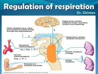
  • 35. Reflex control of respirationReflex control of respiration 1) By receptors inside respiratory system a) Hering Breuer Reflexes due to stretch receptors in muscular wall of bronchi and bronchioles, b) pulmonary irritant receptors, c) J receptors 2) By receptors outside respiratory system A) Those which stimulate respiration - proprioceptors, nociceptors, thermoceptors, emotional stimuli from hypothalamus and limbic system. B)Those which inhibit respiration- Baroreceptors and visceroreceptors
  • 36. Hering Breuer ReflexesHering Breuer Reflexes Located in the muscular portions of the walls of the bronchi and bronchioles throughout the lungs are stretch receptors transmit signals through the vagi into the dorsal respiratory group of neurons when the lungs become overstretched.
  • 37. These signals act in same way as signalsThese signals act in same way as signals from Pneumotaxic centerfrom Pneumotaxic center  when the lungs become overly inflated, the stretch receptors activate an appropriate feedback response that “switches off” the inspiratory ramp and thus stops further inspiration. This is called the Hering - Breuer inflation reflex. This reflex also increases the rate of respiration, as is true for signals from the pneumotaxic center
  • 38. Importance of H-B inflation reflexImportance of H-B inflation reflex 1) This reflex operates only when Tidal volume exceeds 1.5 liters as in exercise and not in normal ventilation and so prevents over inflation of lungs and is thus protective 2) This effect is not seen in man if vagi are cut and also so in transplanted lungs which are denervated.
  • 39. Hering Breuer Deflation reflexHering Breuer Deflation reflex It is seen when there is extreme deflation of lungs as seen in collapse of lungs, pneumothorax, hydrothorax, etc. The receptors are stimulated by change in shape of small airways in collapsed area so also called as compression receptors. So forceful deflation of lungs causes stimulation of receptors → stimulation of inspiration via vagus
  • 40. Effect of irritant receptors in theEffect of irritant receptors in the airwaysairways The epithelium of the trachea, bronchi and bronchioles is supplied with sensory nerve endings called pulmonary irritant receptors and are rapidly adapting and innervated by myelinated vagal nerve fibers. They are stimulated by many incidents like lung hyperinflation, exogenous subs like SO2, smoke and endogenous subs like histamine, PGs. These cause hyperpnoea, coughing and sneezing, bronchial constriction and mucus secretion in diseases as asthma and emphysema.
  • 41. Function of ‘J receptors’Function of ‘J receptors’ A few sensory nerve endings have been described in the alveolar walls in juxtaposition to the pulmonary capillaries—hence the name “J receptors.” they are non myelinated nerve endings (C fibers). They are stimulated especially when the pulmonary capillaries become engorged with blood or when pulmonary edema occurs as in congestive heart failure.  Although the functional role of the J receptors is not clear, their excitation may give the person a feeling of dyspnea
  • 42. J receptors cause the pulmonaryJ receptors cause the pulmonary chemo reflexchemo reflex Apnea followed by Hyperventilation, bradycardia, hypotension, bronchoconstriction, and mucus secretion and skeletal muscle weakness (Seen in Bhopal gas tragedy). This is similar to Coronary Chemo reflex or Bezold Jarisch Reflex
  • 43. Receptors outside respiratory systemReceptors outside respiratory system which stimulate respirationwhich stimulate respiration 1) Proprioceptors – Present in muscles, tendons, joints etc when stimulated reflexly stimulates Inspiratory neurons. This helps to increase ventilation at the start of exercise even before changes in Pco2, Po2 and H ions concentration occur.
  • 44. 2) Nociceptors - Pain receptors 3) Emotional stimuli – all stimulate respiratory system via hypothalamus and limbic system. 4) Chemoreceptors – stimulated by acidosis and hypoxia. Hypotension and shock produce hyperventilation due to stimulation of chemoreceptors.
  • 45. 5) Thermoreceptors – present in hypothalamus are stimulated when there is increase in body temperature as in fever, exercise they send impulses to respiratory center and produce increase in rate of respiration.
  • 46. Those which inhibit respirationThose which inhibit respiration 1) Baroreceptors – inhibit reflexly. Adrenaline apnea is an eg of this. 2) Visceroreceptors – visceral reflexes like vomiting, swallowing, etc reflexly inhibit respiration, Eg is Deglutition apnea
  • 47.
  • 48. Other factors affecting respirationOther factors affecting respiration 1) Sleep- Respiration is less rigorously controlled during sleep than in the waking state, and brief periods of apnea occur in normal sleeping adults. Sleep Apnea Syndrome It is a type of obstructive sleep apnea. It occurs at any age. Cause is failure of Genioglossus muscle to contract during inspiration which causes the tongue to fall backward to obstruct the airways. This muscle normally keeps the tongue forward.
  • 49. S/S are morning headache, fatigue, hypoxemic, Hypercapneic condition Prevention by avoiding sedatives or respiratory depressants prior to sleep, breathing in prone and avoiding sleeping on back, Positive pressurePositive pressure controlled O2 inhalationcontrolled O2 inhalation.
  • 50. Sudden Infant DeathSudden Infant Death SyndromeSyndrome A type of central sleep apnea due to failure of respiratory centers to produce neural impulses so causing total stoppage of respiration.
  • 51. Effect of brain edemaEffect of brain edema The activity of the respiratory center may be depressed or even inactivated by acute brain edema resulting from brain concussion. the head might be struck against some solid object, after which the damaged brain tissues swell, compressing the cerebral arteries against the cranial vault and thus partially blocking cerebral blood supply.
  • 52. TreatmentTreatment Respiratory depression resulting from brain edema can be relieved temporarily by intravenous injection of hypertonic solutions such as highly concentrated mannitol solution. These solutions osmotically remove some of the fluids of the brain, thus relieving intracranial pressure and sometimes re-establishing respiration within a few minutes
  • 53. Effect of Anesthesia- respiratoryEffect of Anesthesia- respiratory depressiondepression The most prevalent cause of respiratory depression and respiratory arrest is over dosage with anesthetics or narcotics.  For instance, sodium pentobarbital depresses the respiratory center considerably more than many other anesthetics, such as halothane.
  • 54. At one time, morphine was used as an anesthetic, but this drug is now used only as an adjunct to anesthetics because it greatly depresses the respiratory center while having less ability to anesthetize the cerebral cortex
  • 55.
  • 56. Chemical regulation of respirationChemical regulation of respiration The ultimate goal of respiration is to maintain proper concentrations of oxygen, carbon dioxide, and hydrogen ions in the tissues. Respiratory activity is highly responsive to changes in each of these. Excess carbon dioxide or excess hydrogen ions in the blood mainly act directly on the respiratory center itself, causing greatly increased strength of both the inspiratory and the expiratory motor signals to the respiratory muscles.
  • 57. Oxygen acts via peripheralOxygen acts via peripheral ChemoreceptorsChemoreceptors  Oxygen, in contrast, does not have a significant direct effect on the respiratory center of the brain in controlling respiration. Instead, it acts almost entirely on peripheral chemoreceptors located in the carotid and aortic bodies, these in turn transmit appropriate nervous signals to the respiratory center for control of respiration
  • 58.
  • 59. CO2 and H ions act directly on centralCO2 and H ions act directly on central chemoreceptorschemoreceptors Chemo sensitive area (CSA) is located bilaterally, lying only 0.2 millimeter beneath the ventral surface of the medulla. This area is highly sensitive to changes in either blood Pco2 or hydrogen ion concentration, and it in turn excites the other portions of the respiratory center
  • 60. Excitation of CSA sensor neurons byExcitation of CSA sensor neurons by HH ions is the primary stimulusions is the primary stimulus Hydrogen ions do not easily cross the blood- brain barrier. changes in hydrogen ion concentration in the blood have considerably less effect in stimulating the chemo sensitive neurons than do changes in blood carbon dioxide, carbon dioxide stimulate these neurons secondarily by changing the hydrogen ion concentration. CO2 has more potent effect than H ions
  • 61. Chemo sensitive areaChemo sensitive area
  • 62.
  • 63. Decreased stimulatory effect of CO2Decreased stimulatory effect of CO2 after first 1 to 2 daysafter first 1 to 2 days Excitation of the respiratory center by carbon dioxide is great for the first few hours after the blood carbon dioxide first increases, but then it gradually declines over the next 1 to 2 days, decreasing to about one fifth the initial effect. It results from renal readjustment of the hydrogen ion concentration in the circulating blood back toward normal after the carbon dioxide first increases the hydrogen concentration. The kidneys achieve this by increasing the blood bicarbonate, which binds with the hydrogen ions in the blood and cerebrospinal fluid to reduce their concentrations
  • 64. Diffusion of Bicarbonate ions- combineDiffusion of Bicarbonate ions- combine with H ions & decreasewith H ions & decrease 2) Over a period of hours, the bicarbonate ions also slowly diffuse through the blood - brain and blood – CSF barriers and combine directly with the hydrogen ions adjacent to the respiratory neurons as well, thus reducing the hydrogen ions back to near normal.
  • 65. A change in blood carbon dioxide concentration therefore has a potent acute effect on controlling respiratory drive but only a weak chronic effect after a few days’ adaptation
  • 66. Unimportance of oxygen for control ofUnimportance of oxygen for control of respiratory centrerespiratory centre Changes in oxygen concentration have virtually no direct effect on the respiratory center itself to alter respiratory drive  although oxygen changes do have an indirect effect, acting through the peripheral chemoreceptors
  • 67. Hb as a good bufferHb as a good buffer The hemoglobin oxygen buffer system delivers almost normal amounts of oxygen to the tissues even, when the pulmonary Po2 changes from a value as low as 60 mm Hg up to a value as high as 600 mm Hg.
  • 68. Chemoreceptors - O2Chemoreceptors - O2 The carotid bodies are located bilaterally in the bifurcations of the common carotid arteries. Their afferent nerve fibers pass through Herring's nerves to the glossopharyngeal nerves and then to the dorsal respiratory area of the medulla.  The aortic bodies are located along the arch of the aorta; their afferent nerve fibers pass through the vagi, also to the dorsal medullary respiratory area.
  • 69.
  • 70. Stimulation of chemoreceptors byStimulation of chemoreceptors by decreased arterial Po2decreased arterial Po2 Shows the effect of different levels of arterial Po2 on the rate of nerve impulse transmission from a carotid body. the impulse rate is particularly sensitive to changes in arterial Po2 in the range of 60 down to 30 mm Hg, a range in which hemoglobin saturation with oxygen decreases rapidly
  • 71. Effect of CO2 and H ion concentrationEffect of CO2 and H ion concentration on chemoreceptorson chemoreceptors Indirect effect- An increase in either carbon dioxide concentration or hydrogen ion concentration also excites the peripheral chemoreceptors and, in this way, indirectly increases respiratory activity. However, the direct effects of both these factors in the respiratory center itself via CSA are much more powerful than peripheral ones.
  • 72. One difference between peripheral andOne difference between peripheral and central effects of CO2 & H ionscentral effects of CO2 & H ions The stimulation by way of the peripheral chemoreceptors occurs as much as five times as rapidly as central stimulation. So that the peripheral chemoreceptors might be especially important in increasing the rapidity of response to carbon dioxide at the onset of exercise. Thus direct effect via CSA is very powerful than indirect via peripheral chemoreceptors. But indirect effect is very rapid than direct effect.
  • 73. Basic mechanism of stimulation of chemoreceptorsBasic mechanism of stimulation of chemoreceptors by o2 deficiency – NT Dopamineby o2 deficiency – NT Dopamine
  • 74. Mechanism of release of NTsMechanism of release of NTs Type I glomus cells have O2-sensitive K+ channels, whose conductance is reduced in proportion to the degree of hypoxia to which they are exposed. This reduces the K+ efflux, depolarizing the cell and causing Ca2+ influx, primarily via L-type Ca2+ channels. The Ca2+ influx triggers action potentials and transmitter release, with consequent excitation of the afferent nerve endings
  • 75. Low Po2 stimulates ventilation whenLow Po2 stimulates ventilation when co2& H ions constantco2& H ions constant Only the ventilatory drive due to the effect of low oxygen on the chemoreceptors is active. There is almost no effect on ventilation as long as the arterial Po2 remains greater than 100 mm Hg. But at pressures lower than 100 mm Hg, ventilation approximately doubles when the arterial Po2 falls to 60 mm Hg
  • 76. AcclimatizationAcclimatization Chronic Breathing of Low Oxygen Stimulates Respiration Even More—The Phenomenon of “Acclimatization” Mountain climbers have found that when they ascend a mountain slowly, over a period of days rather than a period of hours, they breathe much more deeply and therefore can withstand far lower atmospheric oxygen concentrations than when they ascend rapidly.
  • 77. Reason for acclimatizationReason for acclimatization Within 2 to 3 days, the respiratory center in the brain stem loses about four fifths of its sensitivity to changes in Pco2 and hydrogen ions. Therefore, the excess ventilatory blow-off of carbon dioxide that normally would inhibit an increase in respiration fails to occur, and low oxygen can drive the respiratory system to a much higher level of alveolar
  • 78. Instead of the 70 per cent increase in ventilation that might occur after acute exposure to low oxygen, the alveolar ventilation often increases 400 to 500 per cent after 2 to 3 days of low oxygen; this helps greatly in supplying additional oxygen to the mountain climber
  • 79. Effect of exercise on respirationEffect of exercise on respiration regulationregulation In exercise there is rise of co2 formation and more of o2 consumption. Neurogenic stimulation by higher centers and proprioceptors is first most important mechanism to increase ventilation on initiation of exercise even before chemical stimulation develops.
  • 80. If nervous signals are too weak or too strong then chemical factors play an important role in respiratory adjustment in exercise. 1) At the beginning of exercise anticipatory stimulation of respiration occurs thus increasing alveolar ventilation so that it decreases arterial Pco2.
  • 81. 2) After 30 to 40 seconds the amount of co2 released by exercising muscles into blood matches approximately with increase rate of alveolar ventilation and arterial Pco2 returns to normal. Neurogenic factor in exercise shifts the ventilation to higher levels to match the rate of co2 formation and o2 utilization thus keeping Po2 and Pco2 toward normal level.
  • 82. Factors causing Increased ventilationFactors causing Increased ventilation in exercisein exercise 1) Higher centers – motor cortex, hypothalamus, which cause rise of ventilation even before exercise, stimulate VMC increasing BP. 2) Sympathetic stimulation can further stimulate. 3) Increase body temperature 4) Sensory proprioceptors
  • 83. 5) Low P02, high Pco2 and H ions via peripheral chemoreceptors. 6) Increase in plasma K during exercise stimulates peripheral chemoreceptors. 7) Lactic acid liberated in exercise increases co2 which stimulates respiration. 8) Better match of ventilation –
  • 84. Effect of acid base changesEffect of acid base changes Acidosis – H ions conc- stimulation↑ ↑ Alkalosis- H ions conc- stimulation↓ ↓ In metabolic acidosis as seen in diabetes, renal failure, severe muscular exercise, starvation etc more H ions and less HCO3 in blood which stimulate respiration (Kussumaul breathing) which reduces co2 which leads to compensatory fall in blood H ion conc.
  • 85. In metabolic alkalosis as in vomiting with loss of HCL due to pyloric or high intestinal obstruction there is less H ions and more HCO3 so ventilation is depressed and arterial PCO2 increases which brings H ions towards normal.
  • 86. Respiratory acidosis- Here ventilation is depressed due to causes like emphysema, depression of respiratory center, morphine or COPD. Here hypoventilation causes rise of co2 which stimulates central and peripheral chemoreceptors and increases alveolar ventilation and correction of respiratory acidosis.
  • 87. Respiratory alkalosis - as seen in voluntary hyperventilation, chronic lack of o2 at high altitudes, anxiety, hysteria salicylate over dosage etc.
  • 88. Breath holdingBreath holding  Respiration can be voluntarily inhibited for some time, but eventually the voluntary control is overridden. The point at which breathing can no longer be voluntarily inhibited is called the breaking point.  Breaking is due to the rise in arterial PCO2 and the fall in PO2.  Individuals can hold their breath longer after removal of the carotid bodies. Breathing 100% oxygen before breath holding raises alveolar PO2 initially, so that the breaking point is delayed. The same is true of hyperventilating room air, because CO2 is blown off and arterial PCO2 is lower at the start.
  • 89. Reflex or mechanical factors appear to influence the breaking point Subjects who breathe a gas mixture low in CO2 and high in O2 can hold their breath for an additional 20 seconds or more. Psychological factors also play a role, and subjects can hold their breath longer when they are told their performance is very good
  • 90. Hormonal effects on respirationHormonal effects on respiration Ventilation is increased during the luteal phase of the menstrual cycle and during pregnancy Experiments with animals indicate that this is due to activation of estrogen - dependent progesterone receptors in the hypothalamus. However, the physiologic significance of this increased ventilation is unknown.
  • 91. Periodic breathingPeriodic breathing An abnormality of respiration called periodic breathing occurs in a number of disease conditions. The person breathes deeply for a short interval and then breathes slightly or not at all for an additional interval, with the cycle repeating over and over. One type of periodic breathing, Chyne-Stokes breathing, is characterized by slowly waxing and waning respiration occurring about every 40 to 60 seconds.
  • 92. Chyne- Stokes breathingChyne- Stokes breathing Apnea followed by hyperventilationApnea followed by hyperventilation
  • 93. MechanismMechanism Hyperventilation causes - Po2 and wash out of co2↑ – which causes depression of respiration and so apnoea --- which now increases Pco2 and reduces Po2 --- which now stimulates ventilation again.
  • 94. The basic cause of Cheyne-Stokes breathing occurs in everyone. However, under normal conditions, this mechanism is highly “damped.” That is, the fluids of the blood and the respiratory center control areas have large amounts of dissolved and chemically bound carbon dioxide and oxygen.  Therefore, normally, the lungs cannot build up enough extra carbon dioxide or depress the oxygen sufficiently in a few seconds to cause the next cycle of the periodic breathing
  • 95. CausesCauses Physiological Voluntary hyperventilation Healthy infants and adults during sleep High altitude hypoxia Pathological Uremia Cardiac failure(LVF) Brain damage
  • 96. In cardiac failure (LVF)- Slow blood flow- so delayed gas transport from lungs to brain. In LVF - Pulmonary congestion so hampering gas exchange – hypoxia — stimulates resp center --- increase vent – washout of co2 and more o2 – due to sluggish blood flow blood with low co2 takes more time to reach brain resp centers so — apnea.
  • 97. The brain damage often turns off the respiratory drive entirely for a few seconds;  then an extra intense increase in blood carbon dioxide turns it back on with great force. Cheyne-Stokes breathing of this type is frequently a preface to death from brain malfunction.
  • 98. Biot’s breathingBiot’s breathing Respiration characterized by alternate Eupnoea and Apnea. 3-4 cycles of normal breathing followed by abrupt apnea and again abrupt onset of normal respiration.  Seen in meningitis, severe brain damage and diseases affecting medulla.
  • 99. Kussumaul’s breathingKussumaul’s breathing Seen in metabolic acidosis Rapid and deep breathing without uneasiness. Acidosis – H ions conc- stimulation↑ ↑ Alkalosis- H ions conc- stimulation↓ ↓ metabolic acidosis seen in diabetes, renal failure, severe muscular exercise, starvation etc more H ions and less HCO3 in blood which stimulate respiration (Kussumaul's breathing) which reduces co2 which leads to compensatory fall in blood H ion conc.