Coronary circulation
•Download as PPTX, PDF•
14 likes•10,580 views

1. The document describes the anatomy and physiology of the coronary circulation, including the structure and blood supply of the heart muscles and vessels. 2. It discusses how coronary blood flow is regulated by local muscle metabolism and oxygen demand to meet the heart's nutritional needs. 3. The causes, symptoms, and treatments of coronary heart disease like angina and myocardial infarction are explained.

Recommended
More Related Content
What's hot
What's hot (20)
Similar to Coronary circulation
Similar to Coronary circulation (20)
More from DrChintansinh Parmar
More from DrChintansinh Parmar (20)
Recently uploaded
Recently uploaded (20)
Coronary circulation
- 1. Coronary circulation - Dr. Chintan
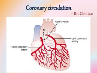
- 3. Physiologic Anatomy - Arteries• the main coronary arteries lie on the surface of the heart and smaller arteries then penetrate from the surface into the cardiac muscle mass • Only the inner 1/10 millimeter of the endocardial surface can obtain significant nutrition directly from the blood inside the cardiac chambers • The left coronary artery supplies mainly the anterior and left lateral portions of the left ventricle • the right coronary artery supplies most of the right ventricle as well as the posterior part of the left ventricle
- 4. Physiologic Anatomy - Veins• Most of the coronary venous blood flow from the left ventricular muscle returns to the right atrium of the heart by way of the coronary sinus — which is about 75 per cent of the total coronary blood flow. • Most of the coronary venous blood from the right ventricular muscle returns through small anterior cardiac veins that flow directly into the right atrium. • A very small amount of coronary venous blood also flows back into the heart through very minute thebesian veins, which empty directly into all chambers of the heart.
- 6. Coronary Blood Flow • The resting coronary blood flow in the human being averages about 225 ml/min - about 4 to 5 % of the total CO. • During strenuous exercise, the heart in the young adult increases its CO – 4 to 7 times • coronary blood flow increases 3 to 4 times to supply the extra nutrients needed by the heart
- 7. Phasic Changes • the coronary capillary blood flow in the left ventricle muscle falls to a low value during systole, which is opposite to flow in vascular beds elsewhere in the body. • The reason for this is strong compression of the left ventricular muscle around the intramuscular vessels during systolic contraction. • During diastole, the cardiac muscle relaxes and no longer obstructs blood flow through the left ventricular muscle capillaries, so that blood flows rapidly during all of diastole. • Blood flow through the coronary capillaries of the right ventricle also undergoes phasic changes - only partial
- 10. • During systole, blood flow through the subendocardial plexus of the left ventricle, where the intramuscular coronary vessels are compressed greatly by ventricular muscle contraction, tends to be reduced
- 11. Control of Coronary Blood Flow• Local Muscle Metabolism Is the Primary Controller of Coronary Flow • local arteriolar vasodilation in response to cardiac muscle need for nutrition • Oxygen Demand as a Major Factor in Local Coronary Blood Flow Regulation • about 70 % of the oxygen in the coronary arterial blood is removed as the blood flows through the heart muscle. • Because not much oxygen is left, very little additional oxygen can be supplied to the heart musculature unless the coronary blood flow ↑
- 12. Control of Coronary Blood Flow• decrease in the oxygen concentration in the heart - release of vasodilator substances from the muscle cells - dilatation the arterioles • In the presence of very low concentrations of oxygen in the muscle cells - a large proportion of the cell’s ATP → AMP → adenosine (potent vasodilator) • Adenosine phosphate compounds, • K ions, H ions, bradykinin, • PGs, NO, CO2
- 13. Control of Coronary Blood Flow• Nervous Control • Stimulation of the autonomic nerves to the heart can affect coronary blood flow both directly and indirectly. • The direct effects result from action of the nervous transmitter substances acetylcholine from the vagus nerves and norepinephrine and epinephrine from the sympathetic nerves on the coronary vessels themselves. • The indirect effects result from secondary changes in coronary blood flow caused by increased or decreased activity of the heart.
- 14. Direct Nervous Control • The distribution of parasympathetic (vagal) nerve fibers to the ventricular coronary system is not very great. • the acetylcholine released by parasympathetic stimulation has a direct effect to dilate the coronary arteries. • There is much more extensive sympathetic innervation of the coronary vessels • The constrictor receptors are called alpha receptors – epicardial • the dilator receptors are called beta receptors - intramuscular
- 15. Indirect Nervous Control • Sympathetic stimulation, which releases NE, E, increases both heart rate and heart contractility as well as increases the rate of metabolism of the heart. • the increased metabolism of the heart sets off local blood flow regulatory mechanisms for dilating the coronary vessels, and the blood flow increases approximately in proportion to the metabolic needs of the heart muscle. • vagal stimulation, with its release of acetylcholine, slows the heart and has a slight depressive effect on heart contractility. • These effects in turn decrease cardiac oxygen consumption and, therefore, indirectly constrict the coronary arteries.
- 16. Control of Coronary Blood Flow• Sympathetic stimulation cause coronary constriction or dilation, but usually constriction. • In some people, the alpha vasoconstrictor effects severe - vasospastic myocardial ischemia during periods of excess sympathetic drive - anginal pain. • Metabolic factors - especially myocardial O2 consumption - are the major controllers of myocardial blood flow. • Whenever the direct effects of nervous stimulation alter the coronary blood flow in the wrong direction, the metabolic control of coronary flow usually overrides the direct coronary nervous effects within seconds.
- 17. Ischemic Heart Disease • Atherosclerosis as a Cause of Ischemic Heart Disease • The most frequent cause of diminished coronary blood flow • people who eat excessive quantities of cholesterol and have a sedentary lifestyle - large quantities of cholesterol gradually become deposited beneath the endothelium at many points in arteries throughout the body. • Gradually, these areas of deposit are invaded by fibrous tissue and frequently become calcified - development of atherosclerotic plaques - protrude into the vessel lumens and either block or partially block blood flow. • A common site for development of atherosclerotic plaques is the first few centimeters of the major coronary arteries.
- 18. Acute Coronary Occlusion• The atherosclerotic plaque can cause a local blood clot called a thrombus, which in turn occludes the artery - coming in direct contact with the flowing blood. • Because the plaque presents an unsmooth surface, blood platelets adhere to it, fibrin is deposited, and RBCs become entrapped to form a blood clot that grows until it occludes the vessel. • The clot breaks away from its attachment and flows to a more peripheral branch of the coronary arterial tree • A thrombus that flows along the artery in this way and occludes the vessel more distally is called a coronary embolus.
- 19. Acute Coronary Occlusion• local muscular spasm of a coronary artery also can occur. • The spasm might result from direct irritation of the smooth muscle of the arterial wall by the edges of an arteriosclerotic plaque, or • it might result from local nervous reflexes that cause excess coronary vascular wall contraction. • The spasm may then lead to secondary thrombosis of the vessel.
- 20. Acute Coronary Occlusion• Lifesaving Value of Collateral Circulation in the Heart
- 21. Myocardial Infarction • Immediately after an acute coronary occlusion, blood flow stops in the coronary vessels beyond the occlusion except for small amounts of collateral flow from surrounding vessels. • The area of muscle that has either zero flow or so little flow that it cannot sustain cardiac muscle function is said to be infarcted. • The overall process is called a myocardial infarction. • Soon after the onset of the infarction, small amounts of collateral blood begin to leak into the infarcted area + progressive dilation of local blood vessels, causes the area to become overfilled with stagnant blood
- 22. Myocardial Infarction • Simultaneously the muscle fibers use the last leftovers of the oxygen in the blood, causing the hemoglobin to become totally de-oxygenated - bluish-brown shade, and the blood vessels of the area appear to be engorged despite lack of blood flow. • In later stages, the vessel walls become highly permeable and leak fluid - local muscle tissue becomes edematous, and the cardiac muscle cells begin to swell because of diminished cellular metabolism. • Within a few hours of almost no blood supply - the cardiac muscle cells die
- 23. Myocardial Infarction • The subendocardial muscle frequently becomes infarcted • The subendocardial muscle has extra difficulty obtaining adequate blood flow because the blood vessels in the sub endocardium are intensely compressed by systolic contraction of the heart • Any condition that compromises blood flow to any area of the heart usually causes damage first in the subendocardial regions, and the damage then spreads outward toward the epicardium.
- 24. • During systole, blood flow through the subendocardial plexus of the left ventricle, where the intramuscular coronary vessels are compressed greatly by ventricular muscle contraction, tends to be reduced
- 25. Causes of Death • (1) decreased cardiac output;
- 26. Systolic Stretch – Cardiac Shock • When the heart becomes incapable of contracting with sufficient force to pump enough blood into the peripheral arterial tree - cardiac failure and death of peripheral tissues ensue as a result of peripheral ischemia. • This condition is called coronary shock, cardiogenic shock, cardiac shock, or low cardiac output failure. • Cardiac shock occurs when more than 40 % of the left ventricle is infarcted. • death occurs in 85 % of patients once they develop cardiac shock.
- 27. Causes of Death • (2) obstructing of blood in the pulmonary blood vessels and then death resulting from pulmonary edema;
- 28. Fibrillation of the Ventricles• 1. Acute loss of blood supply to the cardiac muscle causes rapid depletion of K from the ischemic musculature • ↑ K concentration in the ECF - ↑ irritability of the cardiac musculature • 2. Ischemic musculature often cannot completely repolarize its membranes after a heart beat • electric current flows from this ischemic area of the heart to the normal area - abnormal impulses that can cause fibrillation
- 29. Fibrillation of the Ventricles • 3. Powerful sympathetic reflexes - ↑ irritability of the cardiac muscle • 4. Cardiac muscle weakness caused by MI often causes the ventricle to dilate excessively. • This ↑ the pathway length for impulse conduction in the heart • and frequently causes abnormal conduction pathways all the way around the infarcted area of the cardiac muscle - “circus movement”
- 30. Causes of Death • (4) rupture of the heart. • Systolic stretch – rupture – cardiac tamponade • patient dies of suddenly decreased cardiac output.
- 31. Recovery – rest – coronary steal
- 32. Pain in CHD • Ischemia causes the muscle to release acidic substances, such as lactic acid, or other pain- promoting products, • such as histamine, kinins, or cellular proteolytic enzymes, that are not removed rapidly enough by the slowly moving coronary blood flow. • The high concentrations of these abnormal products then stimulate pain nerve endings in the cardiac muscle, sending pain impulses through sensory afferent nerve fibers into CNS
- 33. Angina Pectoris • In most people who develop progressive constriction of their coronary arteries, cardiac pain, called angina pectoris, begins to appear whenever the load on the heart becomes too great • This pain is usually felt beneath the upper sternum over the heart - often referred to the left arm and left shoulder, to the neck, the side of the face. • The heart originates during embryonic life in the neck, as do the arms. Therefore, both the heart and these surface areas of the body receive pain nerve fibers from the same spinal cord segments.
- 34. Angina Pectoris • Most people who have chronic angina pectoris feel pain when they exercise or when they experience emotions that increase metabolism of the heart or temporarily constrict the coronary vessels because of sympathetic vasoconstrictor nerve signals. • The pain usually lasts for only a few minutes. some patients have such severe and lasting ischemia that the pain is present all the time. • hot, pressing, and constricting; - usually makes the patient stop all unnecessary body activity and come to a complete state of rest.
- 35. Angina Pectoris - Rx • Several vasodilator drugs, when administered during an acute anginal attack, can often give immediate relief from the pain - nitroglycerin and other nitrate drugs. • Chronic - beta blockers – propranolol - block sympathetic beta adrenergic receptors - prevents sympathetic enhancement of heart rate and cardiac metabolism during exercise or emotional episodes. • Therapy with a beta blocker decreases the need of the heart for extra metabolic oxygen during stressful conditions – also reduce the number of anginal attacks as well as their severity.
- 36. Surgical Treatment of CHD • Aortic-Coronary Bypass Surgery - 1960 • removing a section of a subcutaneous vein from an arm or leg and then grafting this vein from the root of the aorta to the side of a peripheral coronary artery beyond the blockage point. • One to five such grafts are usually performed, each of which supplies a peripheral coronary artery beyond a block. • Anginal pain is relieved in most patients - may provide the patient with normal survival expectation • if the heart has already been severely damaged - the bypass procedure is of little value.
- 38. Surgical Treatment of CHD • Coronary Angioplasty – 1980 • A small balloon-tipped catheter is passed under radiographic guidance into the coronary system and pushed through the partially occluded artery until the balloon portion of the catheter connects the partially occluded point. • Then the balloon is inflated with high pressure, which markedly stretches the diseased artery - blood flow through the vessel often increases threefold to fourfold, • Patients are relieved of the coronary ischemic symptoms for at several years - many of the patients eventually require coronary bypass surgery
- 40. Surgical Treatment of CHD • Still newer procedures • a laser beam from the tip of a coronary artery catheter aimed at the atherosclerotic lesion. • The laser dissolves the lesion without damaging the rest of the arterial wall. • a minute metal “stent” placed inside a coronary artery dilated by angioplasty to hold the artery open, thus preventing its restenosis.
- 42. THANQ…