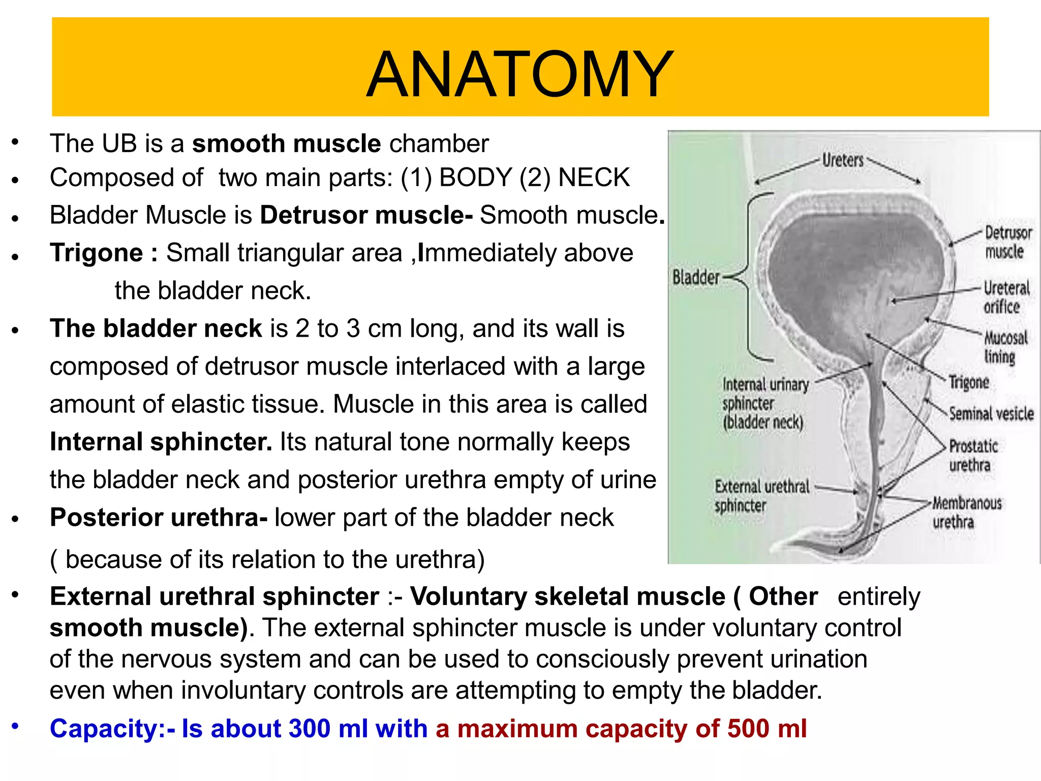
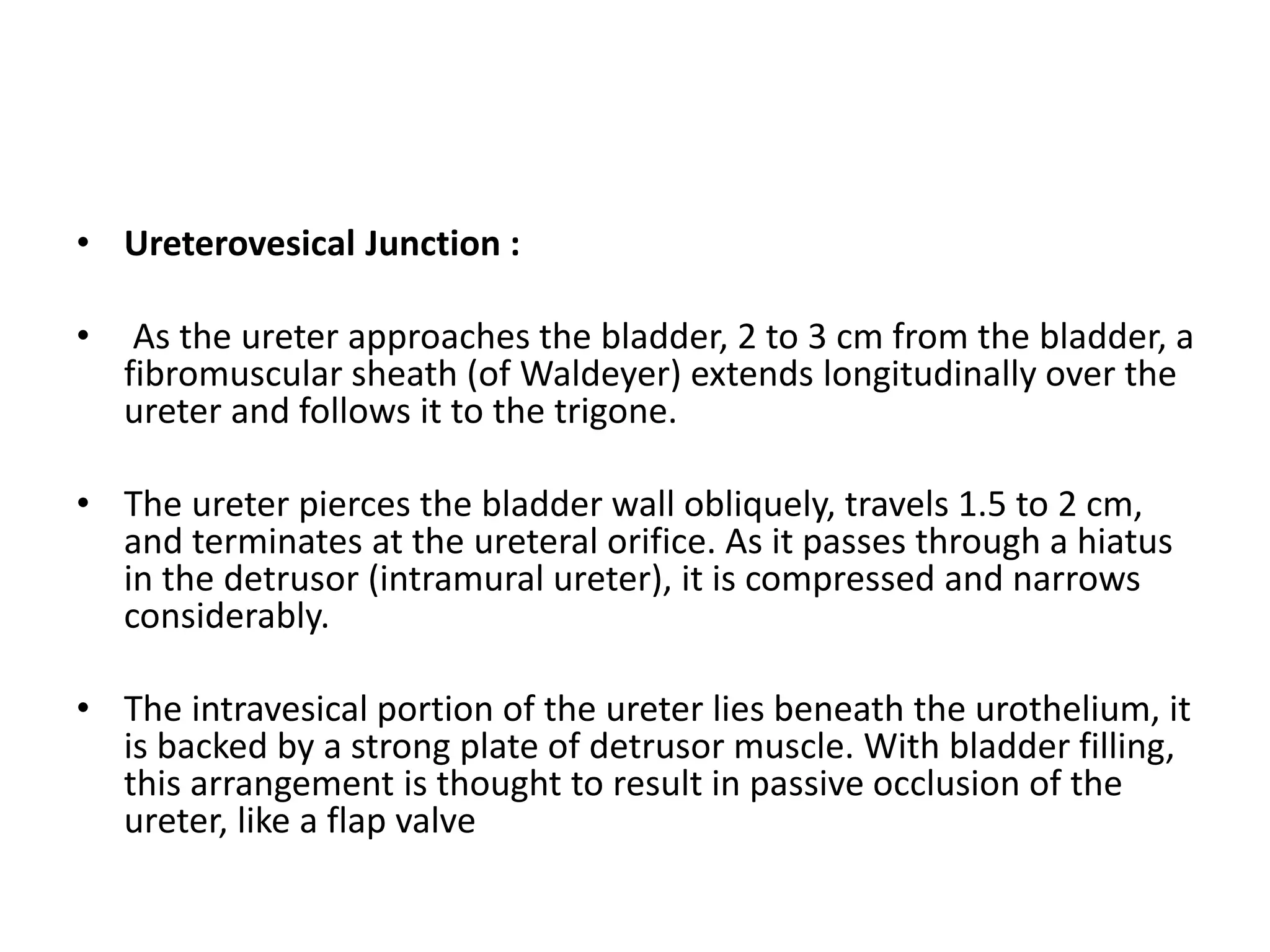
Y2 s1 motor system reflexes basal ganglia 2018 comple lecturevajira54
This document summarizes the motor system, including reflexes, the basal ganglia, and motor functions. It describes the stretch reflex and its components like muscle spindles. It discusses the roles of alpha and gamma motor neurons and how they relate to voluntary and involuntary muscle contractions. Clinical tests of reflexes are used to locate lesions in the motor system.
This document discusses the anatomy and clinical presentation of inguinal hernias. It describes the boundaries and contents of the inguinal canal, as well as structures like the superficial and deep inguinal rings. There are three main types of inguinal hernias - indirect, direct, and sliding. Indirect hernias are more common and involve a defect in the processus vaginalis. Direct hernias involve a weakness in the posterior inguinal wall. Hernia contents can include omentum, intestine, bladder, or other organs. Clinical presentation may include a groin bulge or heaviness, with pain or other symptoms if incarcerated or strangulated.
The inguinal canal is a fibromuscular passage in the lower anterior abdominal wall that extends from the deep inguinal ring to the superficial inguinal ring. It contains the spermatic cord structures in males or the round ligament in females. The main contents include the ductus deferens, testicular vessels, and nerves like the ilioinguinal and genital branch of the genitofemoral nerve. Inguinal hernias occur when abdominal contents like intestine protrude through weak areas in the inguinal canal walls. They can be indirect, occurring through the deep inguinal ring, or direct, through the posterior inguinal wall. Treatment involves surgical hernia repair using techniques
The enteric nervous system (ENS), also known as the intrinsic nervous system, is a complex network of neurons located in the lining of the gastrointestinal tract. It contains around 100-300 million neurons and is capable of independent functioning without input from the central nervous system. The ENS contains two main types of neurons - Dogiel type 1 and 2 - which serve motor and sensory functions respectively. Sensory neurons detect chemicals, pressure, and damage in the gut and initiate peristaltic and secretory reflexes through neurotransmitters like serotonin and acetylcholine. Motor neurons then coordinate contraction and relaxation of muscles and glands through various neurotransmitters to facilitate digestion and movement of contents through the gastrointestinal tract.
The document describes the anatomy of the palmar spaces in the hand. It notes that there are 4 compartments - thenar, hypothenar, intermediate, and adductor. Each compartment contains specific muscles. When infected, pus can collect in the potential spaces between fascial layers. This includes the midpalmar, thenar, and hypothenar spaces. Infections can spread between these spaces and also into the digital web spaces through lumbrical canals. Common infections include flexor tenosynovitis and felons/whitlow. Management involves incision, culture, irrigation, antibiotics, range of motion exercises and splinting.
1) The document discusses the surgical anatomy of inguinal hernias, including the layers of the abdominal wall and contents of the inguinal canal.
2) It describes the history and evolution of techniques for repairing inguinal hernias from ancient times to modern tension-free repairs.
3) Key anatomical structures that relate to inguinal hernias are defined, such as the superficial and deep inguinal rings, inguinal canal, spermatic cord, and the direct and indirect types of inguinal hernias.
The document discusses the history and principles of the Thiersch-Duplay technique for hypospadias repair. It originated in the 19th century with contributions from Thiersch, Duplay, and others. The technique uses the urethral plate as the dorsal wall of the neourethra and has advantages of minimal blood supply disruption and low stricture rates. The document provides details of patient evaluation and the surgical procedure. It finds the technique offers good results, especially for distal hypospadias, though complications like fistula can still occasionally occur. Larger series have shown complication rates generally under 10%.
This document discusses testicular torsion, including:
1. It is a surgical emergency where the spermatic cord twists, compromising blood flow to the testicle. Manual detorsion may be attempted but often requires exploration and fixation to prevent recurrence.
2. Intraoperative assessment of viability involves checking for bleeding after a small incision, and orchiopexy or orchiectomy is performed accordingly.
3. Various fixation techniques are described to prevent recurrent torsion, including tacking the tunica albuginea to the dartos layer with sutures in multiple locations.
This document discusses the inguinal canal, including its boundaries, contents, and defensive mechanisms. It notes that the inguinal canal forms during embryonic development as the testes descend into the scrotum. Hernias can occur if the processus vaginalis remains patent, allowing abdominal contents to enter. There are two main types of inguinal hernias: indirect (oblique) hernias which occur through the deep inguinal ring, and direct hernias which occur through Hesselbach's triangle in the anterior abdominal wall.
Transurethral resection of the prostate (TURP) is a common surgery performed to relieve urinary symptoms caused by an enlarged prostate. Regional anesthesia such as spinal anesthesia is generally preferred over general anesthesia for TURP. Key complications during the procedure include hypotension from sympathetic blockade, hemorrhage, perforation of the bladder or prostate capsule, hypothermia from cold irrigation fluids, and potential for developing TURP syndrome from fluid absorption. Careful patient assessment, fluid management, and monitoring are important to help prevent complications during this common urological procedure.
Defecation..( the guyton and hall physiology)Maryam Fida
Definition:
“Voiding of feces is known as defecation”.
Feces is formed in the large intestine and stored in sigmoid colon.
internal sphincter
Composed of circular smooth muscle
Lies immediately inside the anus
external sphincter
Composed of striated voluntary muscle
Controlled by pudendal nerve. therefore, it is under voluntary, conscious.
reflex pathway
When feces enter rectum
|
Distension of rectal wall
|
Impulses from the nerve endings are transmitted via afferent fibers of pelvic nerve to the defecation center, situated in sacral segments (center) of spinal cord.
|
The center in turn, sends motor impulses to the descending colon, sigmoid colon and rectum via efferent nerve fibers of pelvic nerve.
|
Motor impulses cause strong contraction of descending colon, sigmoid colon and rectum and relaxation of internal sphincter.
Simultaneously, voluntary relaxation of external sphincter occurs. It is due to the inhibition of pudendal nerve.
VOMITING
Definition:
“Vomiting or emesis is the abnormal emptying of stomach and upper part of intestine through esophagus and mouth “.
In this presentation, I have shown how to do open anterior resection both high and low varieties in a step-by-step manner with clear pictures as if reading an atlas of operative surgery.
This document outlines the steps for examining a patient's thyroid gland. It details the relevant history to obtain including symptoms, past medical history, and family history. The physical exam involves inspection of the neck and thyroid, palpation of the gland to assess size, consistency, and mobility, and examination of related structures like eyes, skin, and lymph nodes. Tests are described to evaluate for retrosternal extension and tracheal compression. Examination of cardiovascular, neurological and respiratory systems is also recommended given thyroid abnormalities can impact these areas.
The document summarizes the anatomy of the anal canal. It describes the anal canal as having both a surgical and anatomic component. The surgical anal canal extends from the anorectal junction to the anal verge, while the anatomic canal extends from the dentate line to the anal verge. Key structures discussed include the anorectal ring, internal and external anal sphincters, longitudinal muscle, blood supply, and innervation. The document emphasizes that the anorectal area involves complex anatomical and physiological interactions important for continence and defecation.
This document summarizes the surgical anatomy of the prostate gland. It describes the prostate's location and relations to surrounding structures like the bladder, urethra, and rectum. It details the prostate's blood supply from branches of the internal iliac artery, innervation from pelvic splanchic and pudendal nerves, and lymphatic drainage routes. The document also outlines important surgical structures like the prostatic capsule, neurovascular bundle, and surrounding fascial planes important for nerve-sparing prostatectomy.
The document describes the anatomy of the anterior abdominal wall, including nerves, arteries, veins, and lymph drainage. It also discusses the inguinal canal, spermatic cord, and posterior abdominal wall. Key points include:
- The anterior abdominal wall is supplied by thoracic and lumbar nerves and arteries like the superior and inferior epigastric arteries.
- The inguinal canal allows structures like the spermatic cord to pass from the abdomen into the scrotum in males. It has walls formed by muscles like the internal oblique.
- The spermatic cord contains structures like the vas deferens, testicular vessels, and remnants of the processus vaginalis in males.
rectus sheath, the sheath covering rectus muscle of anterior abdominal wall, formation of the sheath, the muscles involved in ts formation, and the contents the sheath is covering
The rectum is the lower part of the large intestine extending from the sigmoid colon to the anal canal. It is around 5 inches long and located in the pelvis in front of the sacrum and coccyx. It has two flexures that follow the curves of the sacrum and coccyx. The upper third is covered in peritoneum while the lower third has no peritoneal covering. It is supplied by branches of the inferior mesenteric artery and drains into internal iliac and inferior mesenteric lymph nodes. A thorough understanding of rectal anatomy is important for surgical management of rectal conditions and cancer.
Omentum – anatomy, pathological conditions and surgical importanceAravind Endamu
The omentum is a fold of tissue that hangs down from the stomach and extends over other abdominal organs. It has important functions like immunity, absorbing edema, and limiting spread of infection. The greater omentum develops from the dorsal mesogastrium and extends from the stomach to the transverse colon. The lesser omentum connects the stomach and duodenum to the liver. Pathologies of the omentum include cysts, torsion, and tumors. Clinically, the omentum helps drain collections, access the retroperitoneum surgically, and forms adhesions useful for patching perforations.
This document discusses techniques for closing midline laparotomy incisions. It recommends mass closure using continuous slowly absorbable monofilament sutures placed 5-8mm from the wound edge and 4-5mm apart. A suture length to wound length ratio of 4:1 or greater should be used to minimize complications like wound dehiscence and incisional hernia. Proper technique and suture material can reduce surgical site infections, wound failures, and hernia rates.
The document discusses the anatomy of the femoral triangle region. It summarizes that the femoral triangle is bounded laterally by the sartorius muscle, medially by the adductor longus muscle, and superiorly by the inguinal ligament. The femoral triangle contains the femoral vessels and nerve within the femoral sheath in its upper region, and deep inguinal lymph nodes throughout. Femoral hernias occur when abdominal contents protrude through the femoral ring.
This document discusses various surgical methods for treating female stress urinary incontinence, including needle suspension procedures, retropubic colposuspension, pubovaginal slings, and mid-urethral slings. It covers the theories behind these approaches such as the pressure transmission theory and hammock hypothesis. For each method, it provides brief descriptions and highlights complications. Injection therapy is also summarized as a nonsurgical option that aims to improve the urethral seal through injections into the urethral tissues.
The ureter is a tube that carries urine from the kidneys to the bladder. It has 3 parts - the pelvis of the ureter at the kidney, the abdominal part, and the pelvic part. In the abdomen it passes along the posterior abdominal wall. In the pelvis it descends laterally on the pelvic wall before entering the bladder. It has narrowings at the pelviureteric junction, pelvic brim, and bladder entry point where stones can become lodged. In females it has relationships with the uterine artery and ovary, and in males it is crossed by the vas deferens as it enters the bladder.
This is a small handbook on individual surgical disease and its management . I have discussed about Acute Appendicitis and then step by step I explain both open and laparoscopic appendicectomy in this book.
The operative surgery part is very useful for surgical trainees.
This document discusses the anatomy of the mesentery and related structures. It describes the mesentery as the peritoneal fold that suspends the jejunum and ileum from the posterior abdominal wall. It provides blood supply and innervation to the intestines. The root of the mesentery extends from L2 to the right sacroiliac joint and crosses several structures. The superior mesenteric artery supplies the midgut and branches to form the jejunal and ileal arteries. Meckel's diverticulum is described as a remnant of the vitelline duct that can cause complications like perforation or intestinal obstruction.
This document discusses complications of acute pancreatitis, including systemic complications like shock, respiratory failure, and renal failure, as well as local complications affecting the pancreas itself. Local complications include acute fluid collections, pancreatic necrosis, pseudocysts, abscesses, ascites, effusions, pseudoaneurysms, and pancreatic fistulas. The management of these various complications is described, including techniques such as percutaneous drainage, antibiotics, necrosectomy, and surgical interventions.
This document discusses nocturnal enuresis or bedwetting. It begins with the anatomy and physiology of the urinary bladder and micturition process. Nocturnal enuresis is defined as involuntary bladder emptying during sleep at least twice a month after age 5. Causes of nocturnal enuresis include maturational delay, genetics, abnormal antidiuretic hormone levels, defective sleep arousal, and reduced bladder capacity. Diagnosis involves a history, physical exam, urinalysis and ruling out underlying organic causes. Treatment is individualized but may include lifestyle changes, medication, and alarm therapy.
This document provides an overview of neurogenic bladder. It begins with an introduction defining neurogenic bladder as bladder dysfunction due to central nervous system or peripheral nerve disease. It then covers the relevant anatomy and physiology including the innervation of the bladder. The document discusses the central neural control of micturition and the normal voiding process. It also covers development of bladder control in adults and provides descriptions of terminology related to storage and emptying problems.
The document discusses neurogenic bladder and its anatomy, innervation, and types. It provides details on:
1) The urinary bladder is innervated by the parasympathetic, sympathetic, and somatic nervous systems which control storage and voiding functions.
2) There are several types of neurogenic bladder depending on the location of lesions in the central or peripheral nervous system, including loss of supraspinal control, spinal cord lesions above or at the sacral level.
3) Evaluating the type of neurogenic bladder helps determine the symptoms, cystometric findings, and appropriate management.
1. The document discusses the anatomy, physiology, and neurological control of the urinary bladder. It describes the nerve supply, receptors, and micturition pathways.
2. Several pathological types of bladder dysfunction are summarized, including uninhibited, hyperreflexic, and flaccid bladders caused by lesions in different parts of the nervous system.
3. The clinical implications of each type are outlined, such as their symptoms, causes, and complications. Differentiating between neurogenic bladder types helps guide appropriate clinical management of urinary incontinence and retention.
This patient has developed an autonomous bladder as a result of damage to the nerves controlling the bladder during his surgery and resection of the rectum. An autonomous bladder functions independently of the brain with loss of voluntary control.
The document describes the anatomy and physiology of the urinary bladder. It discusses the normal micturition reflex and how it can become dysfunctional. Specifically, it covers three types of abnormal bladder function: irritative symptoms like urgency and frequency, obstructive symptoms like hesitancy, and incontinence. It also discusses various neurological disorders that can cause bladder disturbances, including multiple sclerosis and spinal cord injuries.
The document discusses micturition (urination) including the physiology, anatomy, and abnormalities involved. It begins with the anatomical structures of the bladder including the detrusor muscle and internal and external sphincters. It then describes the micturition reflex initiated by stretch receptors in the bladder wall. This reflex involves afferent and efferent pathways through the pelvic nerves and sacral micturition center. Higher brain centers provide voluntary control. Abnormalities discussed include the atonic bladder due to sensory fiber destruction, and the tabetic bladder caused by syphilis.
This document provides information about the anatomy, physiology, lesions, and management of the urinary bladder. It discusses the bladder's structure, nerve supply, reflex pathways, and normal filling and voiding functions. Common lesions that can affect the bladder include uninhibited neurogenic bladder, hyperreflexic bladder, detrusor-sphincter dyssynergia, and various types of paralytic bladder. Management involves diagnostic evaluation through history, exams, urine tests, ultrasound, and urodynamics to assess storage and voiding functions while minimizing risks of urinary tract infection or damage.
This document discusses neurogenic bladder, which is bladder dysfunction caused by diseases of the central nervous system or peripheral nerves that control urination. It outlines the anatomy and physiology of normal bladder control through nerves and the brain. It describes the different types of neurogenic bladder based on the level of nervous system involvement, including uninhibited, automatic, autonomous, sensory, and motor paralytic bladders. The document discusses how neurogenic bladder should be evaluated through clinical history, examination, urinary tests, urodynamic studies like uroflowmetry and cystometry, and uroneurophysiology tests.
The document summarizes the physiology of micturition (urination). It discusses the anatomy of the ureters and bladder, as well as their innervation. It describes the mechanisms of bladder filling and emptying, including the micturition reflex. It also covers central control of micturition and applied aspects like spinal cord injuries. Recent advances discussed include the sensory role of non-neuronal cells in the bladder and potential new treatments.
The document summarizes the process of micturition (urination). It describes how urine is transported from the kidneys to the urinary bladder via ureters. As the bladder fills, stretch receptors send signals to the brain and spinal cord. When the bladder reaches capacity, the micturition reflex is triggered to relax the internal urethral sphincter and contract the detrusor muscle in the bladder wall to empty urine from the bladder through the urethra. Both voluntary and involuntary neural pathways in the brain, brainstem and spinal cord coordinate the filling and emptying of the bladder.
The process of voiding urine, known as micturition, involves the urinary bladder filling with urine and then emptying through contraction of the detrusor muscle and relaxation of the internal and external urethral sphincters. This is mediated by a micturition reflex initiated by stretch receptors in the bladder wall and regulated by spinal and brain centers. Voluntary control of micturition is achieved through inhibition or facilitation of the reflex by higher brain centers.
The document discusses the anatomy and physiology of the bladder. It describes the two main steps of micturition as the bladder filling progressively until tension rises above a threshold, and then emptying of the bladder. Micturition is regulated by centers in the brain and spinal cord. The bladder is innervated by parasympathetic, sympathetic, and somatic nerves which facilitate filling and emptying. Neurological lesions in different areas can cause different types of neurogenic bladder dysfunction characterized by symptoms like incontinence or retention.
The document discusses the anatomical structure and physiological function of the urinary bladder. It contains the following key points:
1. The urinary bladder is composed of the body, which contains the detrusor muscle responsible for emptying during urination, and the internal and external sphincters that prevent emptying.
2. During filling, the bladder adapts to increasing volume via relaxation, allowing large volumes with minimal pressure rise due to the law of Laplace.
3. Micturition is initiated by a reflex arc when filling reaches 300-400ml. Afferent signals travel to sacral micturition centers which excite detrusor contraction and inhibit sphincters, causing empty
This document provides an overview of neurogenic bladder including:
1. Neurogenic bladder affects 15% of the population and symptoms increase with age. Bladder dysfunction can negatively impact quality of life.
2. The bladder has storage and voiding functions controlled by the brain and spinal cord. Detrusor overactivity, detrusor-sphincter dyssynergia, and detrusor areflexia are types of neurogenic bladder dysfunction.
3. Investigations include post-void residual volume, uroflowmetry, and cystometry to evaluate the bladder and determine appropriate treatment which may include anticholinergics, botulinum toxin injections, clean intermittent catheterization, or surgery
The document summarizes the anatomy and physiology of the urinary system and micturition process. It describes how urine is transported from the kidneys to the bladder through the ureters. It explains that the bladder stores urine through a balance of parasympathetic and sympathetic signals until reaching capacity, at which point a spinal micturition reflex is triggered to initiate voiding through coordinated detrusor contraction and urethral sphincter relaxation under control of the pontine micturition center. Higher brain centers can facilitate or inhibit micturition.
The document discusses the physiology of micturition and bladder dysfunction. It describes the anatomy and innervation of the urinary bladder. Micturition is a reflex that is initiated when the bladder reaches about 300-400 ml in volume. This causes a parasympathetic response that contracts the detrusor muscle and relaxes the internal sphincter, allowing urine to pass into the urethra. Voluntary control is exerted via somatic fibers to the external urethral sphincter. Bladder dysfunctions can occur from lesions that cause deafferentation, denervation, or interrupt signals from facilitatory and inhibitory brain areas.
Micturition is the process of urinating that involves two main steps - the bladder filling with urine until tension triggers the micturition reflex, causing the bladder to empty. This reflex is controlled by the spinal cord but can be inhibited or facilitated by the brain. The urinary bladder stores urine and empties through contraction of the detrusor muscle. Urine enters the bladder via the ureters and exits through the urethra. The micturition reflex maintains control of urination but damage to nerves can cause abnormalities like an atonic bladder with no control or an automatic bladder that empties without brain input.
Similar to types of bladder final Dr Tarun.pptx (20)
IgAN is the commonest GN worldwide with varied presentation without any concrete medical therapy to halt disease progression.So in this slide we will talk about pathogenesis & possible target of future therapies for IgAN
Dermatological complication in chronic kidney diseasetarun kumar
The document discusses several common dermatological manifestations of chronic kidney disease. Pruritus and xerosis are very prevalent in patients with CKD due to dry, itchy skin. Other frequent conditions include pigmentary alterations that cause skin discoloration, acquired perforating dermatosis presenting as crateriform lesions, and calciphylaxis which can lead to necrosis. Conditions like pseudoporphyria and porphyria cutanea tarda may appear similar but can be distinguished by lab tests. Nephrogenic systemic fibrosis is a serious condition that can be prevented by avoiding gadolinium contrast agents in patients with reduced kidney function.
Proteinuria provides diagnostic and prognostic information about renal disease. It is associated with hypertension, obesity, and vascular disease, and can predict risks of chronic kidney disease progression, cardiovascular disease, and mortality. Monitoring proteinuria is important for assessing treatment response, as protein-lowering therapies may be renoprotective. A thorough history, physical exam, urinalysis, and further tests are needed to determine the type and cause of proteinuria and rule out underlying conditions in order to begin appropriate treatment.
Drug resistant tuberculosis is defined as resistance of Mycobacterium tuberculosis to antitubercular drugs. It can be multidrug resistant (MDR), extensively drug resistant (XDR), or have other resistance patterns. Diagnosis involves culture and drug susceptibility testing using solid or liquid media, as well as molecular tests like CBNAAT and line probe assays. Treatment requires specialized regimens using second line drugs for longer periods. Patient follow up assesses treatment response through repeated sputum cultures. Newer drugs like bedaquiline and delamanid are being added to treatment regimens to improve outcomes for drug resistant tuberculosis.
This document provides an overview of atrial fibrillation (AF). It defines AF as a supraventricular arrhythmia characterized by disorganized, rapid, and irregular atrial activation with loss of atrial contraction. Some key points:
- AF prevalence increases with age and is more common in men and whites. It is the most common sustained arrhythmia.
- AF increases the risk of stroke, heart failure, dementia and mortality.
- Causes include hypertension, heart disease, sleep apnea and genetic factors.
- Treatment involves rate control or rhythm control with medications like beta blockers, calcium channel blockers, and antiarrhythmics. Electrical cardioversion and catheter ablation are also
Evolocumab and its clinical outcomes in patients of cardiovascular diseasetarun kumar
This document summarizes a journal club presentation on a clinical trial investigating the PCSK9 inhibitor evolocumab. The trial found that adding evolocumab to statin therapy in patients with cardiovascular disease reduced LDL cholesterol by 59% on average and reduced the risk of the primary composite cardiovascular endpoint of death, heart attack, stroke or revascularization by 15% and the secondary composite of just death, heart attack or stroke by 20% over a median follow up of 26 months. Evolocumab demonstrated a safety profile similar to placebo with no significant differences in adverse events. This trial provides evidence that further lowering of LDL cholesterol beyond standard statin therapy provides cardiovascular benefit.
5-hydroxytryptamine or 5-HT or Serotonin is a neurotransmitter that serves a range of roles in the human body. It is sometimes referred to as the happy chemical since it promotes overall well-being and happiness.
It is mostly found in the brain, intestines, and blood platelets.
5-HT is utilised to transport messages between nerve cells, is known to be involved in smooth muscle contraction, and adds to overall well-being and pleasure, among other benefits. 5-HT regulates the body's sleep-wake cycles and internal clock by acting as a precursor to melatonin.
It is hypothesised to regulate hunger, emotions, motor, cognitive, and autonomic processes.
Osteoporosis - Definition , Evaluation and Management .pdfJim Jacob Roy
Osteoporosis is an increasing cause of morbidity among the elderly.
In this document , a brief outline of osteoporosis is given , including the risk factors of osteoporosis fractures , the indications for testing bone mineral density and the management of osteoporosis
DECLARATION OF HELSINKI - History and principlesanaghabharat01
This SlideShare presentation provides a comprehensive overview of the Declaration of Helsinki, a foundational document outlining ethical guidelines for conducting medical research involving human subjects.
Travel Clinic Cardiff: Health Advice for International TravelersNX Healthcare
Travel Clinic Cardiff offers comprehensive travel health services, including vaccinations, travel advice, and preventive care for international travelers. Our expert team ensures you are well-prepared and protected for your journey, providing personalized consultations tailored to your destination. Conveniently located in Cardiff, we help you travel with confidence and peace of mind. Visit us: www.nxhealthcare.co.uk
low birth weight presentation. Low birth weight (LBW) infant is defined as the one whose birth weight is less than 2500g irrespective of their gestational age. Premature birth and low birth weight(LBW) is still a serious problem in newborn. Causing high morbidity and mortality rate worldwide. The nursing care provide to low birth weight babies is crucial in promoting their overall health and development. Through careful assessment, diagnosis,, planning, and evaluation plays a vital role in ensuring these vulnerable infants receive the specialize care they need. In India every third of the infant weight less than 2500g.
Birth period, socioeconomical status, nutritional and intrauterine environment are the factors influencing low birth weight
Kosmoderma Academy, a leading institution in the field of dermatology and aesthetics, offers comprehensive courses in cosmetology and trichology. Our specialized courses on PRP (Hair), DR+Growth Factor, GFC, and Qr678 are designed to equip practitioners with advanced skills and knowledge to excel in hair restoration and growth treatments.
The skin is the largest organ and its health plays a vital role among the other sense organs. The skin concerns like acne breakout, psoriasis, or anything similar along the lines, finding a qualified and experienced dermatologist becomes paramount.
Breast cancer: Post menopausal endocrine therapyDr. Sumit KUMAR
Breast cancer in postmenopausal women with hormone receptor-positive (HR+) status is a common and complex condition that necessitates a multifaceted approach to management. HR+ breast cancer means that the cancer cells grow in response to hormones such as estrogen and progesterone. This subtype is prevalent among postmenopausal women and typically exhibits a more indolent course compared to other forms of breast cancer, which allows for a variety of treatment options.
Diagnosis and Staging
The diagnosis of HR+ breast cancer begins with clinical evaluation, imaging, and biopsy. Imaging modalities such as mammography, ultrasound, and MRI help in assessing the extent of the disease. Histopathological examination and immunohistochemical staining of the biopsy sample confirm the diagnosis and hormone receptor status by identifying the presence of estrogen receptors (ER) and progesterone receptors (PR) on the tumor cells.
Staging involves determining the size of the tumor (T), the involvement of regional lymph nodes (N), and the presence of distant metastasis (M). The American Joint Committee on Cancer (AJCC) staging system is commonly used. Accurate staging is critical as it guides treatment decisions.
Treatment Options
Endocrine Therapy
Endocrine therapy is the cornerstone of treatment for HR+ breast cancer in postmenopausal women. The primary goal is to reduce the levels of estrogen or block its effects on cancer cells. Commonly used agents include:
Selective Estrogen Receptor Modulators (SERMs): Tamoxifen is a SERM that binds to estrogen receptors, blocking estrogen from stimulating breast cancer cells. It is effective but may have side effects such as increased risk of endometrial cancer and thromboembolic events.
Aromatase Inhibitors (AIs): These drugs, including anastrozole, letrozole, and exemestane, lower estrogen levels by inhibiting the aromatase enzyme, which converts androgens to estrogen in peripheral tissues. AIs are generally preferred in postmenopausal women due to their efficacy and safety profile compared to tamoxifen.
Selective Estrogen Receptor Downregulators (SERDs): Fulvestrant is a SERD that degrades estrogen receptors and is used in cases where resistance to other endocrine therapies develops.
Combination Therapies
Combining endocrine therapy with other treatments enhances efficacy. Examples include:
Endocrine Therapy with CDK4/6 Inhibitors: Palbociclib, ribociclib, and abemaciclib are CDK4/6 inhibitors that, when combined with endocrine therapy, significantly improve progression-free survival in advanced HR+ breast cancer.
Endocrine Therapy with mTOR Inhibitors: Everolimus, an mTOR inhibitor, can be added to endocrine therapy for patients who have developed resistance to aromatase inhibitors.
Chemotherapy
Chemotherapy is generally reserved for patients with high-risk features, such as large tumor size, high-grade histology, or extensive lymph node involvement. Regimens often include anthracyclines and taxanes.
Histololgy of Female Reproductive System.pptxAyeshaZaid1
Dive into an in-depth exploration of the histological structure of female reproductive system with this comprehensive lecture. Presented by Dr. Ayesha Irfan, Assistant Professor of Anatomy, this presentation covers the Gross anatomy and functional histology of the female reproductive organs. Ideal for students, educators, and anyone interested in medical science, this lecture provides clear explanations, detailed diagrams, and valuable insights into female reproductive system. Enhance your knowledge and understanding of this essential aspect of human biology.
10 Benefits an EPCR Software should Bring to EMS Organizations Traumasoft LLC
The benefits of an ePCR solution should extend to the whole EMS organization, not just certain groups of people or certain departments. It should provide more than just a form for entering and a database for storing information. It should also include a workflow of how information is communicated, used and stored across the entire organization.
Are you looking for a long-lasting solution to your missing tooth?
Dental implants are the most common type of method for replacing the missing tooth. Unlike dentures or bridges, implants are surgically placed in the jawbone. In layman’s terms, a dental implant is similar to the natural root of the tooth. It offers a stable foundation for the artificial tooth giving it the look, feel, and function similar to the natural tooth.
Lecture 6 -- Memory 2015.pptlearning occurs when a stimulus (unconditioned st...AyushGadhvi1
learning occurs when a stimulus (unconditioned stimulus) eliciting a response (unconditioned response) • is paired with another stimulus (conditioned stimulus)
2. OUTLINE
• Introduction
• Applied anatomy and physiology
• Neuraxis and circuitry
• Common symptoms of neurogenic bladder
• Levels of bladder dysfunction
• Investigations
• Treatment available
3. • The bladder is the most anterior element of the
pelvic viscera. Situated in the pelvic cavity when
empty, but expands superiorly into the
abdominal cavity when full.
• The urinary bladder is abdominal at birth,
positioned at the extraperitoneal area of the
lower abdominal wall.
• Around the 5th or 6th year of age the bladder
gradually descends into the area of the true
(minor) pelvis.
INTRODUCTION
4. • Urinary bladder functions as a storage organ that
can empty to completion at appropriate time and
place.
• Problems related to bladder are often obvious
like enuresis, incontinence or may not be
apparent like recurrent UTIs, day time urgency
frequency syndrome.
• Early intervention may prevent renal damage
from retrograde effects of high bladder pressures.
5. ANATOMY
•
•
•
•
•
•
•
•
The UB is a smooth muscle chamber
Composed of two main parts: (1) BODY (2) NECK
Bladder Muscle is Detrusor muscle- Smooth muscle.
Trigone : Small triangular area ,Immediately above
the bladder neck.
The bladder neck is 2 to 3 cm long, and its wall is
composed of detrusor muscle interlaced with a large
amount of elastic tissue. Muscle in this area is called
Internal sphincter. Its natural tone normally keeps
the bladder neck and posterior urethra empty of urine
Posterior urethra- lower part of the bladder neck
( because of its relation to the urethra)
External urethral sphincter :- Voluntary skeletal muscle ( Other entirely
smooth muscle). The external sphincter muscle is under voluntary control
of the nervous system and can be used to consciously prevent urination
even when involuntary controls are attempting to empty the bladder.
Capacity:- Is about 300 ml with a maximum capacity of 500 ml
6. • Ureterovesical Junction :
• As the ureter approaches the bladder, 2 to 3 cm from the bladder, a
fibromuscular sheath (of Waldeyer) extends longitudinally over the
ureter and follows it to the trigone.
• The ureter pierces the bladder wall obliquely, travels 1.5 to 2 cm,
and terminates at the ureteral orifice. As it passes through a hiatus
in the detrusor (intramural ureter), it is compressed and narrows
considerably.
• The intravesical portion of the ureter lies beneath the urothelium, it
is backed by a strong plate of detrusor muscle. With bladder filling,
this arrangement is thought to result in passive occlusion of the
ureter, like a flap valve
7. • This anatomic arrangement helps prevent reflux during bladder
filling by fixing and applying tension to the ureteral orifice. As the
bladder fills, its lateral wall telescopes outward on the ureter,
thereby increasing intravesical ureteral length.
• Vesicoureteral reflux is thought to result from insufficient
submucosal ureteral length and poor detrusor backing.
• Chronic increases in intravesical pressure resulting from bladder
outlet obstruction can cause herniation of the bladder mucosa
through the weakest point of the hiatus above the ureter and
produce a “Hutch diverticulum” and reflux.
9. Efferent innervation NERVE ACTION FUNCTION
Parasympathetic Pelvic nerve(nervi Detrusor muscle Voiding
S2,3,4 erigentes) – hypogastric contraction
plexus Internal sphincter
relaxation
Sympathetic Hpogastric nerves Detrusor muscle Storage
T11-L2 ---inferior hypogastric relaxation
ganglion Internal sphincter
Contraction
Somatic :FROM Pudendal nerve Voluntary innervations Voluntary control
AHC- S-2,3,4 (ventral rami) initiate or inhibits
micturition through
cortical control
Afferent innervation
Parasympathetic Pudendal nerve –enter Sensation of pain and Carried normal
S= 2,3,4 through posterior rami and distension conveyed sensation
terminate in anterolateral from bladder wall and
column internal capsule
Sympathetic (T9 L2) Hypogastric plexus:enter Sensation of painful Carried painful
through posterior rami and distension conveyed sensation
terminate in from bladder wall
anteromediolateral column
T9-L2
10. RECEPTORS & INNERVATION
•
•
•
• Detrusor - intermediolateral
gray column of S2,3,4
parasympathetic – pelvic n
(M2 receptors)
External urethral sphincter -
innervated by somatomotor
S2,3,4 nucleus (Onuf’s
Nucleus)-pudendal n
(Nicotinic receptor)
Trigone and internal
sphincter innervated by
Sympathetic T10,11,12 (less
important)
SNS acts through B3 and A1
receptors
•
•
•
Afferent Pathways
Sensations of pain, temp, urgency is
follows the anterolateral white
columns.
Conscious sensations (bladder
distention, ongoing micturition, tactile
pressure) follow the posterior columns
A-delta fibers – Micturition reflex,
stretch and fullness sensation
• C-fibers – Noxious sensation
11. NEURAXIS
•
•
•
•
Frontal lobe- Sends inhibitory signals
Pons (Pontine Micturition Center=PMC)
– Major relay/excitatory center
– Coordinates urinary sphincters and the bladder
– Affected by emotions
Spinal cord (S2-4)-Intermediary between upper and lower control
Peripheral nervous system-
Parasympathetic (S2-S4)-Pelvic nerves
Excitatory to bladder, relaxes sphincter
Somatic (S2-S4)-Pudendal nerves -Excitatory to external sphincter
Sympathetic (T10-L2)
– Hypogastric nerves to pelvic ganglia
– Inhibitory to bladder body, excitatory to bladder base/urethra
12. • Afferents to Spinal cord : sphincter relaxation
• Afferents to Pons : Contraction of detrusor
• Spinal center: Reflex ill-sustained contractions of
detrusor leads to incomplete evacuation
• Pontine center: Coordinating center. Synchronization
and maintenance of sustained contractions to
complete evacuation.
• Cortical Center: Controls pontine center till a suitable
socially acceptable situation for micturition is available.
13. Peripheral Nervous System
• Parasympathetic (S2-S4)
– Pelvic nerves
• Excitatory to bladder,
relaxes sphincter
• Somatic (S2-S4)
– Pudendal nerves
• Excitatory to external
sphincter
• Sympathetic (T10-L2)
– Hypogastric nerves to pelvic
ganglia
– Inhibitory to bladder body,
excitatory to bladder
base/urethra
• Afferents through Pelvic, pudendal,
hypogastric by
A-delta fibers – Micturition reflex,
stretch and fullness sensation
C-fibers – Noxious sensation
14. Normal Voiding
Normal Voiding
• SNS primarily controls bladder and the IUS
–
–
–
Bladder increases capacity but not pressure
Internal urinary sphincter to remain tightly closed
Parasympathetic stimulation inhibited
• PNS:-Immediately prior to PNS stimulation,
SNS is suppressed
Stimulates detrusor to contract
Pudendal nerve is inhibited external sphincter opens
facilitation of voluntary urination
• Somatics (pudendal N) regulate EUS,Pelvic diaphragm
Delaying voiding or voluntary voiding:
•
• When an individual cannot find a bathroom nearby, the brain inhibit PMC to
prevent detrusor contractions & actively contract the levator muscles to
keep the external sphincter closed
Thus , voiding process requires coordination of both the ANS and somatic
nervous system, which are in turn controlled by the PMC located in the
brainstem.
15. MICTURITION(VOIDING) REFLEX
Sensation of bladder fullness via
pelvic and pudendal nerves to
S 2,3,4
Frontal lobe decides social appropriateness
Periaqueductal gray matter
RECIPROCAL ACTIVITY BETWEEN SPHINCTER & DETRUSSOR
Medial Pontine micturition center
Onuf’s nucleus to pudendal nerves
Detrussor center (S 2,3,4) to pelvic nerves
Micturition
17. DEVELOPMENT
•
•
•
In child Controlled by
Sacral spinal cord reflex
Newborns void 20 x/day with
only a slight decrease during
the 1st year of life
Bladder capacity increases &
voiding frequency decrease
with growth
Bladder capacity in Ounces
(30ml) = Age (yrs) +2
•
•
•
•
•
1-2 yrs: conscious
sensation of bladder
fullness develops
3yrs: Ability to initiate or
inhibit voiding voluntarily
develops
4yrs: Voiding comes
under reliable voluntary
control
By age 4 Micturition spinal
reflex fully modulated by
CNS micturition center via a
spinobulbospinal tact
Initially child has better
control over external
sphincter than bladder
Cortical
Diencephalic
Mechanism
PMC
Spinal efferent
Mechanism
Ganglia
Perineal
stimulation
18. • Neurogenic bladder refers to dysfunction of the
urinary bladder due to disease of the central
nervous system or peripheral nerves involved in
the control of micturition .
• Non Neurogenic bladder refers to dysfunction of
the urinary bladder due to dynamic disturbance
of genitourinary system.
• Complaints about bladder function are common
in patients with neurological disease
• 98% of lifetime bladder is in storage phase
Bladder Disorders
19. Description of Terminology
Storage - At low pressure until such time as it is convenient and socially
acceptable to void
Voiding - Initiated by inhibition of the striated sphincter and pelvic floor,
followed some seconds later by a contraction of the detrusor muscle.
• Storage Problem: Failure to Store normal volumes of urine at low pressure
& without leakage
–
–
Non compliant bladder -Irritable bladder
Inadequate sphincter tone during filling
• Emptying Problem: Failure to empty completely, on command, efficiently at
low pressures
–
–
Failure of neurological control of bladder -Bladder muscle failure
Failure of sphincter relaxation during voiding
• Storage symptoms
Frequency
Urgency
Urge incontinence
Nocturia
Voiding symptoms:
Hesitency
Slow stream
Straining to void
Terminal dribbling
Feeling of incomplete emptying.
20. Description of Terminology
Hesitency: Difficulty to initiate micturition
Urinary retention: Is the inability of the urinary bladder to
empty. The cause may be neurologic or nonneurologic .
Urinary frequency: Voiding more than 7 times during day
and more than once in night
Urgency: extreme desire to void
Urinary incontinence: Involuntary loss of urine that is
objectively demonstrable & is a social or hygenic
problem
Nocturia : Interruption of sleep by urge to void
21. Description of Terminology
• Overflow incontinence: Involuntary passage of urine at a greater than
normal bladder capacity. Due to impaired detrusor contractility OR
•
•
•
•
•
A frequent dribble of urine as a result of inefficient bladder emptying
: drugs, peripheral nerve injury, old age, myogenic injury
Stress incontinence: Incontinence because of increase in intra abdominal
pressure Causes: trauma after birth, pelvic surgery, vaginal wall
hypermobility,irradiation , meningomyelocele
DETRUSOR HYPEREFLEXIA(DH): OAB ;involuantary detrusor contraction
symptoms due to a suprapontine neurologic disorder. The detrusor &
sphincter function incoordination.
DETRUSOR SPHINCTER DYSSYNERGIA(DSD)-: overactive bladder
symptoms due to neurologic UMN disorder of the suprasacral spinal cord.
Paradoxically, the patient is in urinary retention; they are in dyssynergy (lack
of coordination).
DETRUSOR AREFLEXIA :Is complete inability of the detrusor to empty due
to a lower motor neuron lesion ( eg , sacral cord or peripheral nerves injury)
AUTONOMIC DYSREFLEXIA: Is an exaggerated sympathetic response to
any stimuli below the level of the lesion
22. TYPE AND LOCALIZATION OF
BLADDER
•
•
•
•
•
•
1.LOSS OF SUPRASPINAL CONTROL (UNINHIBITED
BLADDER)
2.SPINAL CORD LESION ABOVE SACRAL LEVEL
REFLEX NEUROGENIC BLADDER (AUTOMATIC)
3.SPINAL CORD LESION INVOLVING SACRAL
LEVEL AUTONOMOUS BLADDER
4.LESION INVOLVING AFFERENT SENSORY
NEURONS SENSORY NEUROGENIC BLADDER
5.LESION INVOLVING EFFERENT MOTOR
NEURONS MOTOR PARALYTIC BLADDER
6.OTHERS: Stroke/Dementia/NPH/PD/MSA/MS
Diabetic cystopathy/Spinal shock
23. Reflexic –Spastic
/uninhibited/UMN
Areflexic -
autonomous/flaccid/LMN
Sensory
characteristics characteristics characteristics
No inhibitions influence Bladder acts as if there lack of sensation of need
time & place of voiding
Bladder empties in
response to stretching of
bladder wall
were paralysis of all motor
functions
to urinate
clinical manifestations clinical manifestations Clinical manifestations
-Incontinence Fills without emptying Poor bladder sensation ,
-frequency Retention Infrequent voiding of large
-urgency
-voiding is unpredictable
and incomplete
Dribbling incontinence residual volume
causes :- corticospinal causes:- lower motor causes:- damage to
tract lesion neuron lesion cuased by sensory limb of bladder
observed in trauma involving S2-S4 spinal reflex arc seen in
SCI/stroke/multiple lesions of cauda multiple
sclerosis/brain
tumor/brain trauma
equina/pelvic nerves sclerosis/diabetes
mellitus
24. Type of Urinary Incontinence
1.Stress-Urine loss during activities such as coughing, sneezing,
laughing or lifting.
2.Urge-A sudden need to urinate, occasionally with large volume urine
loss. Can also exist without incontinence (Urgency).
3.Overflow- A frequent dribble of urine as a result of inefficient bladder
emptying symptoms are similar to stress incontinence.
4.Mixed- stress + urge forms.
5.Functional-
Urine loss not associated with
any pathology or problem in
the urinary system.
25. CORTICAL BLADDER (UNINHIBITED BLADDER):
Physiologic: Newborns and infants – periodic
complete evacuation.
Pathologic:
• Lesion in paracentral lobule (cerebral palsy,
multiple sclerosis, trauma, infarcts)
• Uncontrolled evacuation in socially unacceptable
situations.
• Since pontine arc is intact evacuation is complete,
no residual urine and coordination is good, no
detrusor sphincter dyssynergia.
• No VUR, “Safe bladder.”
• Associated with dementia (frontal lobe).
26. UMN/ AUTOMATIC /HYPERREFLEXIC
BLADDER
• Detrusor- sphincter dyssynergia is a rule
• Bladder sensation variably interrupted
• Bladder tone increased, capacity reduced
• Small residual urine
• Urgency, frequency and urge incontinence
• In incomplete lesions Inability to initiate
voluntary micturition
• Cystometrogram shows uninhibited contractions
of detrusor in response to small volume of fluid
• Causes: spine cord trauma, compressive
myelopathy, myeilitis, syringomyelia
27. AUTONOMOUS BLADDER:
• Combined involvement of both sensory and motor
limbs (Cauda equina lesions, spina bifida)
• Local vesical plexus takes over the control and
functions as autonomous bladder
• Continuous dribbling
• Incomplete evacuation
• High residual volumes
28. SENSORY NEUROGENIC BLADDER
• Afferent sensory limb is lost
• No bladder sensation
• Overflow incontinence
• Can void with straining in a
timetablefashion, but emptying is
incomplete.
• Bulbocavernosus & anal reflexes absent
• Causes: Tabes dorsalis Neuropathies
mainly small fibers: DM, Amyloidosis
29. MOTOR PARALYTIC BLADDER:
• Lesion involving Efferent motor limb
• Bladder tone flaccid, sensation intact
• Painful retention of urine or impaired
bladder emptying
• Bladder capacity and residual urine
markedly increased, infection risk high
• Bulbocavernosus & anal reflexes absent
• Causes: Lumbosacral meningomyelocele,
tethered cord syndrome,Extensive pelvic
surgery or trauma,Lumber spinal stenosis,GBS.
31. Diabetic cystopathy:
• 10 or more years after the onset of DM
•
•
•
•
•
•
•
D/t autonomic and peripheral neuropathy
No exact data on the prevalence, incidence, and risk factors diabetic
cystopathy are available
Most patients with a diabetic neurogenic bladder show prominent
signs of other long-term diabetic complications
Bladder dysfunction appears to be related to the severity of
diabetes, not to its duration
C/f –Initially loss of sensation of bladder filling followed by loss of
motor function
Urodynamics-elevated residual urine, decreased bladder sensation,
impaired detrusor contractility, and, eventually, detrusor areflexia
Rx- long-term indwelling catheterization, or urinary diversion.
32. Clinical evaluation - History:
• Urinary symptoms:
1.Onset: Etiology help
2.Sense of bladder filling: Motor/Sensory/Cortical
3.Can they feel urine passing: Afferent Neuraxis
4.Can they stop urine passing in midstream at will: Efferent Neuraxis
5.Does bladder leak continually or suddenly pass large volume:
OI/DSD/Sensory
6.Frequency: NON NEUROGENIC/NEUROGENIC
7.Stream: NON NEUROGENIC/NEUROGENIC
8.Initiation: CORTEX/OUTLET
9.Termination : CORTEX/OUTLET
10.Ablity to stop on command : CORTEX
11.Volume of urine passed : LMN/UMN
12.H/O of spinal injury or surgery and meningomyelocele, Low backache, lower limb
paresis, sensory sympt. PD, CVA, MS Drugs: anticholinergics and α adrenergics
Sexual and bowel dysfunction & Other autonomic symptoms Genitourinary symp:
UTI, reflux, stones,surgery Obstetric history: no. of deliveries, prolapse uterus
33. Laboratory Studies
• Urinalysis and urine culture- UTI can cause
irritative voiding symptoms and urge
incontinence.
• Urine cytology- carcinoma-in-situ of the urinary
bladder causes symptoms of urinary frequency
and urgency BUN and creatinine are checked if
compromised renal function is suspected.
• MRI spine and brain
• Radiological evaluation of upper urinary tract
34. ASSESMENT OF LOWER URINARY TRACT :
• Urodynamic studies are necessary to document
type of bladder dysfunction
• Measurement of urine flow rate
• Measurement of post-void residual(PVR) volume
• Cystometry during filling and voiding
• Video-cystometry
• Urethral pressure profile measurement
• Assessment of pelvic floor neurophysiology
35. INVESTIGATIONS
•
•
•
•
•
•
• Cystoscopy Indicated for people complaining of persistent irritative
voiding symptoms or hematuria
It can diagnose obvious causes of bladder overactivity, such as
cystitis, stone, and tumor, easily
Determine etiology of the incontinence and may influence treatment
decisions
Videourodynamics
When cystometry is carried out using a contrast filling medium and
the procedure is visualized radiographically
Useful to see Reflux into the ureters
Thickening of the bladder wall and bladder diverticula.
In detecting sphincter or bladder neck incompetence in genuine
stress incontinence.
Inspect the outflow tract during voiding in patients with
suspected obstruction
37. NON-INVASIVE CONSERVATIVE TREATMENT
•
•
•
• Electrical stimulation:
Stimulation of levator ani muscles using painless electric shocks
Electrical stimulation of pelvic floor muscles produces a contraction
of the levator ani muscles and EUS while inhibiting bladder
contraction.
Depends on a preserved reflex arc through the intact sacral
micturition center Can be used in conjunction with biofeedback or
pelvic floor muscle exercises.
Effective in : Stress incontinence, as well as urge and mixed
incontinence
Stimulation for a minimum of 4 weeks
Decreasing bladder outlet resistance
Alpha-blockers (non-selective and selective) have been partially
successful for decreasing bladder outlet resistance, residual urine
and autonomic dysreflexia.
38. Drugs for detrusor overactivity
• Anticholinergic are the most useful medications available for neurogenic
detrusor overactivity.
GENERIC NAME
Oxybutynin
DOSE (mg)
2.5-5
Tolterodine ( selective) 2
FREQUENCY
tds
bd
• Trospium chloride 20 bd
• Propiverin
• Solifenacin
• Darifenacin
25-150 tds
39. • Beta-3 receptor Agonist :
Mirabegron (25-50mg OD)
Virabegron ( FDA approved 2020)
• Combination Therapy :Combination therapy with an
anti-muscarinic and a beta-3 adrenoceptor agonist for OAB
refractory to monotherapy with either anti-muscarinics or
beta-3 adrenoceptor agonists can be considered.
40. Drugs for detrusor underactivity
• Cholinergic drugs, such as bethanechol chloride and
•
•
•
distigmine bromide , have been considered to enhance
detrusor contractility and promote bladder emptying.
The available studies do not support the use of
parasympathomimetics because of possible serious
possible side effects
Combination therapy with a cholinergic drug and an
alpha-blocker appears to be more useful than
monotherapy
There is no drug with evidence of efficacy for underactive
detrusor(LOE 2a, Gr of recom B).
41. Catheters:
• 3 types
– Indwelling urethral catheters
– Suprapubic catheters
– Intermittent catheterization
Catheterization usually used for
•
•
Atonic bladder with overflow incontinence
Overactive bladder with detrusor sphincter dyssynergia
42. Intermittent catheterization
• Intermittent self- or third-party catheterization is the gold
•
•
standard for the Mx of neurogenic bladder, Performed
using a short, rigid, plastic catheter
Drain the bladder at timed Intervals (eg,awakening,
every 3-6 hours during the day, and before bed) or
based on bladder vol
The average adult empties the bladder 4-5 times a day.
Thus, catheterization should occur 4-5 times a day
Patients should wash their hands with soap and water.
Sterile gloves are not necessary Intermittent
catheterization
43. GUIDELINES FOR CATHETERIZATION
•
•
•
•
•
•
•
•
1. Intermittent catheterization is the standard treatment for patients
who are unable to empty their bladder
2. Patients should be well instructed in the technique and risks of IC.
3. Aseptic IC is the method of choice
4. The catheter size should be 12-14 Fr
5. The frequency of IC is 4-6 times per day
6. The bladder volume should remain below 400 mL
7.Indwelling transurethral and suprapubic catheterization should be
used only exceptionally, under close control, and the catheter
should be changed frequently.
Silicone catheters are preferred and should be changed every 2-4
weeks, while (coated) latex catheters need to be changed every 1-2
weeks.
44. OTHER TREATMENT
• Botulinum toxin injections in the bladder most
effective minimally invasive treatment to reduce
neurogenic detrusor overactivity
• Repeated injections seem to be possible without
loss of efficacy
• Sphincterotomy is the standard treatment for
DSD. Bladder outlet resistance can be
reduced without completely losing the
closure function of the urethra
• The laser technique is advantageous
Sphincterotomy
45. TAKE HOME MESSAGE
•
•
•
•
•
•
• Complaints about bladder function are common in
patients with neurological disease
Neurological evaluation is important to diagnose type of
neurogenic bladder.
Urodynamic studies are important to diagnose detrusor
hyperreflexia (DH), detrusor sphincter dyssynergia
(DSD), detrusor areflexia and organic outlet obstruction
For DH, anticholinergics are primary T/t.
For DSD, anticholinergics with α - blocker may be tried
along with CIC
For detrusor areflexia best therapy is CIC
Long term use of indwelling catheters should be avoided