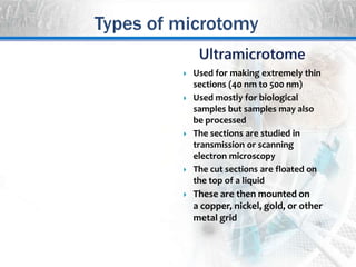
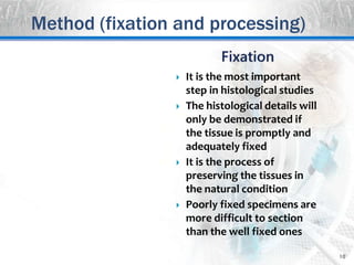
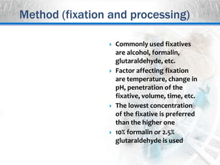
Biological Techniques
2
 Microtomy. Technique by
which tissues can be sliced into
thin sections and then
attached to a surface for
microscopy
 Microtomes. These are
instruments used to cut
uniformly thin sections of the
tissues for further study.
 Microtomes use sharp glass or
steel blades/diamond knives to
cut the tissues into thin
sections of uniform thickness
Introduction
 Thickness of the sections is
predetermined at regular
distance on the microtome
 The sections are attached to a
surface like glass slides for
staining
 Tissues like muscles, bones, hair
are cut of thickness from
50 nm and 100 µm
 Thickness of the sections may be
fixed according to the need of
the study and material involved
 The stained sections can be
studied by light or electron
microscopy
Introduction
Biological Techniques
Microtomy-
Types of
microtomy
Types of microtomy
 Main types include
◦ Rotary microtome
◦ Cryotome
◦ Ultra microtome
◦ Laser microtome
 Use depends on the material
under study and the nature of
the work
 Simple studies involve rotary
microtome while detailed
studies may involve ultra
microtome, etc.
 Similarly cryomicrotome
involves sectioning of the
frozen tissues.
 The most common type of the
microtome
 Rotary action involves the
sectioning process at
predetermined thickness on
every rotation of the flywheel
 Blade is fixed at horizontal
position
 The sample holder moves the
sample ahead by the fixed
distance for cutting
 Flywheel of the instrument may
be automatic or manual
 Section thickness may vary from
0.5 µm to 60 µm
Types of microtomy
Types of microtomy
 Used for making extremely thin
sections (40 nm to 500 nm)
 Used mostly for biological
samples but samples may also
be processed
 The sections are studied in
transmission or scanning
electron microscopy
 The cut sections are floated on
the top of a liquid
 These are then mounted on
a copper, nickel, gold, or other
metal grid
Biological Techniques
Microtomy-
Method (fixation
and processing)
 It consists of following
main steps
 Fixation
 Processing
 Dehydration
 Clearing
 Embedding
 Section cutting
 Staining
 Deparaffinization
Method (fixation and processing)
10
 It is the most important
step in histological studies
 The histological details will
only be demonstrated if
the tissue is promptly and
adequately fixed
 It is the process of
preserving the tissues in
the natural condition
 Poorly fixed specimens are
more difficult to section
than the well fixed ones
Method (fixation and processing)
 Commonly used fixatives
are alcohol, formalin,
glutaraldehyde, etc.
 Factor affecting fixation
are temperature, change in
pH, penetration of the
fixative, volume, time, etc.
 The lowest concentration
of the fixative is preferred
than the higher one
 10% formalin or 2.5%
glutaraldehyde is used
Method (fixation and processing)
12
 It is a process in which tissues
are treated to make the thin
sections
 A specimen is generally
processed as follows
 Dehydration. It is the removal
of water from the tissues by
immersing serially in 50%, 70%,
85% and 100% alcohol for some
time
 Clearing. It is removal of the
dehydrant from the tissues by
immersing in paraffin miscible
medium like xylene
Method (fixation and processing)
Biological Techniques
Microtomy-
Method
(embedding)
14
 It is the process of placing the
processed samples into molds
like paraffin blocks, etc.
 The blocks are then used on
the microtome to make thin
sections
 There are a number of
techniques that can be used to
make sections from difficult
blocks.
 If the block is hard or friable,
the exposed surface can be
soaked in cold water or a
softening agent
Method (embedding)
15
 Embedding is an important
step that always requires a
care
 Any excess wax on the
outside of a cassette
should be removed to
ensure the block is firmly
held during the sectioning
 Specimen orientation is
very important
 Paraffin embedded
specimens may be stored
indefinitely for future use
Method (embedding)
Biological Techniques
Microtomy-
Method
(sectioning and
staining)
17
 It is process of cutting
the embedded samples
into thin sections
 It requires great care as
tissues of the diagnostic
importance can easily be
lost or the block surface
may be damaged.
 We should be sure that
all of the clamping
mechanisms are
tightened securely
Method (sectioning and staining)
18
 The thickness of the
sections is normally
between 10 μm to 30 μm.
 The cuts sections are then
floated on warm water (at
40 °C) bath that helps to
remove wrinkles
 These are then placed on
the glass microscopic
slides or some other
support for staining, etc.
Method (sectioning and staining)
19
 The slides must always
be grease and dust free
and stored and handled
correctly.
 It involves reversal of the
process of embedding to
remove paraffin and
then staining by water
soluble stains
 The slides are treated
with xylene and alcohol
to remove the paraffin
Method (sectioning and staining)
20
 Some of the stains need
mordants (Iodine acts as a
mordant in Gram staining
of bacteria)
 The stains used depend on
their differential affinity
for various cellular
organelles
 Some of the special stains
are used in special cases
 Slides must always be
properly labeled
Method (sectioning and staining)

microtomy

  • 1.
  • 2.
    2  Microtomy. Techniqueby which tissues can be sliced into thin sections and then attached to a surface for microscopy  Microtomes. These are instruments used to cut uniformly thin sections of the tissues for further study.  Microtomes use sharp glass or steel blades/diamond knives to cut the tissues into thin sections of uniform thickness Introduction
  • 3.
     Thickness ofthe sections is predetermined at regular distance on the microtome  The sections are attached to a surface like glass slides for staining  Tissues like muscles, bones, hair are cut of thickness from 50 nm and 100 µm  Thickness of the sections may be fixed according to the need of the study and material involved  The stained sections can be studied by light or electron microscopy Introduction
  • 4.
  • 5.
    Types of microtomy Main types include ◦ Rotary microtome ◦ Cryotome ◦ Ultra microtome ◦ Laser microtome  Use depends on the material under study and the nature of the work  Simple studies involve rotary microtome while detailed studies may involve ultra microtome, etc.  Similarly cryomicrotome involves sectioning of the frozen tissues.
  • 6.
     The mostcommon type of the microtome  Rotary action involves the sectioning process at predetermined thickness on every rotation of the flywheel  Blade is fixed at horizontal position  The sample holder moves the sample ahead by the fixed distance for cutting  Flywheel of the instrument may be automatic or manual  Section thickness may vary from 0.5 µm to 60 µm Types of microtomy
  • 7.
    Types of microtomy Used for making extremely thin sections (40 nm to 500 nm)  Used mostly for biological samples but samples may also be processed  The sections are studied in transmission or scanning electron microscopy  The cut sections are floated on the top of a liquid  These are then mounted on a copper, nickel, gold, or other metal grid
  • 8.
  • 9.
     It consistsof following main steps  Fixation  Processing  Dehydration  Clearing  Embedding  Section cutting  Staining  Deparaffinization Method (fixation and processing)
  • 10.
    10  It isthe most important step in histological studies  The histological details will only be demonstrated if the tissue is promptly and adequately fixed  It is the process of preserving the tissues in the natural condition  Poorly fixed specimens are more difficult to section than the well fixed ones Method (fixation and processing)
  • 11.
     Commonly usedfixatives are alcohol, formalin, glutaraldehyde, etc.  Factor affecting fixation are temperature, change in pH, penetration of the fixative, volume, time, etc.  The lowest concentration of the fixative is preferred than the higher one  10% formalin or 2.5% glutaraldehyde is used Method (fixation and processing)
  • 12.
    12  It isa process in which tissues are treated to make the thin sections  A specimen is generally processed as follows  Dehydration. It is the removal of water from the tissues by immersing serially in 50%, 70%, 85% and 100% alcohol for some time  Clearing. It is removal of the dehydrant from the tissues by immersing in paraffin miscible medium like xylene Method (fixation and processing)
  • 13.
  • 14.
    14  It isthe process of placing the processed samples into molds like paraffin blocks, etc.  The blocks are then used on the microtome to make thin sections  There are a number of techniques that can be used to make sections from difficult blocks.  If the block is hard or friable, the exposed surface can be soaked in cold water or a softening agent Method (embedding)
  • 15.
    15  Embedding isan important step that always requires a care  Any excess wax on the outside of a cassette should be removed to ensure the block is firmly held during the sectioning  Specimen orientation is very important  Paraffin embedded specimens may be stored indefinitely for future use Method (embedding)
  • 16.
  • 17.
    17  It isprocess of cutting the embedded samples into thin sections  It requires great care as tissues of the diagnostic importance can easily be lost or the block surface may be damaged.  We should be sure that all of the clamping mechanisms are tightened securely Method (sectioning and staining)
  • 18.
    18  The thicknessof the sections is normally between 10 μm to 30 μm.  The cuts sections are then floated on warm water (at 40 °C) bath that helps to remove wrinkles  These are then placed on the glass microscopic slides or some other support for staining, etc. Method (sectioning and staining)
  • 19.
    19  The slidesmust always be grease and dust free and stored and handled correctly.  It involves reversal of the process of embedding to remove paraffin and then staining by water soluble stains  The slides are treated with xylene and alcohol to remove the paraffin Method (sectioning and staining)
  • 20.
    20  Some ofthe stains need mordants (Iodine acts as a mordant in Gram staining of bacteria)  The stains used depend on their differential affinity for various cellular organelles  Some of the special stains are used in special cases  Slides must always be properly labeled Method (sectioning and staining)