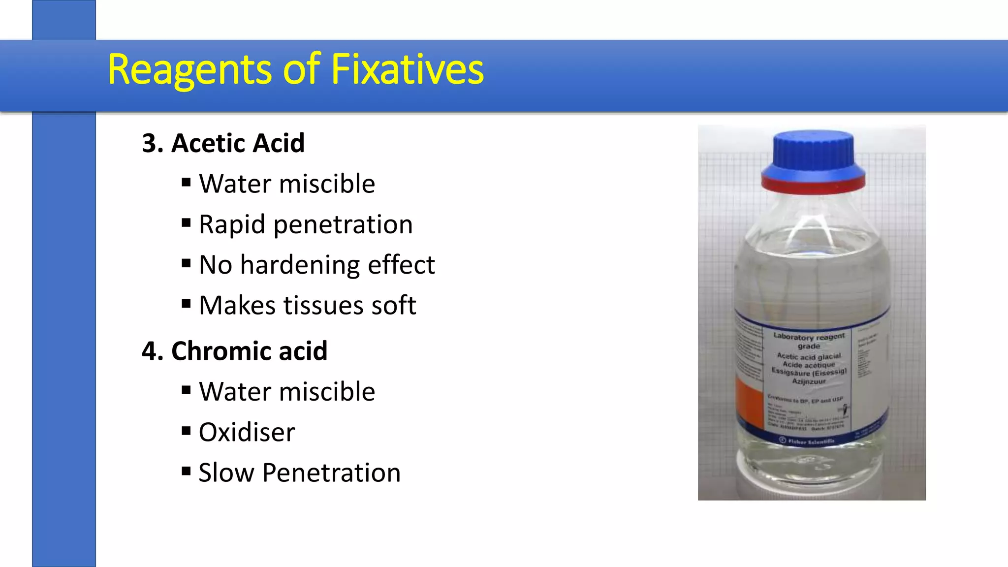
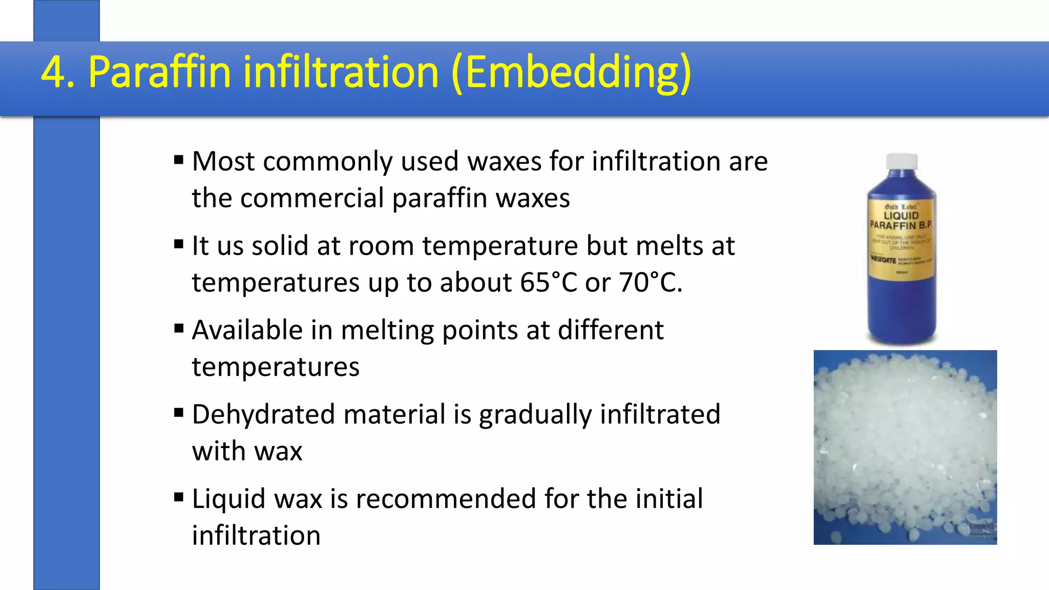
PREPARATION OF BIOLOGICAL SPECIMENS
FOR LIGHT MICROSCOPY
Dr. Ratheesh Chandra P.
 Sample collection
 Killing and Fixation
 Dehydration
 Clearing
 Paraffin Embedding
 Microtomy
 Staining
 Observation
Steps
1. Killing and Fixation
 Essential requirement
 Performed by fixative
 Killing – Sudden stoppage of life processes
 Fixation - Preservation of a “life-like” state
 Purposes –
 Preservation of natural form
 Modifying RI
 Making material resistant and hard
 Preparing material for improving staining
Reagents of Fixatives
 No single reagent for the purpose
 Combinations of reagents
 Principle – Keep balance between properties
 1. Ethyl alcohol
 Water soluble
 Reducing agent
 Rapid Penetrability
 Shrink tissues
 Hardening effect
 Makes tissues difficult to stain
2. Formalin
 Aqueous Formaldehyde
 Reducing agent
 Water miscible
 Slow Penetration
 Causes shrinkage
 Great Hardening effect
 Makes staining difficult
Reagents of Fixatives
3. Acetic Acid
 Water miscible
 Rapid penetration
 No hardening effect
 Makes tissues soft
4. Chromic acid
 Water miscible
 Oxidiser
 Slow Penetration
Reagents of Fixatives
Killing and Fixing Fluids (Fixatives)
 Many groups based on their ingredients
 Selection depends on specific requirement
 Some stable
 Some unstable
 Some known by their ingredients
 Some by their investigators
1. Farmer’ s Formula
 Glacial Acetic acid- 5m ml
 Absolute Alcohol - 15 ml
 Ideal for cytological preparations – Root tips, Anther
 Fixation time – Root tips – 15 m, Anthers – 1 h
 Washing and storage in 70% alcohol
2. Carnoy’s Formula
 Absolute alcohol - 10 ml
 Chloroform - 15 ml
 Glacial Acetic acid- 5 ml
 Ideal for cytological preparations
 Fixation time – 10- 15 m.
 Washing and storage in 85% alcohol
Killing and Fixing Fluids (Fixatives)
Acetic
Acid
Alcohol
Mixtures
Killing and Fixing Fluids (Fixatives)
1. Rawlin’ Formula
 95% Ethyl alcohol - 50 ml
 Glacial Acetic acid - 5 ml
 Formalin - 10 ml
 Water - 35 ml
 For delicate materials
 Good hardening action and
 Materials may be stored in this for years even
 For hard woody materials decrease acid and increased formalin
 Fixation time: 18 hrs
 Wash in alcohol and store in same
Formalin
Acetic
acid
Alcohol
(FAA)
Mixtures
Killing and Fixing Fluids (Fixatives)
1. Chromo acetic acid ( Weak)
Chromic acid 1% - 50ml
Acetic acid 1% - 50 ml
2. Chromo acetic (Medium)
Chromic acid 1% - 70ml
Acetic acid 1% - 20 ml
Water - 10 ml
3. Chromoic acetic : Strong
Chromic acid 1% - 97 ml
Acetic acid 1% - 3 ml
Chromo
Acetic
Acid
Mixtures
Killing and Fixing Fluids (Fixatives)
 Recommended for delicate objects like
filamentous and thalloid plants, root tips, Floral
organs and small sections of leaves or stems
 Fixation time: Few minutes for algae, 12 hours
for small leaf and root tips
 24 hours for larger pieces of tissue
 Wash well in running water for 24 hours and
then in distilled water for 12 hours
Chromo
Acetic
Acid
Mixtures
1. Navaschin’s Formula
Sol. A: Chromic acid (1%) - 15 ml
Glacial acetic acid - 10 ml
Distilled water - 90 ml
Sol. B: Formalin - 40 ml
Distilled water - 60 ml
 Mix equal quantities of A and B just before use
 Fixation time : 12 hours
 Washing in water not required
 Navashin’s original formula has been modified by
many investigators and the name CRAF has been
coined for such types
Chromo
Acetic
Acid
Formalin
Mixtures
Killing and Fixing Fluids (Fixatives)
 Craf I
 Chromic acid 1% 20 ml
 Acetic acid 1% 75 ml
 Formalin 5 ml
 Craf II
Chromic acid 1 % 20 ml
Acetic acid 10% 10 ml
Formalin 5 ml
Distilled water 65 ml
Chromo
Acetic
Acid
Formalin
Mixtures
Killing and Fixing Fluids (Fixatives)
CRAF
 In all these formalin should be added just before use
 A few hours after the addition of formalin a color change
takes place.
 Subsequently the color become olive green
 This fluid now acts as a preservative
 Fixation time 12 hours
 No washing in water
Chromo
Acetic
Acid
Formalin
Mixtures
Killing and Fixing Fluids (Fixatives)
2. Dehydration
 Chemical removal of water and fixative from the specimen
 Replace them with dehydrating fluid - dehydrant
 Many dehydrants are alcohols. Several are hydrophilic so attract water from tissue.
 Practiced in graded series
 Progressively decreasing concentration of water
 Progressively increasing concentration of dehydrant
Dehydrants – Reagents in dehydration
 Some merely removes water
 Some acts also as solvents of mounting media
 Common dehydrants are ethyl alcohol, acetone, normal butyl alcohol , tertiary
butyl alcohol Glycerine, Dioxan etc.
Ethyl Alcohol/Isopropyl alcohol
 Most common
 Progressively increasing concentrations – 10%, 20%, 30%, 40% …… 100%
 Begin with a grade same as the water content in the tissue
 Time required – soft tissues ~30 minutes – Hard/ large tissue- ~6-12 hrs.
Normal Butyl Alcohol
 Advantage – solvents of paraffin – directly followed to impregnation
 Grades are prepared in combination with ethyl alcohol
Series No. 95 % Ethyl
Alcohol (ml)
Normal butyl
alcohol (ml)
Distilled water
(ml)
1.
2.
3.
4.
5.
6.
7.
8.
20
25
30
30
25
20
15
0
10
15
25
40
55
70
85
100
70
60
45
30
20
10
0
0
1 hour
2 hour
Tertiary Butyl Alcohol (TBA)
Series No. Absolute Alcohol (ml) 95% Ethyl
Alcohol (ml)
TBA
(ml)
Dist. Water
(ml)
1.
2.
3.
4.
5.
0
0
0
0
25
50
50
50
50
0
10
20
35
50
75
100
40
30
15
0
0
Dehydrate first in ethyl alcohol upto 50%
Three changes in absolute TBA
3. Clearing (Dealcoholization)
 Removal of alcohol from the tissues
 Replacing the dehydrating fluid with a fluid that is totally miscible with
both the dehydrating fluid and the embedding medium- Paraffin
 Transition step between dehydration and infiltration
 Only needed when the dehydrants are not solvents of wax
 Clearing agents- Xylene, Toluene, Chloroform, Benzene, Petrol etc.
Reagents in Clearing - Xylene
 Xylene- Conventional reagent in dealcoholization
 Practiced in graded series (30 min 1hr in each)
Series No. Ethyl alcohol (ml) Xylene (ml)
1
2
3
4
5
6
7
8
9
10
90
80
70
60
50
40
30
20
10
0
10
20
30
40
50
60
70
80
90
100
4. Paraffin infiltration (Embedding)
 Most commonly used waxes for infiltration are
the commercial paraffin waxes
 It us solid at room temperature but melts at
temperatures up to about 65°C or 70°C.
 Available in melting points at different
temperatures
 Dehydrated material is gradually infiltrated
with wax
 Liquid wax is recommended for the initial
infiltration
Paraffin Series
Paraffin Series Interval
50% TBA + 50 % Liquid Paraffin
100 % Liquid paraffin
1- 6 hours
1- 6 hours
20 %
40 %
60 %
80 %
100 %
1- 6 hours
1- 6 hours
1- 6 hours
1- 6 hours
1- 6 hours
at 70⁰ C
Oven
Paraffinwax
Paraffin Embedding
 Three changes in 100 % wax
 Paraffin block-material preparation
 Attachment of the block into the holder of the
microtome
 Sectioning with microtome
Steps involved
1. Killing and fixation
2. Dehydration
3. Clearing
4. Paraffin infiltration
5. Casting of wax impregnated material into blocks
6. Attachment of the block into the holder of the microtome
7. Microtomy
8. Affixing paraffin ribbon on glass slides
9. Removal of wax
10.Staining and mounting
Sections
 Sectioning allows light pass through the material
 FREE HAND SECTIONS
 SERIAL SECTIONS
FREE HAND SECTIONS
 Can be done if the material is hard
 Thin sections - 10 µM can be taken
 Sectioning with razor
Serial sections
 Serial sections are produced by paraffin method
 Paraffin infiltrated material are affixed on
wooden blocks
 Objects are cut into a series of sections
 Serial sections are placed on adhesive smeared
glass slides
 Serial sections enables the reconstruction of
structure of organ
 Orientation of vasculature, cellular organization
etc. can be studied
5. Microtomy
Stains and Staining
 Staining - Use of dyes to provide color to various tissue constituents
 Different tissue constituents react differently to dyes – contrast
 Chromogen
 Chromophore
 Auxochrome – acid/ alkali radicals. Responsible for solubility
Stains - classification
Principle Chemical Nature
Chemical Nature Basic : Colored organic base+ uncolored acetate, chloride or sulphate radical
(safranin, methylene blue, crystal violet)
Acidic : Metallic base (Na, K) + Colored organic radical (Aniline Blue, Eosin,
Orange G )
Neutral : Combinations of acidic and basic dyes (Giesma stain, Sudan black B)
Affinity to different
plant parts
Nuclear : Nucleus
Cytoplasmic: Cytoplasm
Microtechnical
purposes
Histological: defines tissues (xylem, phloem etc.)
Cytological : Define cell components (nucleus, chromosomes etc.)
Stains
Natural Dyes – dyes obtained from plant/ animal (Brazilin, Hematoxylin, Carmine)
Synthetic dyes – made from Coal tar – (Orange G, Safranine, Fast Green)
 Brazilin (Timber of Caesalpinia crista, C. echinata)
 Hematoxylin Hematoxylon campechianum
 Carmine Insect Dactylopius coccus
Staining Methods
1. Progressive staining
2. Regressive (Retrogressive staining)
3. Counter staining
4. Double, triple and quadruple staining
Methods of Staining
Progressive Staining
 Useful for beginners
 Tissues are understained first
 Gradually more stain is added until the desired intensity attained
 Staining interval required is determined by trial
Regressive (Retrogressive) Staining
 Overstained first
 Then destained until the desired intensity is attained
 Destaining agent – 70% alcohol with 1% acetic acid
 Proper washing after differentiation
Counterstaining
 Staining certain part of cells/ tissues with one stain
 Other parts with a contrasting color
Double/ Triple/ Quadruple staining
 Use of 2, 3, 4 colors on same section
 Double staining - Safranin O and Fast Green
 Triple staining - Safranin O, Gentian Violet and Orange G
 Quadruple Staining - Safranin O, Methyl violet, Fast Green and Orange G
Methods of Staining
Whole Mounts
 Used to preserve and retain natural color, form and shape of
whole plants/ plant parts
 Microscopic museum materials preserved in ethyl alcohol,
formalin
Water – 72 ml
Formaldehyde – 5 ml
Glacial acetic acid – 3 ml
Glycerine - 20 ml
 Temporary whole mounts – small filamentous algae- in 10%
glycerine/ coverslip
Whole Mounts
 Permanent whole mounts – Microscopic Material
 Constant handling requires preparation of permanent nature
1. Killing and fixation
2. Washing in water
3. Staining with hematoxylin for 30 min -1 hour
4. Destaining in 0.1% HCl
5. Transfer to glass slide
6. Covering with DPX and cover slip
Cytological Methods
 Used to study the minute details of the cell structure – nucleus
 Smear and squash methods are the most common.
 Smear – Smearing material on glass slide (Acetocarmine
method, Feulgen method)
 Squashes - component parts separate and are not studied
intact.
MACERATION
 Separation of cells of fixed plant or animal material through
hydrolysis
 Useful to visualize the 3d nature of structural elements
 Reagents used depends – nature of middle lamella
Maceration
Middle lamella
 Herbaceous – Pectin (boiling in water)
 Woody - Lignin (alkali/ acid/ enzyme treatment)
 3 common methods in practice
1. Schultze’ s Method
 Treatment with con. H2SO4+KClO3 and warming
 After thorough bleaching washing in water
2. Jeffrey’ s Method
 Treatment in equal vol 10% HNO3+K2CrO4 at 30° C - 40° C for 1 -2 days
 Thorough washing
3. Harlow’ s Method
 Treatment in chorine water –2 hours
 Washing in running water
 Boiling in 3% Na2SO3 – 15 min
 Washing
 Staining in Safranin
 Washing in water
 Dehydration with hygrobutol
 Infiltration with Canada balsum
 After placing the material on glass slides tease with needles
 Mount with cover slip
Maceration
?

PREPARATION OF BIOLOGICAL SPECIMENS FOR LIGHT MICROSCOPY

  • 1.
    PREPARATION OF BIOLOGICALSPECIMENS FOR LIGHT MICROSCOPY Dr. Ratheesh Chandra P.
  • 2.
     Sample collection Killing and Fixation  Dehydration  Clearing  Paraffin Embedding  Microtomy  Staining  Observation Steps
  • 3.
    1. Killing andFixation  Essential requirement  Performed by fixative  Killing – Sudden stoppage of life processes  Fixation - Preservation of a “life-like” state  Purposes –  Preservation of natural form  Modifying RI  Making material resistant and hard  Preparing material for improving staining
  • 4.
    Reagents of Fixatives No single reagent for the purpose  Combinations of reagents  Principle – Keep balance between properties  1. Ethyl alcohol  Water soluble  Reducing agent  Rapid Penetrability  Shrink tissues  Hardening effect  Makes tissues difficult to stain
  • 5.
    2. Formalin  AqueousFormaldehyde  Reducing agent  Water miscible  Slow Penetration  Causes shrinkage  Great Hardening effect  Makes staining difficult Reagents of Fixatives
  • 6.
    3. Acetic Acid Water miscible  Rapid penetration  No hardening effect  Makes tissues soft 4. Chromic acid  Water miscible  Oxidiser  Slow Penetration Reagents of Fixatives
  • 7.
    Killing and FixingFluids (Fixatives)  Many groups based on their ingredients  Selection depends on specific requirement  Some stable  Some unstable  Some known by their ingredients  Some by their investigators
  • 8.
    1. Farmer’ sFormula  Glacial Acetic acid- 5m ml  Absolute Alcohol - 15 ml  Ideal for cytological preparations – Root tips, Anther  Fixation time – Root tips – 15 m, Anthers – 1 h  Washing and storage in 70% alcohol 2. Carnoy’s Formula  Absolute alcohol - 10 ml  Chloroform - 15 ml  Glacial Acetic acid- 5 ml  Ideal for cytological preparations  Fixation time – 10- 15 m.  Washing and storage in 85% alcohol Killing and Fixing Fluids (Fixatives) Acetic Acid Alcohol Mixtures
  • 9.
    Killing and FixingFluids (Fixatives) 1. Rawlin’ Formula  95% Ethyl alcohol - 50 ml  Glacial Acetic acid - 5 ml  Formalin - 10 ml  Water - 35 ml  For delicate materials  Good hardening action and  Materials may be stored in this for years even  For hard woody materials decrease acid and increased formalin  Fixation time: 18 hrs  Wash in alcohol and store in same Formalin Acetic acid Alcohol (FAA) Mixtures
  • 10.
    Killing and FixingFluids (Fixatives) 1. Chromo acetic acid ( Weak) Chromic acid 1% - 50ml Acetic acid 1% - 50 ml 2. Chromo acetic (Medium) Chromic acid 1% - 70ml Acetic acid 1% - 20 ml Water - 10 ml 3. Chromoic acetic : Strong Chromic acid 1% - 97 ml Acetic acid 1% - 3 ml Chromo Acetic Acid Mixtures
  • 11.
    Killing and FixingFluids (Fixatives)  Recommended for delicate objects like filamentous and thalloid plants, root tips, Floral organs and small sections of leaves or stems  Fixation time: Few minutes for algae, 12 hours for small leaf and root tips  24 hours for larger pieces of tissue  Wash well in running water for 24 hours and then in distilled water for 12 hours Chromo Acetic Acid Mixtures
  • 12.
    1. Navaschin’s Formula Sol.A: Chromic acid (1%) - 15 ml Glacial acetic acid - 10 ml Distilled water - 90 ml Sol. B: Formalin - 40 ml Distilled water - 60 ml  Mix equal quantities of A and B just before use  Fixation time : 12 hours  Washing in water not required  Navashin’s original formula has been modified by many investigators and the name CRAF has been coined for such types Chromo Acetic Acid Formalin Mixtures Killing and Fixing Fluids (Fixatives)
  • 13.
     Craf I Chromic acid 1% 20 ml  Acetic acid 1% 75 ml  Formalin 5 ml  Craf II Chromic acid 1 % 20 ml Acetic acid 10% 10 ml Formalin 5 ml Distilled water 65 ml Chromo Acetic Acid Formalin Mixtures Killing and Fixing Fluids (Fixatives)
  • 14.
    CRAF  In allthese formalin should be added just before use  A few hours after the addition of formalin a color change takes place.  Subsequently the color become olive green  This fluid now acts as a preservative  Fixation time 12 hours  No washing in water Chromo Acetic Acid Formalin Mixtures Killing and Fixing Fluids (Fixatives)
  • 15.
    2. Dehydration  Chemicalremoval of water and fixative from the specimen  Replace them with dehydrating fluid - dehydrant  Many dehydrants are alcohols. Several are hydrophilic so attract water from tissue.  Practiced in graded series  Progressively decreasing concentration of water  Progressively increasing concentration of dehydrant
  • 16.
    Dehydrants – Reagentsin dehydration  Some merely removes water  Some acts also as solvents of mounting media  Common dehydrants are ethyl alcohol, acetone, normal butyl alcohol , tertiary butyl alcohol Glycerine, Dioxan etc. Ethyl Alcohol/Isopropyl alcohol  Most common  Progressively increasing concentrations – 10%, 20%, 30%, 40% …… 100%  Begin with a grade same as the water content in the tissue  Time required – soft tissues ~30 minutes – Hard/ large tissue- ~6-12 hrs.
  • 17.
    Normal Butyl Alcohol Advantage – solvents of paraffin – directly followed to impregnation  Grades are prepared in combination with ethyl alcohol Series No. 95 % Ethyl Alcohol (ml) Normal butyl alcohol (ml) Distilled water (ml) 1. 2. 3. 4. 5. 6. 7. 8. 20 25 30 30 25 20 15 0 10 15 25 40 55 70 85 100 70 60 45 30 20 10 0 0 1 hour 2 hour
  • 18.
    Tertiary Butyl Alcohol(TBA) Series No. Absolute Alcohol (ml) 95% Ethyl Alcohol (ml) TBA (ml) Dist. Water (ml) 1. 2. 3. 4. 5. 0 0 0 0 25 50 50 50 50 0 10 20 35 50 75 100 40 30 15 0 0 Dehydrate first in ethyl alcohol upto 50% Three changes in absolute TBA
  • 19.
    3. Clearing (Dealcoholization) Removal of alcohol from the tissues  Replacing the dehydrating fluid with a fluid that is totally miscible with both the dehydrating fluid and the embedding medium- Paraffin  Transition step between dehydration and infiltration  Only needed when the dehydrants are not solvents of wax  Clearing agents- Xylene, Toluene, Chloroform, Benzene, Petrol etc.
  • 20.
    Reagents in Clearing- Xylene  Xylene- Conventional reagent in dealcoholization  Practiced in graded series (30 min 1hr in each) Series No. Ethyl alcohol (ml) Xylene (ml) 1 2 3 4 5 6 7 8 9 10 90 80 70 60 50 40 30 20 10 0 10 20 30 40 50 60 70 80 90 100
  • 21.
    4. Paraffin infiltration(Embedding)  Most commonly used waxes for infiltration are the commercial paraffin waxes  It us solid at room temperature but melts at temperatures up to about 65°C or 70°C.  Available in melting points at different temperatures  Dehydrated material is gradually infiltrated with wax  Liquid wax is recommended for the initial infiltration
  • 22.
    Paraffin Series Paraffin SeriesInterval 50% TBA + 50 % Liquid Paraffin 100 % Liquid paraffin 1- 6 hours 1- 6 hours 20 % 40 % 60 % 80 % 100 % 1- 6 hours 1- 6 hours 1- 6 hours 1- 6 hours 1- 6 hours at 70⁰ C Oven Paraffinwax
  • 23.
    Paraffin Embedding  Threechanges in 100 % wax  Paraffin block-material preparation  Attachment of the block into the holder of the microtome  Sectioning with microtome
  • 24.
    Steps involved 1. Killingand fixation 2. Dehydration 3. Clearing 4. Paraffin infiltration 5. Casting of wax impregnated material into blocks 6. Attachment of the block into the holder of the microtome 7. Microtomy 8. Affixing paraffin ribbon on glass slides 9. Removal of wax 10.Staining and mounting
  • 25.
    Sections  Sectioning allowslight pass through the material  FREE HAND SECTIONS  SERIAL SECTIONS FREE HAND SECTIONS  Can be done if the material is hard  Thin sections - 10 µM can be taken  Sectioning with razor
  • 26.
    Serial sections  Serialsections are produced by paraffin method  Paraffin infiltrated material are affixed on wooden blocks  Objects are cut into a series of sections  Serial sections are placed on adhesive smeared glass slides  Serial sections enables the reconstruction of structure of organ  Orientation of vasculature, cellular organization etc. can be studied
  • 27.
  • 28.
    Stains and Staining Staining - Use of dyes to provide color to various tissue constituents  Different tissue constituents react differently to dyes – contrast  Chromogen  Chromophore  Auxochrome – acid/ alkali radicals. Responsible for solubility
  • 29.
    Stains - classification PrincipleChemical Nature Chemical Nature Basic : Colored organic base+ uncolored acetate, chloride or sulphate radical (safranin, methylene blue, crystal violet) Acidic : Metallic base (Na, K) + Colored organic radical (Aniline Blue, Eosin, Orange G ) Neutral : Combinations of acidic and basic dyes (Giesma stain, Sudan black B) Affinity to different plant parts Nuclear : Nucleus Cytoplasmic: Cytoplasm Microtechnical purposes Histological: defines tissues (xylem, phloem etc.) Cytological : Define cell components (nucleus, chromosomes etc.)
  • 30.
    Stains Natural Dyes –dyes obtained from plant/ animal (Brazilin, Hematoxylin, Carmine) Synthetic dyes – made from Coal tar – (Orange G, Safranine, Fast Green)  Brazilin (Timber of Caesalpinia crista, C. echinata)  Hematoxylin Hematoxylon campechianum  Carmine Insect Dactylopius coccus Staining Methods 1. Progressive staining 2. Regressive (Retrogressive staining) 3. Counter staining 4. Double, triple and quadruple staining
  • 31.
    Methods of Staining ProgressiveStaining  Useful for beginners  Tissues are understained first  Gradually more stain is added until the desired intensity attained  Staining interval required is determined by trial Regressive (Retrogressive) Staining  Overstained first  Then destained until the desired intensity is attained  Destaining agent – 70% alcohol with 1% acetic acid  Proper washing after differentiation
  • 32.
    Counterstaining  Staining certainpart of cells/ tissues with one stain  Other parts with a contrasting color Double/ Triple/ Quadruple staining  Use of 2, 3, 4 colors on same section  Double staining - Safranin O and Fast Green  Triple staining - Safranin O, Gentian Violet and Orange G  Quadruple Staining - Safranin O, Methyl violet, Fast Green and Orange G Methods of Staining
  • 33.
    Whole Mounts  Usedto preserve and retain natural color, form and shape of whole plants/ plant parts  Microscopic museum materials preserved in ethyl alcohol, formalin Water – 72 ml Formaldehyde – 5 ml Glacial acetic acid – 3 ml Glycerine - 20 ml  Temporary whole mounts – small filamentous algae- in 10% glycerine/ coverslip
  • 34.
    Whole Mounts  Permanentwhole mounts – Microscopic Material  Constant handling requires preparation of permanent nature 1. Killing and fixation 2. Washing in water 3. Staining with hematoxylin for 30 min -1 hour 4. Destaining in 0.1% HCl 5. Transfer to glass slide 6. Covering with DPX and cover slip
  • 35.
    Cytological Methods  Usedto study the minute details of the cell structure – nucleus  Smear and squash methods are the most common.  Smear – Smearing material on glass slide (Acetocarmine method, Feulgen method)  Squashes - component parts separate and are not studied intact. MACERATION  Separation of cells of fixed plant or animal material through hydrolysis  Useful to visualize the 3d nature of structural elements  Reagents used depends – nature of middle lamella
  • 36.
    Maceration Middle lamella  Herbaceous– Pectin (boiling in water)  Woody - Lignin (alkali/ acid/ enzyme treatment)  3 common methods in practice 1. Schultze’ s Method  Treatment with con. H2SO4+KClO3 and warming  After thorough bleaching washing in water 2. Jeffrey’ s Method  Treatment in equal vol 10% HNO3+K2CrO4 at 30° C - 40° C for 1 -2 days  Thorough washing
  • 37.
    3. Harlow’ sMethod  Treatment in chorine water –2 hours  Washing in running water  Boiling in 3% Na2SO3 – 15 min  Washing  Staining in Safranin  Washing in water  Dehydration with hygrobutol  Infiltration with Canada balsum  After placing the material on glass slides tease with needles  Mount with cover slip Maceration
  • 38.