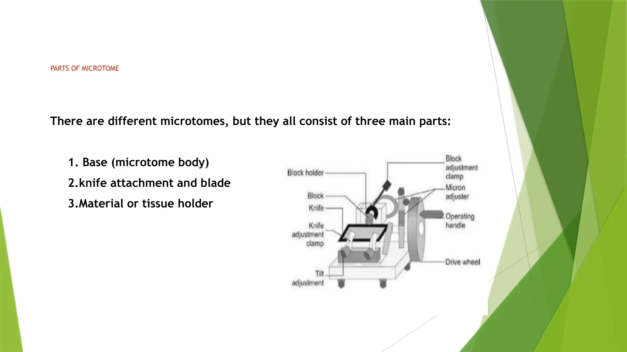
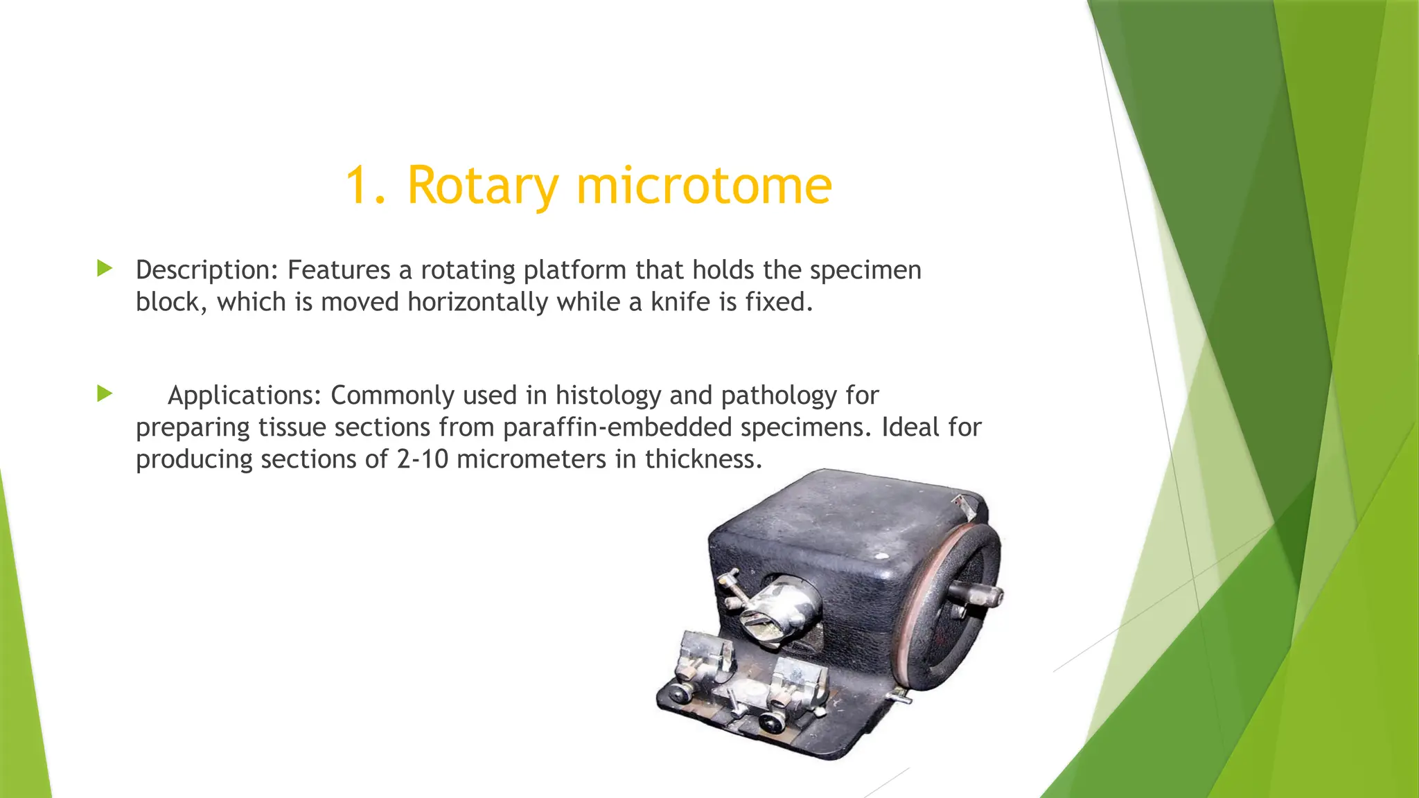
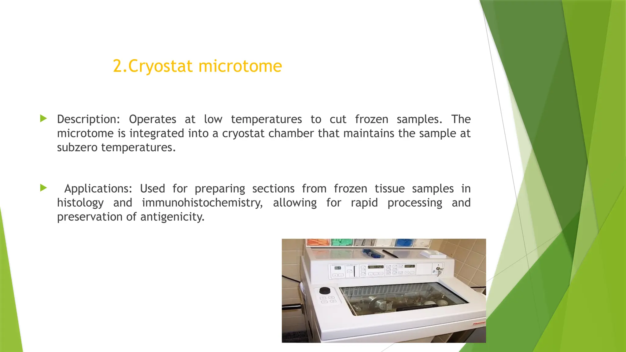
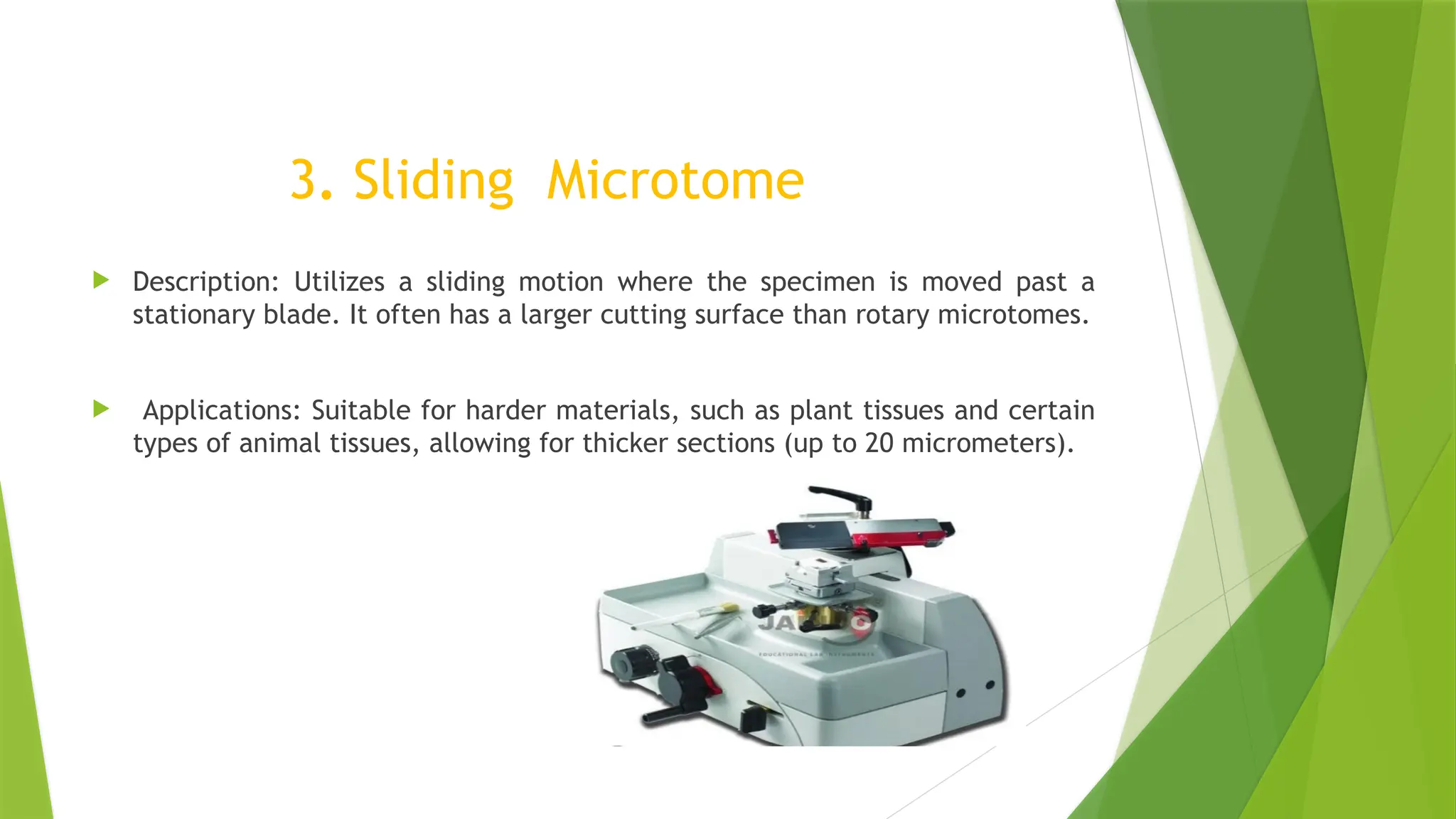
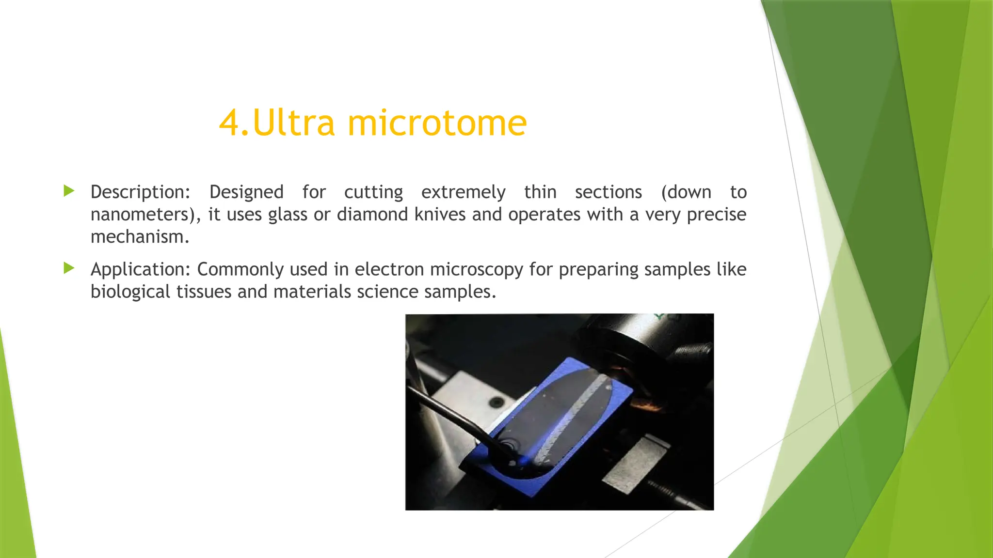
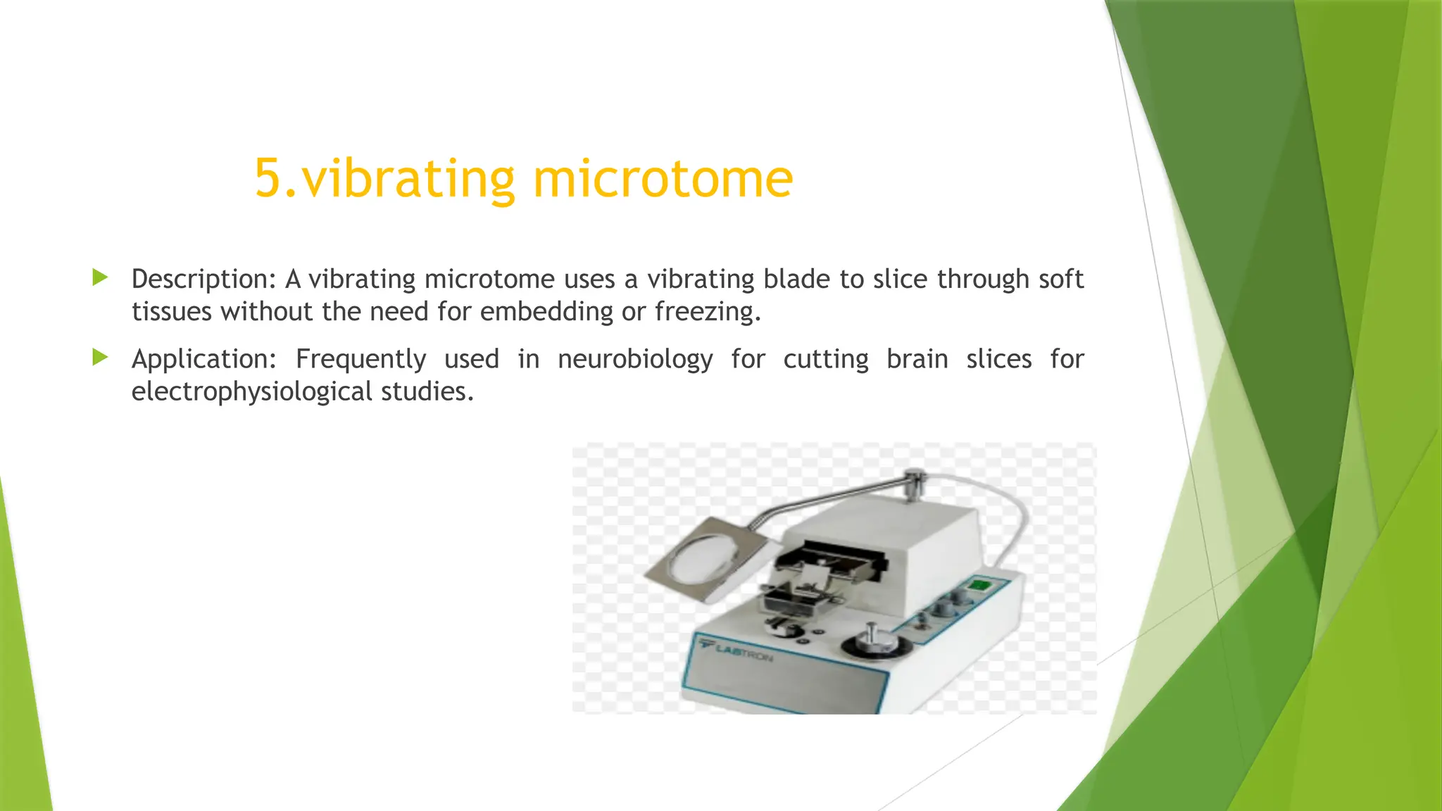
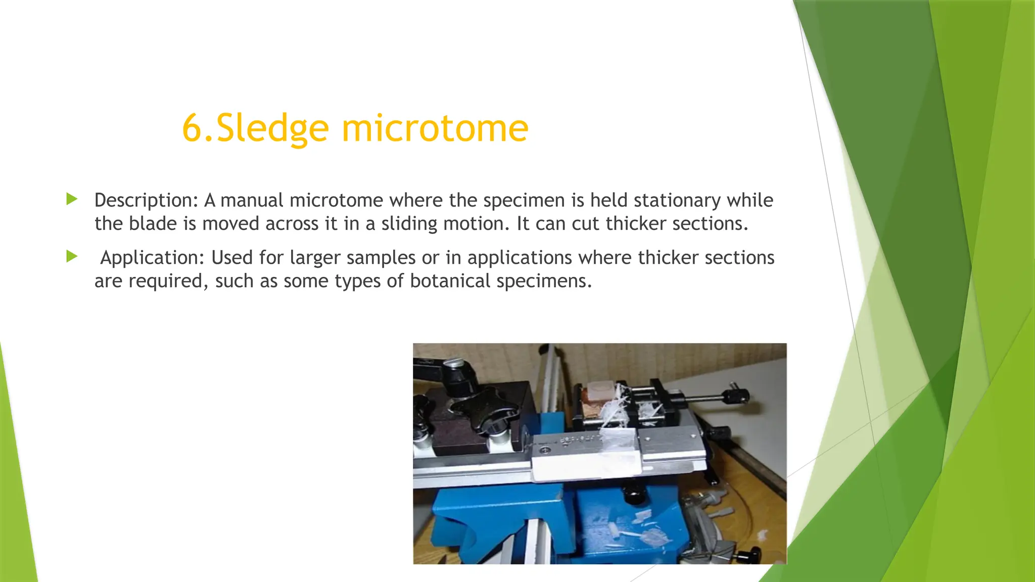
DEPARTMENT OF MICROBIOLOGY
SUBJECT- CLINICAL LABORATORY TECHNOLOGY
TOPIC-MICROTOMES AND MOUNTING SECTIONS
VIVEKANANDHA ARTS AND SCIENCE
COLLEGE FOR WOMEN
VEERACHIPALAYAM -636007,
SANKAGIRI, TAMILNADU
SUBJECT INCHARGE:
DR.V.BHARATHI
ASSISTANT PROFESSOR
DEPARTMENT OF
MICROBIOLOGY
VIAAS, SANKAGIRI
PRESENTED BY:
K.KAVYA
II B.SC MICROBIOLOGY
DEPARTMENT OF
MICROBIOLOGY
VIAAS, SANKAGIRI
MICROTOMES AND
MOUNTING SECTIONS
AGENDA
 Introduction
 History
 Most common application of microtomes
 Parts of microtomes
 Various types of microtomes
 Mounting sections
 Types of mounting sections
INTRODUCTION
 Microtomes are precision instruments used to cut extremely thin
sections of material such as tissue samples for microscopic
examination. Different types of microtomes are used depending on
the nature of the sample and the specific requirements of the
examination.
 A microtome is a cutting tool used to product extremely thin slices
of material known as sections. Importanence of microtomes are used
in microscopy ,allowing for the preparation of samples for
observation under transmitted light or electron radiation.
HISTORY
 Microtome was invented by WILHELM HIS at 1865. He invented the
microtome , a mechanical device used to slice thin tissue sections for
microscopic examination . He was the author of Anatomie menschlicher
Embryonen 3 vol (1880-85;Human Embryonic Anatomy), considered the
first accurate and exhaustive study of the development of the human
embryo.
 WILHELM HIS (born on July 9 ,1831, Basel, switz.—died May 1,
1904,Leipzig, Germany).was a Swiss –born German anatomist and
embryologist who created the science of histogenesis,or the study of the
Embryonic origins of different types of animal tissue.
THE MOST COMMON APPLICATION OF MICROTOMES ARE;
 Traditional Histology Technique.
 Cryosectioning Technique.
 Electron Microscopy Technique.
 Botanical Microtomy Technique.
PARTS OF MICROTOME
There are different microtomes, but they all consist of three main parts:
1. Base (microtome body)
2.knife attachment and blade
3.Material or tissue holder
VARIOUS TYPES OF MICROTOMES
 Rotary microtome
 Cryostat microtome
 Sliding microtome
 Ultra microtome
 Vibrating microtome
 Sledge microtome
1. Rotary microtome
 Description: Features a rotating platform that holds the specimen
block, which is moved horizontally while a knife is fixed.
 Applications: Commonly used in histology and pathology for
preparing tissue sections from paraffin-embedded specimens. Ideal for
producing sections of 2-10 micrometers in thickness.
2.Cryostat microtome
 Description: Operates at low temperatures to cut frozen samples. The
microtome is integrated into a cryostat chamber that maintains the sample at
subzero temperatures.
 Applications: Used for preparing sections from frozen tissue samples in
histology and immunohistochemistry, allowing for rapid processing and
preservation of antigenicity.
3. Sliding Microtome
 Description: Utilizes a sliding motion where the specimen is moved past a
stationary blade. It often has a larger cutting surface than rotary microtomes.
 Applications: Suitable for harder materials, such as plant tissues and certain
types of animal tissues, allowing for thicker sections (up to 20 micrometers).
4.Ultra microtome
 Description: Designed for cutting extremely thin sections (down to
nanometers), it uses glass or diamond knives and operates with a very precise
mechanism.
 Application: Commonly used in electron microscopy for preparing samples like
biological tissues and materials science samples.
5.vibrating microtome
 Description: A vibrating microtome uses a vibrating blade to slice through soft
tissues without the need for embedding or freezing.
 Application: Frequently used in neurobiology for cutting brain slices for
electrophysiological studies.
6.Sledge microtome
 Description: A manual microtome where the specimen is held stationary while
the blade is moved across it in a sliding motion. It can cut thicker sections.
 Application: Used for larger samples or in applications where thicker sections
are required, such as some types of botanical specimens.
MOUNTING SECTIONS
 Mounting sections involves transferring the thin tissue slices cut by the
microtome onto slides for microscopic examination . The process varies
slightly depending on the microtome type and the nature of the tissue, but
here’s a general overview;
TYPES:
1.Mounting paraffin sections .
2.Mounting frozen sections .
3.Mounting ultrathin sections.
4.Mounting vibratome sections.
1.MOUNTING PARAFFIN SECTIONS
 WATER BATH METHOD: After cutting, the paraffin sections are floated on a
warm water bath around 40°C .The warmth allows the sections to flatten out
and removes wrinkles .
 SLIDE TRANSFER: The flattened section is then carefully picked up using a
glass microscope slide . It adheres to the slides as the water evaporates.
 DRYING: The slide is dried ,often on a warming tray, to ensure the section is
securely attached to the slide before staining.
2.MOUNTING FROZEN SECTIONS
 DIRECT TRANSFER : Frozen sections are typically transferred directly onto a
slide immediately after cutting.
 FIXATION : After mounting, the sections may need to be fixed , usually with
cold acetone or alcohol, to preserve the tissue’s integrity before staining.
3.MOUNTING ULTRATHIN SECTIONS
 GRID MOUNTING: For ultrathin sections, the sections are typically floated
onto a water surface and then picked up onto tiny metal grids used in
electron microscopy.
 STAINING AND DRYING : The grids are then stained with heavy metals (eg.
uranyl acetate , leaf citrate) and dried before examination under the electron
microscope.
 DIRECT PLACEMENT: Sections are typically transferred directly from the
blade or water bath onto a slide or into a petri dish containing a buffer
solution.
 USE OF ADHESIVE: Sometimes, a small amount of adhesive like albumin is
used to help the section adhere to the slide , especially for thicker or less
pliable tissues.
4.MOUNTING VIBRATOME SECTIONS
REFERENCE
 https://en.m.wikipedia.org/wiki/Microtome
 https://www.britannica.com/biography/Wilhelm-His-Swiss-anatomist
 https://web.archive.org/web/20090425234311/http://
encarta.msn.com/encyclopedia_761573144/Histology.html
 Bancroft, John; Stevens, Alan, eds. (1982). The Theory and Practice of
Histological Techniques (2nd
ed.). Longman group Limited.
THANK YOU….

Clinical laboratory technology-Microtomes and mounting sections

  • 1.
    DEPARTMENT OF MICROBIOLOGY SUBJECT-CLINICAL LABORATORY TECHNOLOGY TOPIC-MICROTOMES AND MOUNTING SECTIONS VIVEKANANDHA ARTS AND SCIENCE COLLEGE FOR WOMEN VEERACHIPALAYAM -636007, SANKAGIRI, TAMILNADU SUBJECT INCHARGE: DR.V.BHARATHI ASSISTANT PROFESSOR DEPARTMENT OF MICROBIOLOGY VIAAS, SANKAGIRI PRESENTED BY: K.KAVYA II B.SC MICROBIOLOGY DEPARTMENT OF MICROBIOLOGY VIAAS, SANKAGIRI
  • 2.
  • 3.
    AGENDA  Introduction  History Most common application of microtomes  Parts of microtomes  Various types of microtomes  Mounting sections  Types of mounting sections
  • 4.
    INTRODUCTION  Microtomes areprecision instruments used to cut extremely thin sections of material such as tissue samples for microscopic examination. Different types of microtomes are used depending on the nature of the sample and the specific requirements of the examination.  A microtome is a cutting tool used to product extremely thin slices of material known as sections. Importanence of microtomes are used in microscopy ,allowing for the preparation of samples for observation under transmitted light or electron radiation.
  • 5.
    HISTORY  Microtome wasinvented by WILHELM HIS at 1865. He invented the microtome , a mechanical device used to slice thin tissue sections for microscopic examination . He was the author of Anatomie menschlicher Embryonen 3 vol (1880-85;Human Embryonic Anatomy), considered the first accurate and exhaustive study of the development of the human embryo.  WILHELM HIS (born on July 9 ,1831, Basel, switz.—died May 1, 1904,Leipzig, Germany).was a Swiss –born German anatomist and embryologist who created the science of histogenesis,or the study of the Embryonic origins of different types of animal tissue.
  • 6.
    THE MOST COMMONAPPLICATION OF MICROTOMES ARE;  Traditional Histology Technique.  Cryosectioning Technique.  Electron Microscopy Technique.  Botanical Microtomy Technique.
  • 7.
    PARTS OF MICROTOME Thereare different microtomes, but they all consist of three main parts: 1. Base (microtome body) 2.knife attachment and blade 3.Material or tissue holder
  • 8.
    VARIOUS TYPES OFMICROTOMES  Rotary microtome  Cryostat microtome  Sliding microtome  Ultra microtome  Vibrating microtome  Sledge microtome
  • 9.
    1. Rotary microtome Description: Features a rotating platform that holds the specimen block, which is moved horizontally while a knife is fixed.  Applications: Commonly used in histology and pathology for preparing tissue sections from paraffin-embedded specimens. Ideal for producing sections of 2-10 micrometers in thickness.
  • 10.
    2.Cryostat microtome  Description:Operates at low temperatures to cut frozen samples. The microtome is integrated into a cryostat chamber that maintains the sample at subzero temperatures.  Applications: Used for preparing sections from frozen tissue samples in histology and immunohistochemistry, allowing for rapid processing and preservation of antigenicity.
  • 11.
    3. Sliding Microtome Description: Utilizes a sliding motion where the specimen is moved past a stationary blade. It often has a larger cutting surface than rotary microtomes.  Applications: Suitable for harder materials, such as plant tissues and certain types of animal tissues, allowing for thicker sections (up to 20 micrometers).
  • 12.
    4.Ultra microtome  Description:Designed for cutting extremely thin sections (down to nanometers), it uses glass or diamond knives and operates with a very precise mechanism.  Application: Commonly used in electron microscopy for preparing samples like biological tissues and materials science samples.
  • 13.
    5.vibrating microtome  Description:A vibrating microtome uses a vibrating blade to slice through soft tissues without the need for embedding or freezing.  Application: Frequently used in neurobiology for cutting brain slices for electrophysiological studies.
  • 14.
    6.Sledge microtome  Description:A manual microtome where the specimen is held stationary while the blade is moved across it in a sliding motion. It can cut thicker sections.  Application: Used for larger samples or in applications where thicker sections are required, such as some types of botanical specimens.
  • 15.
    MOUNTING SECTIONS  Mountingsections involves transferring the thin tissue slices cut by the microtome onto slides for microscopic examination . The process varies slightly depending on the microtome type and the nature of the tissue, but here’s a general overview; TYPES: 1.Mounting paraffin sections . 2.Mounting frozen sections . 3.Mounting ultrathin sections. 4.Mounting vibratome sections.
  • 16.
    1.MOUNTING PARAFFIN SECTIONS WATER BATH METHOD: After cutting, the paraffin sections are floated on a warm water bath around 40°C .The warmth allows the sections to flatten out and removes wrinkles .  SLIDE TRANSFER: The flattened section is then carefully picked up using a glass microscope slide . It adheres to the slides as the water evaporates.  DRYING: The slide is dried ,often on a warming tray, to ensure the section is securely attached to the slide before staining.
  • 17.
    2.MOUNTING FROZEN SECTIONS DIRECT TRANSFER : Frozen sections are typically transferred directly onto a slide immediately after cutting.  FIXATION : After mounting, the sections may need to be fixed , usually with cold acetone or alcohol, to preserve the tissue’s integrity before staining.
  • 18.
    3.MOUNTING ULTRATHIN SECTIONS GRID MOUNTING: For ultrathin sections, the sections are typically floated onto a water surface and then picked up onto tiny metal grids used in electron microscopy.  STAINING AND DRYING : The grids are then stained with heavy metals (eg. uranyl acetate , leaf citrate) and dried before examination under the electron microscope.
  • 19.
     DIRECT PLACEMENT:Sections are typically transferred directly from the blade or water bath onto a slide or into a petri dish containing a buffer solution.  USE OF ADHESIVE: Sometimes, a small amount of adhesive like albumin is used to help the section adhere to the slide , especially for thicker or less pliable tissues. 4.MOUNTING VIBRATOME SECTIONS
  • 20.
    REFERENCE  https://en.m.wikipedia.org/wiki/Microtome  https://www.britannica.com/biography/Wilhelm-His-Swiss-anatomist https://web.archive.org/web/20090425234311/http:// encarta.msn.com/encyclopedia_761573144/Histology.html  Bancroft, John; Stevens, Alan, eds. (1982). The Theory and Practice of Histological Techniques (2nd ed.). Longman group Limited.
  • 21.