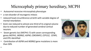
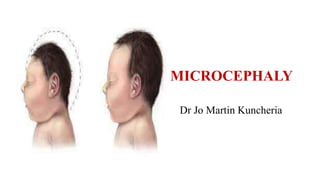
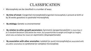
This document defines microcephaly and discusses its classification, etiology, pathogenesis, and diagnosis. Microcephaly is defined as an occipitofrontal circumference more than 3 standard deviations below the mean for age and gender. It can be classified as congenital or postnatal, genetic or environmental, symmetric or asymmetric, and isolated or syndromic. Causes include genetic conditions like primary microcephaly and chromosomal abnormalities, as well as congenital infections, metabolic disorders, and environmental factors. Neuroimaging can aid diagnosis by identifying brain abnormalities and calcifications.
The majority of children have a head size that is appropriate for age and gender. But a few have a too-large head at birth or may be of postnatal acceleration. Macrocephaly is used when the head size exceeds the mean by more than two standard devotions of age and gender. In addition, Macrocephaly is seen in association with several cranio-skeletal dysplastic conditions.
This presentation discusses cranial hemorrhage in a newborn baby. We have included extracranial and intracranial bleed discussion in neonates. Intraventricular hemorrhage (IVH) is further discussed in details in terms of pathophysiology, management strategies and clinical studies related to it.
Hope this presentation is helpful for the knowledge and practice of medical students, pediatricians and neonatologists and helps in practical management of your NICU babies as well.
This presentation is about Malnutrition in Pediatrics; Epidemiology, Risk factors, etiology, Clinical Evaluation, plotting on Growth charts and Management are Covered.
This file is one of my medical PPT series ,mainly intended for medical students. Information and pictures are highly organized to serve this aim. All the credits of info & pics are reserved for their owners.
Neural tube defects are birth defects of the brain, spine, or spinal cord. They happen in the first month of pregnancy, often before a woman even knows that she is pregnant. The two most common neural tube defects are spina bifida and anencephaly.
NVBDCP.pptx Nation vector borne disease control programSapna Thakur
NVBDCP was launched in 2003-2004 . Vector-Borne Disease: Disease that results from an infection transmitted to humans and other animals by blood-feeding arthropods, such as mosquitoes, ticks, and fleas. Examples of vector-borne diseases include Dengue fever, West Nile Virus, Lyme disease, and malaria.
micro teaching on communication m.sc nursing.pdfAnurag Sharma
Microteaching is a unique model of practice teaching. It is a viable instrument for the. desired change in the teaching behavior or the behavior potential which, in specified types of real. classroom situations, tends to facilitate the achievement of specified types of objectives.
Basavarajeeyam is an important text for ayurvedic physician belonging to andhra pradehs. It is a popular compendium in various parts of our country as well as in andhra pradesh. The content of the text was presented in sanskrit and telugu language (Bilingual). One of the most famous book in ayurvedic pharmaceutics and therapeutics. This book contains 25 chapters called as prakaranas. Many rasaoushadis were explained, pioneer of dhatu druti, nadi pareeksha, mutra pareeksha etc. Belongs to the period of 15-16 century. New diseases like upadamsha, phiranga rogas are explained.
Here is the updated list of Top Best Ayurvedic medicine for Gas and Indigestion and those are Gas-O-Go Syp for Dyspepsia | Lavizyme Syrup for Acidity | Yumzyme Hepatoprotective Capsules etc
Ozempic: Preoperative Management of Patients on GLP-1 Receptor Agonists Saeid Safari
Preoperative Management of Patients on GLP-1 Receptor Agonists like Ozempic and Semiglutide
ASA GUIDELINE
NYSORA Guideline
2 Case Reports of Gastric Ultrasound
These lecture slides, by Dr Sidra Arshad, offer a quick overview of the physiological basis of a normal electrocardiogram.
Learning objectives:
1. Define an electrocardiogram (ECG) and electrocardiography
2. Describe how dipoles generated by the heart produce the waveforms of the ECG
3. Describe the components of a normal electrocardiogram of a typical bipolar lead (limb II)
4. Differentiate between intervals and segments
5. Enlist some common indications for obtaining an ECG
6. Describe the flow of current around the heart during the cardiac cycle
7. Discuss the placement and polarity of the leads of electrocardiograph
8. Describe the normal electrocardiograms recorded from the limb leads and explain the physiological basis of the different records that are obtained
9. Define mean electrical vector (axis) of the heart and give the normal range
10. Define the mean QRS vector
11. Describe the axes of leads (hexagonal reference system)
12. Comprehend the vectorial analysis of the normal ECG
13. Determine the mean electrical axis of the ventricular QRS and appreciate the mean axis deviation
14. Explain the concepts of current of injury, J point, and their significance
Study Resources:
1. Chapter 11, Guyton and Hall Textbook of Medical Physiology, 14th edition
2. Chapter 9, Human Physiology - From Cells to Systems, Lauralee Sherwood, 9th edition
3. Chapter 29, Ganong’s Review of Medical Physiology, 26th edition
4. Electrocardiogram, StatPearls - https://www.ncbi.nlm.nih.gov/books/NBK549803/
5. ECG in Medical Practice by ABM Abdullah, 4th edition
6. Chapter 3, Cardiology Explained, https://www.ncbi.nlm.nih.gov/books/NBK2214/
7. ECG Basics, http://www.nataliescasebook.com/tag/e-c-g-basics
Rasamanikya is a excellent preparation in the field of Rasashastra, it is used in various Kushtha Roga, Shwasa, Vicharchika, Bhagandara, Vatarakta, and Phiranga Roga. In this article Preparation& Comparative analytical profile for both Formulationon i.e Rasamanikya prepared by Kushmanda swarasa & Churnodhaka Shodita Haratala. The study aims to provide insights into the comparative efficacy and analytical aspects of these formulations for enhanced therapeutic outcomes.
ABDOMINAL TRAUMA in pediatrics part one.drhasanrajab
Abdominal trauma in pediatrics refers to injuries or damage to the abdominal organs in children. It can occur due to various causes such as falls, motor vehicle accidents, sports-related injuries, and physical abuse. Children are more vulnerable to abdominal trauma due to their unique anatomical and physiological characteristics. Signs and symptoms include abdominal pain, tenderness, distension, vomiting, and signs of shock. Diagnosis involves physical examination, imaging studies, and laboratory tests. Management depends on the severity and may involve conservative treatment or surgical intervention. Prevention is crucial in reducing the incidence of abdominal trauma in children.
DISSERTATION on NEW DRUG DISCOVERY AND DEVELOPMENT STAGES OF DRUG DISCOVERYNEHA GUPTA
The process of drug discovery and development is a complex and multi-step endeavor aimed at bringing new pharmaceutical drugs to market. It begins with identifying and validating a biological target, such as a protein, gene, or RNA, that is associated with a disease. This step involves understanding the target's role in the disease and confirming that modulating it can have therapeutic effects. The next stage, hit identification, employs high-throughput screening (HTS) and other methods to find compounds that interact with the target. Computational techniques may also be used to identify potential hits from large compound libraries.
Following hit identification, the hits are optimized to improve their efficacy, selectivity, and pharmacokinetic properties, resulting in lead compounds. These leads undergo further refinement to enhance their potency, reduce toxicity, and improve drug-like characteristics, creating drug candidates suitable for preclinical testing. In the preclinical development phase, drug candidates are tested in vitro (in cell cultures) and in vivo (in animal models) to evaluate their safety, efficacy, pharmacokinetics, and pharmacodynamics. Toxicology studies are conducted to assess potential risks.
Before clinical trials can begin, an Investigational New Drug (IND) application must be submitted to regulatory authorities. This application includes data from preclinical studies and plans for clinical trials. Clinical development involves human trials in three phases: Phase I tests the drug's safety and dosage in a small group of healthy volunteers, Phase II assesses the drug's efficacy and side effects in a larger group of patients with the target disease, and Phase III confirms the drug's efficacy and monitors adverse reactions in a large population, often compared to existing treatments.
After successful clinical trials, a New Drug Application (NDA) is submitted to regulatory authorities for approval, including all data from preclinical and clinical studies, as well as proposed labeling and manufacturing information. Regulatory authorities then review the NDA to ensure the drug is safe, effective, and of high quality, potentially requiring additional studies. Finally, after a drug is approved and marketed, it undergoes post-marketing surveillance, which includes continuous monitoring for long-term safety and effectiveness, pharmacovigilance, and reporting of any adverse effects.
Title: Sense of Taste
Presenter: Dr. Faiza, Assistant Professor of Physiology
Qualifications:
MBBS (Best Graduate, AIMC Lahore)
FCPS Physiology
ICMT, CHPE, DHPE (STMU)
MPH (GC University, Faisalabad)
MBA (Virtual University of Pakistan)
Learning Objectives:
Describe the structure and function of taste buds.
Describe the relationship between the taste threshold and taste index of common substances.
Explain the chemical basis and signal transduction of taste perception for each type of primary taste sensation.
Recognize different abnormalities of taste perception and their causes.
Key Topics:
Significance of Taste Sensation:
Differentiation between pleasant and harmful food
Influence on behavior
Selection of food based on metabolic needs
Receptors of Taste:
Taste buds on the tongue
Influence of sense of smell, texture of food, and pain stimulation (e.g., by pepper)
Primary and Secondary Taste Sensations:
Primary taste sensations: Sweet, Sour, Salty, Bitter, Umami
Chemical basis and signal transduction mechanisms for each taste
Taste Threshold and Index:
Taste threshold values for Sweet (sucrose), Salty (NaCl), Sour (HCl), and Bitter (Quinine)
Taste index relationship: Inversely proportional to taste threshold
Taste Blindness:
Inability to taste certain substances, particularly thiourea compounds
Example: Phenylthiocarbamide
Structure and Function of Taste Buds:
Composition: Epithelial cells, Sustentacular/Supporting cells, Taste cells, Basal cells
Features: Taste pores, Taste hairs/microvilli, and Taste nerve fibers
Location of Taste Buds:
Found in papillae of the tongue (Fungiform, Circumvallate, Foliate)
Also present on the palate, tonsillar pillars, epiglottis, and proximal esophagus
Mechanism of Taste Stimulation:
Interaction of taste substances with receptors on microvilli
Signal transduction pathways for Umami, Sweet, Bitter, Sour, and Salty tastes
Taste Sensitivity and Adaptation:
Decrease in sensitivity with age
Rapid adaptation of taste sensation
Role of Saliva in Taste:
Dissolution of tastants to reach receptors
Washing away the stimulus
Taste Preferences and Aversions:
Mechanisms behind taste preference and aversion
Influence of receptors and neural pathways
Impact of Sensory Nerve Damage:
Degeneration of taste buds if the sensory nerve fiber is cut
Abnormalities of Taste Detection:
Conditions: Ageusia, Hypogeusia, Dysgeusia (parageusia)
Causes: Nerve damage, neurological disorders, infections, poor oral hygiene, adverse drug effects, deficiencies, aging, tobacco use, altered neurotransmitter levels
Neurotransmitters and Taste Threshold:
Effects of serotonin (5-HT) and norepinephrine (NE) on taste sensitivity
Supertasters:
25% of the population with heightened sensitivity to taste, especially bitterness
Increased number of fungiform papillae
2. DEFINITION
Defined as a occipitofrontal circumference that measures more than
3 SD below the mean for age, gender and gestation
3.
4. • Birth 34 cm
• 6 months 43 cm
• 1 year 46 cm
• 2 year 48cm “HC usually 1 -2 cm larger than CC”
• 3 year 49cm
• 4 year 50cm
• 12 year 52 cm
Head growth velocity In cm
At birth 34cm
First 0-3 months +2cm/month
Next 3 months (3-6months) +1cm/month
Next 6 months (6-12 months) +0.5cm/month
1-3 year of age +0.25cm/month
5. Standardized charts
•
• FENTON chart for preterm babies from 22 weeks of gestational age
• The Centers for Disease Control and Prevention (CDC) National Center for Health
Statistics head circumference charts for children 0 to 36 months of age
• The World Health Organization (WHO) head circumference charts for children zero
to 60 months of age
• Nellhaus head circumference charts
• Fels head circumference charts
6. PATHOGENESIS
Two major mechanisms involved are:
● Lack of brain development or abnormal brain development during
neurogenesis leading to reduction in the number of neurons
● Injury or insult to a previously normal brain leading to reduction in the
number of dendritic processes and synaptic connections
7. CLASSIFICATION
• Microcephaly can be classified in a number of ways:
• By time of onset: Congenital microcephaly(Congenital microcephaly is present at birth or
by 36 weeks gestation) vs postnatal microcephaly
• By etiology: Genetic vs environmental
• By relation to other growth parameters: Symmetric (proportionate)(OFC is more than 2
to 3 standard deviations (SD) below the mean, but proportionate to weight and length (or height)
which also are below the mean) or asymmetric (disproportionate)
• By association with other anomalies: isolated (or pure) microcephaly(not associated with
any other anomalies) vs syndromal (or complex) microcephaly
8. ETIOLOGY
• Subdivided into 2 main groups:
Primary (genetic/isolated) microcephaly and
Secondary (non-genetic) microcephaly
• A precise diagnosis is important for genetic counseling and for
prediction for future pregnancies
9. Isolated microcephaly
•Also called microcephaly vera, primary microcephaly or true microcephaly. Characteristic clinical
features of this phenotype include:
• Group of conditions which-
Do no usually have associated malformations
Follow mendelian pattern of inheritance or have association with specific genetic syndrome
• Infants are usually identified at birth
•Is present at birth and uncomplicated by anomalies outside the brain
•The brain may have normal architecture but is small (more than 3 standard deviations [SD] below the
mean)
• The more common types include familial and autosomal dominant microcephaly and a series of
chromosomal syndromes .Also have autosomal recessive inheritance
10. Microcephaly primary hereditary, MCPH
• Autosomal recessive microcephaly phenotype
• a rare disorder of neurogenic mitosis
• reduced head circumference at birth with variable degree of
mental retardation.
• brain size reduced to almost one-third of its original volume
due to reduced number of generated cerebral cortical
neurons
• Seven genetic loci (MCPH1-7) with seven corresponding
genes MCPH1, WDR62, ASPM, CDK5RAP2, CEP152, CENPJ,
and STIL identified
• Contribution of ASPM and WDR62 gene mutations is more
than 50%
•
11. AUTOSOMAL DOMINANT
MICROCEPHALY
X LINKED MICROCEPHALY
Microcephaly with normal intelligence,
occasional mild mental retardation
Microcephaly with epilepsy and significant development delay
Short stature with broad thumbs and great
toes
Affected males are thin build with a long and thin face, epicanthic folds,
almond-shaped eyes, upslanting palpebral fissures, micrognathia and
behavioral problems
Philtrum fullness
- Maxillary overbite
Carrier females have normal physical appearance and intelligence
X linked: This means women in this family are likely to be at 50% risk of
having affected sons.
The females who have had affected sons are likely to have a 50% chance of
having a further affected son.
Upslanting palpebral fissures
- Horizontal nystagmus
- Alternating esotropia
- Hypotelorism
Prominent ears
12. Syndromic microcephaly
• Syndromic microcephaly is associated with a large number of conditions:
SYNDROME Clinical features
Trisomy 21 Hypotonia 80%, Poor Moro reflex 85%, Hyperflexibility of joints 80%, Excess skin on back of neck
80%, Flat facial profile 90%, Slanted palpebral fissures 80%, Anomalous auricles 60%, Dysplasia of
pelvis 70%, Dysplasia of midphalanx of fifth finger 60% and Simian crease 45%
Trisomy 18 Prominent occiput, narrow bifrontal diameter, hypoplastic supraorbital ridge, short
palpebral fissures, micrognathia, structural cardiac lesions (ventricular septal defect, atrial
septal defect, patent ductus arteriosus)
Trisomy 13 Holoprosencephaly, wide sagittal suture, cleft lip, cleft palate, loose skin, transverse
palmar crease, polydactyly, posterior prominence of heel; structural cardiac lesions
(ventricular septal defect, patent ductus arteriosus, atrial septal defect, dextrocardia)
Cri-du-chat
(5p15.2 deletion)
Round face, hypertelorism, micrognathia, epicanthal folds, hypotonia, high-pitched cry
13. SYNDROME Clinical features
Fetal alcohol syndrome Pre- and postnatal growth retardation, short palpebral fissures, flat philtrum, thin upper lip
Seckel syndrome Pre- and postnatal growth retardation, proportionate short stature; micrognathia, facial
asymmetry, downslanting palpebral fissures, prominent beaked nose; limb hypoplasia
Smith-Lemli-Opitz Ptosis, broad nasal tip, anteverted nostrils, cleft palate, micrognathia, congenital heart defects,
syndactyly of second and third toes, postaxial polydactyly, hypospadias or cryptorchidism (in boys)
Cornelia de Lange Pre- and postnatal growth retardation, generalized hirsutism, fusion of eyebrows (synophrys),
arched brows, long eyelashes, small upturned nose, thin lips, midline beaking of the upper lip;
limb reduction defects, missing fingers, syndactyly of second and third toes
Miller-Dieker
lissencephaly (17p13.3
deletion)
Bitemporal narrowing, upturned nose, small jaw, vertical furrowing of forehead, micrognathia,
genitourinary anomalies
Wolf-Hirschhorn (4p
deletion)
Congenital heart disease, hearing loss, prominent glabella, hypertelorism, wide nasal bridge,
beaked nose, short philtrum, down-turned upper lip
Rubinstein-Taybi Postnatal short stature, low anterior hairline, hypoplastic maxilla, micrognathia, heavy eyebrows,
long eyelashes, broad thumbs and big toes
Aicardi-Goutières Congenital microcephaly, abnormal eye movements, hepatosplenomegaly, cerebral calcification,
thrombocytopenia, spasticity, seizures
15. HOLOPROSENCEPHALY SPECTRUM
Incomplete development and septation of the midline
central nervous system structures
Hypotelorism
Facial clefts
Nasal malformations
The clinical manifestations range from an isolated single
maxillary incisor to cebocephaly (eg, small mouth, single
nostril, and close-set eyes or cyclopia.
16. Lissencephaly
• impaired migration of
neurons from the germinal
matrix lining the ventricles
• Smooth brain, loss or
reduction of sulci
• Microcephaly develops in all
patients with lissencephaly
by the first year; a minority
is microcephalic at birth
17. • Atelencephaly (or aprosencephaly) is a rare brain malformation
without any telencephalon derived brain structures (the cerebrum
and related structures)
• It can be differentiated from anencephaly by the presence of a skull
and an intact scalp
FETAL BRAIN DISRUPTION SEQUENCE
• OFC 5.8 SD below the mean, overlapping cranial sutures, prominence of the
occipital bone, and scalp rugae
• It is thought to result from destruction or necrosis of the brain tissue
secondary to prenatal insult(vascular disruption, intrauterine infection,gene
mutations)
18. SCHIZENCEPHALY
• Asymmetric infolding of cortical
gray matter along the primary
brain cleft in the perisylvian
region
POLYMICROGYRIA
Malformation characterized by
excessive gyri on the surface of the
brain
19. PACHYGYRIA
• Developmental malformation
characterized by a reduction in
the number of sulci of the
cerebrum
• Is often seen in lissencephaly
HYDRANENCEPHALY
• Vascular insult to the brain in
which fluid-filled cavities replace
the cerebral hemispheres
• Cerebellum, midbrain, thalami,
and basal ganglia are usually
preserved
20. Metabolic disorders
1. Various metabolic disorders may be associated with
microcephaly
2. Usually causes postnatal or acquired microcephaly
24. ACQUIRED MICROCEPHALY
• Results from a large number of noxious agents that can affect a fetus in utero or an infant
during periods of rapid brain growth
• These terms refer to the brain that had normal or near-normal size at birth and grows
abnormally slowly thereafter.
• Particularly the 1st 2 yr of life
• Eg: Rett, Seckel, and Angelman syndromes and in encephalopathy syndromes associated with
severe seizure disorders
25. Diagnosis
Neuroimaging
• Calcifications are better seen in CT than MRI
• primary microcephaly include Simplified gyral pattern(shallow sulci &few
gyri), foreshortened frontal lobe, enlarged ventricles, thin corpus callosum,
delayed myelination & enlarged subarachnoid space
• Metabolic cause produce abnormalities of white matter ,basal ganglia,
corpus callosum & cerebellum
• Acquired perinatal ischemic insult produce white matter loss, thin corpus
callosum,ventricular dilatation , necrosis of basal ganglia
26. Diagnosis
Genetic evaluation (H/o genetic phenotype, family history, consanguinity)
FISH – subtelomeric deletion
Array comparative genome hybridization (array CGH) analysis
Chromosomal breakage analysis
Karyotyping, chromosomal microarray ,clinical exome sequencing – non
syndromic primary microcephaly( if normal do whole exome sequencing)
28. Prognosis & Management
• Lifelong condition with no curative treatment
• Comprehensive management by a team of geneticist, neurologist,
clinical psychologist, physiotherapist, occupational therapist, speech
therapist
• Early intervention will improve final motor , cognitive , behavioural ,
epilepsy, movement disorder, vision & hearing abnormalities
• Life expectancy is reduced & obtaining normal development is meager
• Genetic counseling to prevent future pregnancies being affected.