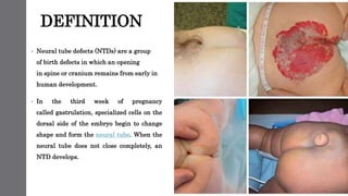
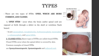
This document discusses neural tube defects (NTDs), which are birth defects where an opening remains in the spine or skull early in development. The main types are open defects like spina bifida and anencephaly, and closed defects. NTDs can be caused by folic acid deficiency, genetics, medications, and other factors. Symptoms vary depending on the specific defect. Diagnosis may involve ultrasounds and alpha-fetoprotein testing. Treatment ranges from surgery to palliative care, while prevention focuses on adequate folic acid intake before and during pregnancy.
2. DEFINITION
• Neural tube defects (NTDs) are a group
of birth defects in which an opening
in spine or cranium remains from early in
human development.
• In the third week of pregnancy
called gastrulation, specialized cells on the
dorsal side of the embryo begin to change
shape and form the neural tube. When the
neural tube does not close completely, an
NTD develops.
3. INCIDENCE
• NTDs are one of the most common birth defects, affecting
over 300,000 births each year worldwide.
• For example, spina bifida affects approximately 1,500
births annually in the United States, or about 3.5 in every
10,000 (0.035% of US births), which has decreased from
around 5 per 10,000 (0.05% of US births) since folate
fortification of grain products was started.
• The number of deaths in the US each year due to neural
tube defects also declined from 1,200 before folate
fortification was started to 840
4. ETIOLOGY
1. FOLIC ACID DEFECIENCY
• Inadequate levels of folate (vitamin B9)
and vitamin B12 during pregnancy have been
found to lead to increased risk of NTDs.
• Folate is required for the production and
maintenance of new cells, for DNA synthesis
and RNA synthesis.
• Folate is needed to carry one carbon groups for
methylation and nucleic acid synthesis
2. GENE ENVIRONMENT
INTERACTION
• The association seen between reduced neural tube
defects and folic acid supplementation is due to a gene-
environment interaction
• Supplementing folic acid during pregnancy reduces the
prevalence of NTDs by not exposing this otherwise
sub-clinical mutation to aggravating conditions.
• Other potential causes can include folate
antimetabolites (such as methotrexate), mycotoxins in
contaminated corn meal, arsenic, hyperthermia in
early development, and radiation.
• Maternal obesity has also been found to be a risk
factor for NTDs
3.INTAKE OF ANTI EPILEPTIC MEDICATIONS
IN PREGNANCY
5. PATHOPHYSIOLOGY
Neural plate appears on 17 th day from the ectoderm
On 18 th day the neural plate folds along the midline
By the end of 24 day the neural plate fuses to form neural tube
Fusion begins in the cephalic end and proceeds to the caudal end
Then the anterior and posterior ends fuses to form spinal cord
Depending on the location of the defect the type of NTD shows up
6. TYPES
• There are two types of NTDs: OPEN, WHICH ARE MORE
COMMON, AND CLOSED.
1. OPEN NTDS : occur when the brain and/or spinal cord are
exposed at birth through a defect in the skull or vertebrae (back
bones).
ď‚– Ex:are anencephaly, encephaloceles, hydranencephaly, iniencephal
y, schizencephaly, and spina bifida.
2. CLOSED NTD’s: Rarer types of NTDs are called closed NTDs.
Closed NTDs occur when the spinal defect is covered by skin.
Common examples of closed NTDs
are lipomyelomeningocele, lipomeningocele and tethered cord.
7. TYPES OF OPEN NTD
• Anencephaly
• Anencephaly (without brain) is a neural tube defect
that occurs when the head end of the neural tube
fails to close,
• usually during the 23rd and 26th days of pregnancy,
resulting in an absence of a major portion of the
brain and skull.
• Infants born with this condition are born without the
main part of the forebrain—the largest part of the
cerebrum—and are usually blind, deaf and
unconscious. The lack of a functioning cerebrum will
ensure that the infant will never gain consciousness.
• Infants are either stillborn or usually die within a
few hours or days after birth.
• Encephaloceles
• Encephaloceles are characterized by protrusions of
the brain through the skull that are sac-like and
covered with membrane.
• They can be a groove down the middle of the upper
part of the skull, between the forehead and nose, or
the back of the skull. Encephaloceles are often
obvious and diagnosed immediately.
8. TYPES OF OPEN NTD
• Iniencephaly:
• Iniencephaly is a rare neural tube defect that
results in extreme bending of the head to the
spine.
• The diagnosis can usually be made on antenatal
ultrasound scanning, but if not will undoubtedly
be made immediately after birth because the
head is bent backwards and the face looks
upwards. Usually the neck is absent.
• The skin of the face connects directly to the chest
and the scalp connects to the upper back. The
infant will usually not survive more than a few
hours.
• Hydranencephaly:
• Hydranencephaly is a condition in
which the cerebral hemispheres are
missing and instead filled with sacs of
cerebrospinal fluid.
9. SPINA BIFIDAS
• Spina bifida cystica
• This includes meningocele and myelomeningocele.
• Meningocele is less severe and is characterized by
herniation of the meninges, but not the spinal cord,
through the opening in the spinal canal.
• Myelomeningocele involves herniation of the meninges
as well as the spinal cord through the opening.
• Spina bifida occulta:
• In this type of neural tube defect, the
meninges do not herniate through the
opening in the spinal canal.
• By definition, spina bifida
occulta means hidden split spine.
• The most frequently seen form of spina
bifida occulta is when parts of the bones of
the spine, called the spinous process, and
the neural arch appear abnormal on a
radiogram, without involvement of the spinal
cord and spinal nerves. The risk of
recurrence in those who have a first degree
relative (a parent or sibling) is 5–10 times
greater compared to the general population.
10. CLINICAL FEATURES
• The symptoms associated with NTDs vary depending on the specific type of defect.
• Symptoms include physical problems (such as paralysis and urinary and bowel control
problems),
• blindness,
• deafness,
• intellectual disability, lack of consciousness, and, in some cases, death.
• Some people with NTDs have no symptoms.
• Most children with certain types of NTDs die or experience serious disability.
11. DIAGNOSTIC TESTS
• Tests for neural tube defects include ultrasound examination and measurement of
maternal serum alpha-fetoprotein (MSAFP).
• Often, these defects are apparent at birth, but acute defects may not be diagnosed until
much later in life. An elevated MSAFP measured at 16–18 weeks gestation is a good
predictor of open neural tube defects, however the test has a very high false positive rate
• MSAFP screening combined with routine ultrasonography has the best detection rate
although detection by ultrasonography is dependent on operator training and the quality
of the equipment.
12. TREATMENT
• As of 2008, treatments of NTDs depends on the severity of the complication.
• No treatment is available for anencephaly and infants usually do not survive more than a few
hours.
• Aggressive surgical management has improved survival and the functions of infants with spina
bifida, meningoceles and mild myelomeningoceles. The success of surgery often depends on the
amount of brain tissue involved in the encephalocele.
• The goal of treatment for NTDs is to allow the individual to achieve the highest level of
function and independence. Fetal surgery in utero before 26 weeks gestation has been
performed with some hope that there is benefit to the final outcome including a reduction
in Arnold–Chiari malformationb-oth the cerebellum and the brain stem extend into the
foramen magnum. and thereby decreases the need for a ventriculoperitoneal shunt
13. PREVENTION
• In 1996, the United States Food and Drug Administration published regulations requiring
the addition of folic acid to enriched breads, cereals, flour and other grain products.
• It is important to note that during the first four weeks of pregnancy (when most women do
not even realize that they are pregnant), adequate folate intake is essential for proper
operation of the neurulation process.
• Therefore, women who could become pregnant are advised to eat foods fortified with folic
acid or take supplements in addition to eating folate-rich foods to reduce the risks of serious
birth defects.
• Women who may become pregnant are advised to get 400 micrograms of folic acid daily.
Women who have previously given birth to a child with a neural tube defect may benefit
from a supplement containing 4.0 mg/5.0 mg daily, following advice provided by their doctor.