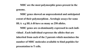
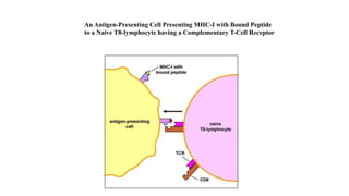
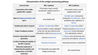
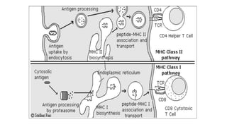
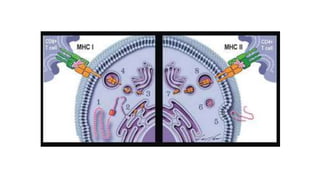
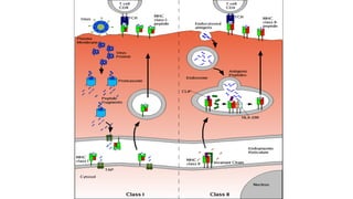
DEFINITION
Major histocompatibility complex (MHC) is a
large genomic region or gene family found on the short
arm of chromosome 6, they encode MHC molecules in
most vertebrates .
MHC antigens are alloantigens ( from the Greek
“allos” different ) distinguishing individuals within a
given species .There is an extremely remote chance that
two unrelated individuals will be found to share an
individual set of MHC antigen.
Discovery of MHC loci
MHC genes were first identified in inbred mice strains.
Clarence Little transplanted tumors across different strains and
found rejection of transplanted tumors according to strains of
host versus donor. George Snell in 1940s selectively bred two
mouse strains, attained a new strain nearly identical to one of the
progenitor strains, but differing crucially in histocompatibility
that is, tissue compatibility upon transplantation .The genetic
region that controlled graft rejection and contained several
linked genes was named MHC and thereupon identified an
MHC locus . For this work, Snell was awarded the 1980 Nobel
Prize in Physiology or Medicine.
MHC molecules are responsible for:
1- Transplant (graft rejection ).
2- Induction and regulation of the immune response .
3- Autoimmune diseases .
4- Defense against infection.
MHC genes that control immune responses are called
Immune response (Ir) genes .
MHC molecules are expressed on all nucleated cells of all
mammalian species .
They help the immune system to distinguish self from nonself
peptides .
MHC genes are diverse ,they vary greatly from individual to
another i.e. MHC alleles have polymorphism (diversity) .
MHC are the most polymorphic genes present in the
genome.
MHC genes showed an unprecedented and anticipated
extent of their polymorphism . Serologic assays for some
HLA e.g HLA-B have as many as 250 alleles.
MHC genes are co-dominantly expressed in each indi-
vidual . Each individual expresses the alleles that are
inherited from each of the 2 parents which maximises the
number of MHC molecules available to bind peptides for
presentation to T cells.
Thymic Education
T-lymphocytes belonging to one specific individual present
a property called MHC Restriction they only can detect an
antigen if it is displayed by an MHC molecule from the same
individual .
This is due to the fact that each T lymphocyte has a dual
specificity .The T cell receptor (TCR) recognizes at the same
time
1 Some residues from the peptide and
2 Some residues from the displaying MHC molecule .
The physiologic function of MHC molecules is the
presentation of peptide antigens to T cells . For most of the time
MHC molecules are presenting self peptides which T cells must
ignore while foreign polypeptides originating from viruses,
bacteria… must be appropriately attacked by T cells.
MHC abbreviation is often used to refer to Human Leukocyte
Antigen (HLA ) gene product ( Leukocyte because the antibodies
where tested by binding to the leukocytes of other individuals and
Antigens because the molecules where recognized by antibodies).
MHC complex consists of more than 70 genes . They are
classified into class I, II, III MHC.
MHC STRUCTURE
Class I gene complex contains 3 loci A,B,C, each of which codes
of α chain polypeptides.
Class II gene complex contains at least 3 loci ,DP ,DQ and DR.
Each of these loci codes for one α and a variable number of β
chain polypeptides .
Class III region encodes for other immune components, such as
complement components (C2,C4,factor B ).Some encode
cytokines e.g. TNF α and also hsp. They do not share the same
function as classI and classII molecules ,they are located between
them in the short arm of chromosome 6.
MHC and HLA classes : two classes have been
identified (HLA class I and II) ,they are composed of
6 different loci.
MHC (HLA) class I includes :
a - 3 major HLA loci: (HLA-A, HLA-B and HLA-C).
b - 3 minor loci (HLA-E, F, G ) .
Multiple alleles are present on each locus .
MHC (HLA) class II includes : 3Major loci .
HLA-DP
α-chain encoded by HLA-DPA1 locus
β-chain encoded by HLA-DPB1locus
HLA-DQ
α-chain encoded by HLA-DQA1 locus
β-chain encoded by HLA-DQB1 locus
HLA-DR
α-chain encoded by HLA-DRA locus
4 β-chains (only 3 possible per person), encoded by HLA-
DRB1, DRB3, DRB4, DRB5 loci
3 MINOR LOCI
(HLA- DM , DN and DO) .
The other MHC class II proteins, DM and DO, are
1 used in the internal processing of antigens,
2 loading the antigenic peptides generated
from pathogens onto the HLA molecules of antigen-
presenting cell.
MHC genes are expressed in co-dominant fashion . The alleles
inherited for both progenitors are expressed in equivalent way.
Each chromosome is found twice (diploid ) so
12 HLA antigens are found in normal tissue type
of an individual. New recombinant haplotypes
results from occasional crossing over between two
parental chromosomes.
Expression of MHC Molecules
Class I molecules are expressed on all nucleated cells . Their expression is
linked to the functions of class I restricted CD 8+T cells. Their function
is to kill cells infected with intracellular organisms such as viruses ,as well
as tumors that express tumor antigens .
Class II molecules are expressed only on dendritic cells , B lymphocytes
and macrophages. Class II- restricted CD 4+ helper T lymphocytes have to
recognize only antigens presented by these cells.
CD4+Th cells function is:
1 – To activate (or help) macrophages to eliminate extracellular microbes
that have been phagocytosed .
2- To activate B cells to make antibodies that eliminate extracellular
miccrobes .
Haplotypes
Human leukocyte antigens (HLA) are the MHC
antigens of humans.
Definition of haplotype :
A haplotype, a contraction of the phrase "haploid
genotype", is a set of closely linked genetic markers present
on one chromosome which tend to be inherited together (not
easily separable by recombination)”. Another way to think
about it is that a haplotype is half of a genotype..
A set of MHC alleles on each chromosome is
called an MHC haplotype .
MHC genes are inherited as a group ( haplotype
loci and alleles) , one from each parent one paternal
and one maternal haplotype each containing three
Class I (B,C,A) and three Class II (DP , DQ , DR ).
These haplotypes can be used to trace migrations
in the human population because they are often
much like a fingerprint of an event that has occurred
in evolution.
Allele
It is one of different forms of a gene present at a
particular chromosomal locus.
An individual who is heterozygous at a locus has 2
different alleles, each on a different member of a pair of
chromosomes one paternal and one maternal.
If a particular gene in a population has many different
alleles ,the gene locus is said to be polymorphic. The
MHC locus is extremely polymorphic .
HLA nomenclature .
HLA is identified by a letter for locus and a number
denoting the allele (A1,B5).
Not all HLA specificities have been defined.
Some individuals express unknown specificities at
some loci (usually class II) which the typing
laboratory reports as “blank”. Investigation of these
“blank” specificities often leads to the discovery of
new HLA antigens.
To avoid unnecessary confusion, they are assigned a
numerical designation by regularly held workshops of the
World Health Organization . At first, the designation is
preceded by a w, indicating a provisional assignment. For
example , DQw3 designates an antigenic specificity of
the DQ locus that has been tentatively designated as
w3 by a workshop. When worldwide agreement is
reached about the fact that this is a new specificity, the w
is dropped.
IMPORTANT ASPECTS OF MHC
1- Although there is a high degree of polymorphism
for a species, an individual has maximum of six
different class I MHC products and only slightly
more class II MHC products (considering only the
major loci).
2- Each MHC molecule has only one binding
site. The different peptides a given MHC molecule
can bind all to the same site, but only one at a time.
3- Because each MHC molecule can bind many
different peptides, binding is termed degenerate.
4- MHC molecules are membrane-bound;
recognition by T cells requires cell-cell contact .
5- Alleles for MHC genes are co-dominant. Each
MHC gene product is expressed on the cell surface of an
individual nucleated cell.
6- A peptide must associate with a given MHC of
that individual, otherwise no immune response can
occur. That is one level of control.
7- Mature T cells must have a T cell receptor that
recognizes the peptide associated with MHC. This is the
second level of control.
8-Cytokines (especially interferon-γ) increase level of
expression of MHC.
9- Peptides from the cytosol associate with class I MHC
and are recognized by Tc cells. Peptides from within vesicles
associate with class II MHC and are recognized by Th cells.
10-Polymorphism in MHC is important for survival of the
species.
Schematic presentation of the structure of MHC class I and class II molecules.
PBR = peptide bound region.
Polypeptide bound
to MHC groove
Transmembrane
region
Cytoplasmic
region
MHC class I MHC class II
An Antigen-Presenting Cell Presenting MHC-I with Bound Peptide
to a Naive T8-lymphocyte having a Complementary T-Cell Receptor
Fine structural differences can be detected in the
α1 and α2 domains of all class I molecules and in
α1 and β1 domains of all class II molecules
The variations found , are due to differences
in the amino acids sequence ,these variations
could be detected serologically.
There is no identical MHC molecules in
different individuals, it is only present in identical
twins .
There are a number of different MHC class I
and class II molecules with those of each class
having a similar basic structure e.g. all HLA-A
molecules will be very similar but they will exhibit
differences from HLA-B and HLA-C molecules .
Functions of HLA antigens
In infectious disease
In graft rejection
In autoimmunity
In cancer
IN INFECTIOUS DISEASE
Proteins produced inside the cells (native and foreign e.g. viruses
proteins and mutated proteins) are displayed on HLA antigens (MHC
class I) on the cell surface .CD8+ CTLs can also recognize
phagocytized microbes if the later or their antigens can escape from
phagocytic vesicles into the cytosol. CD8+ T cells can recognize the
infected cells and destroy them .
APCS engulf foreign pathogens , e. g. bacteria ,by phagocytosis .
Bacterial protein is digested to small peptides which are loaded onto
HLA antigens (MHC class II ) and presented on the cell surface to be
recognized by CD4+ T cells to eliminate them .
IN AUTOIMMUNITY
As HLA antigens are inherited some of them play a role in
autoimmune diseases such as SLE and Sjögren’s syndrome . HLA
typing has led to some improvement and acceleration in the
diagnosis of some autoimmune diseases e.g. Type I diabetes and
Celiac disease .
IN GRAFT REJECTION
Any cell displaying some other HLA type is considered non-
self or invader resulting in the rejection of the tissues bearing those
cells. HLA typing is important in transplantation to prevent tissue
rejection .
IN CANCER
Some HLA mediated diseases are related to cancer e.g.
Enteropathy - associated T-cell Lymphoma in gluten
sensitive enteropathy. On the other hand HLA molecules
play a protective role in recognizing the increase in
antigens that where not tolerated because of low levels in
normal state. Abnormal cells may be targeted for apoptosis
mediating many cancers before clinical diagnosis.
MHC class I
MHC class I (HLA- A, B and C) are expressed
on all nucleated cells (except the neurons) .
They display peptides generated from endoge-
nously produced proteins (antigens) e.g. viral
proteins or cancer antigen and present them to
CD8+ T cells. MHC class I play an important role
in tissue transplant rejection .
MHC class I can be grouped as:
Classic molecules HLA- A, B and C whose function
is the presentation of intracellular antigen peptides to
cytotoxic T lymphocytes (CD8).
Non classic molecules (named MHC class I-B) in this
group we find HLA -E, F and G, they interact with
inhibitory receptors in NK cells . They do not present
antigens to T lymphocytes .
Class I MHC molecules are composed of two polypeptide chains, a
long α chain and a short β chain called β2-microglobulin.
The α chain has four regions.
1- A cytoplasmic region, containing sites for phosphorylation and
binding to cytoskeletal elements.
2- A transmembrane region containing hydrophobic amino acids by
which the molecule is anchored in the cell membrane.
3- A highly conserved α3 immunoglobulin-like domain to which
CD8 binds. The β2- microglobulin associates with the α3 chain and helps
maintain the proper conformation of the molecule.
4- A highly polymorphic peptide binding region formed from the α1
and α2 domains.
MHC class I encodes non identical pairs of
peptide-binding proteins. They are expressed
on all nucleated cells. MHC class I proteins
contain an α heavy chain and a β2 micro-
globulin (not part of the MHC encoded by
chromosome15). The α chain is subdivided
into 3 regions (α1,α2 & α3) these are exposed
to the extracellular space , they are linked to
the cellular membrane through a trans-
membrane region .The β2-microglobulin is
associated to the α3 chain . Without the β2
microglobulin , the class I antigen will not be
expressed on the cell surface .
Individuals with defective β2 microglobulin gene have a
deficiency of cytotoxic T cells .
The antigenic peptides (composed of 9 amino acids) is
located in a peptide-binding groove (cleft) formed between
the α1and α2 regions in the heavy chain.
Antigenic peptides fit into these 'slots' much like a hand
fits into a glove or a key fits into a lock. The α1and α2
domains show polymorphism (great variability in amino
acids).
The specific binding sites (anchor sites) where the specific
binding of a peptide molecule in the peptide-binding groove
of MHC requires the peptide to have one or more specific
amino acid at a fixed position .The other amino acids can be
variable so that each MHC molecules can bind many dif-
ferent peptides.
The α1 & α2 domains (show polymorphism) bind TCR of
CD8 T lymphocytes.CD8 molecules present on CD8 T lym-
phocytes binds to the highly conserved region of α3.β2micro
globulin is non-covalently bound to the constant α3 domain.
CD8 Cytotoxic T lymphocytes
can recognize peptide antigen
only when presented along
MHC I molecule.
.
Peptide processing for peptides associated to
MHC-I molecules :
Proteins present in the cytosol are degraded
by the proteasome and the resulting peptides
are internalized by the Transporter Associated
Protein (TAP) channel in the endoplasmic
reticulum ,where they become associated with
MHC -I molecules freshly synthesized .The
MHC-I peptide complexes enter in the Golgi
apparatus, where they are glycosylated ,and
from there they enter in secreting vesicles
which fuse with the cell membrane. In this way
the complexes become exposed to the outside
of the cell, allowing the contact with circulating
T lymphocytes.
1 intracellular or endogenously produced
proteins
2 Proteins are processed by proteasomes
into peptides(8-12 a a)and transported to
ER by transporter associated proteins(TAP)
3 In the endoplasmic reticulum the peptides
bind to MHC class I molecules , (heavy chain
and β2 microglobulin).Molecular chaperons
(calnexin) associate with partially assembled
class I complex. Tapasin forms a bridge between
TAP and the class I molecules waiting to be
loaded with peptide.
4 the complex is transported through the
Golgi apparatus to the cell surface.
All nucleated cells express class I MHC. Proteins are fragmented in the cytosol by
proteasome (a complex of proteins having proteolytic activity) or by other
proteases. The fragments are then transported across the membrane of the
endoplasmic reticulum by transporter proteins. (The transporter proteins and some
components of the proteosome have their genes in the MHC complex). Synthesis
and assembly of class I heavy chain and beta2 microglobulin occurs in the
endoplasmic reticulum. Within the endoplasmic reticulum, the MHC class I heavy
chain, beta2microglobulin and peptide form a stable complex that is transported to
the cell surface.
TAP function as a transporter that translocates
peptides into the lumen of the ER .
Tapasin , an encoded protein in the ER, appears to
form a bridge between class I and TAP , until peptide is
associated with the class I molecule .Peptide loading
involves other proteins such as calnexin , calreticuline
and ERP57.These chaperones promote and guide the
assembly of stable class I-β2 microglobulin -peptide
complexes .
MHC class II
MHC molecules encodes heterodimeric peptide-binding
proteins that modulate antigen loading onto MHC class II
proteins in the lysosomal compartment such as MHC II DM,
MHCII DQ ,MHCII DR and MHCII DP. They are expressed
on most immune system cells specially on antigen presen-
ting cells (macrophages, dendritic cells, B cells and epithe-
lium of thymus ). MHC class II proteins contain α and β
chains and they present antigen fragments to T-helper cells
by binding to CD4 receptor on the T-helper cells.
MHC class II in human can be grouped in :
Classic molecules :HLA-DP, HLA-DQ, HLA-DR. They
represent peptides to T helper lymphocytes by binding to
CD4 receptor on T- helper cells .
Non classic molecules : HLA-DM,HLA-DN, HLA-DO.
They have intracellular function (used in the internal
processing of antigens) they are not exposed in the cellular
membrane. They load the antigenic peptides generated from
pathogens on the classic MHC-II molecules .
MHC class II is formed of two
chains ,one α and one β with two
domains for each one α1 & α2 ,
β1 & β2 . Each chain is linked to
the cell membrane through a
transmembrane region .The anti-
genic peptide is located in a cleft
formed by α1 and β1 peptides .
Class II MHC molecules are composed of two polypeptide chains
an α and a β chain of approximately equal length . Both chains have
four regions:
1-A cytoplasmic region containing sites for phosphorylation and
binding to cytoskeletal elements
2-A transmembrane region containing hydrophic amino acids by
which the molecule is anchored in the cell membrane
3-A highly conserved α2 domain and a highly conserved β2
domain to which CD4 binds
4-A highly polymorphic peptide binding region formed from the
α1 and β1 domains
Because class II MHC is loaded with extracellular
proteins, it is mainly concerned with presentation of
extracellular pathogens (for example, bacteria that might
be infecting a wound or the blood). Class II molecules
interact exclusively with CD4+ "helper" T cells (THC).
The helper T cells then help to trigger an appropriate
immune response which may include localized inflam-
mation and swelling due to recruitment of phagocytes or
may lead to a full-force antibody immune response due to
activation of B-cells .
MHC class II are important in humoral immunity .
MHC class II is only present on antigen presenting cells
(B cells and phagocytes).
They present foreign antigen derived from extra-
cellular proteins to helper T lymphocytes (CD4), which
stimulate B-cells to elicit an antibody response. The
antigenic peptide is present in a cleft formed by α1 and β1
peptides.
Peptides associated with class II MHC are 13-25
amino acid molecules long. The MHC class II-dependent
pathway of antigen presentation is called the endocytic
or exogenous pathway .
Extracellular proteins are endocytosed , digested in
lysosomes , and bound by the class II molecule prior
to the molecule’s migration to the plasma membrane .
CD4 Helper T lymphocytes
can recognize peptide antigen
only when presented along
MHC II molecules .
5 Extracellular or exogenous antigens are
taken up in APC s by endocytosis
6 The antigen is degraded in a lysosome
into immunogenic peptides .
7 MHC class II molecules formed in ER
enter the MHC vesicle .
8 The peptide antigens are joined to
MHC class II molecules in these vesicles
to be transported to the cell surface .
The nascent MHC class II protein in the rough ER has its peptide-binding cleft blocked by
the invariant chain (Ii; a trimer) to prevent it from binding cellular peptides or peptides from
the endogenous pathway. The invariant chain also facilitates MHC class II's export from the
ER in a vesicle which fuses with a late endosome containing the endocytosed degraded
proteins. The invariant chain then broken down in stages, leaving only a small fragment
called CLIP which still blocks the peptide binding cleft . An MHC class II-like structure,
HLA-DM, removes CLIP and replaces it with a peptide from the endosome. The stable
MHC class-II is then presented on the cell surface.
Characteristics of the antigen processing pathway
GOOD LUCK

Presentation MHC.pptx

  • 2.
    DEFINITION Major histocompatibility complex(MHC) is a large genomic region or gene family found on the short arm of chromosome 6, they encode MHC molecules in most vertebrates . MHC antigens are alloantigens ( from the Greek “allos” different ) distinguishing individuals within a given species .There is an extremely remote chance that two unrelated individuals will be found to share an individual set of MHC antigen.
  • 3.
    Discovery of MHCloci MHC genes were first identified in inbred mice strains. Clarence Little transplanted tumors across different strains and found rejection of transplanted tumors according to strains of host versus donor. George Snell in 1940s selectively bred two mouse strains, attained a new strain nearly identical to one of the progenitor strains, but differing crucially in histocompatibility that is, tissue compatibility upon transplantation .The genetic region that controlled graft rejection and contained several linked genes was named MHC and thereupon identified an MHC locus . For this work, Snell was awarded the 1980 Nobel Prize in Physiology or Medicine.
  • 4.
    MHC molecules areresponsible for: 1- Transplant (graft rejection ). 2- Induction and regulation of the immune response . 3- Autoimmune diseases . 4- Defense against infection. MHC genes that control immune responses are called Immune response (Ir) genes . MHC molecules are expressed on all nucleated cells of all mammalian species . They help the immune system to distinguish self from nonself peptides . MHC genes are diverse ,they vary greatly from individual to another i.e. MHC alleles have polymorphism (diversity) .
  • 5.
    MHC are themost polymorphic genes present in the genome. MHC genes showed an unprecedented and anticipated extent of their polymorphism . Serologic assays for some HLA e.g HLA-B have as many as 250 alleles. MHC genes are co-dominantly expressed in each indi- vidual . Each individual expresses the alleles that are inherited from each of the 2 parents which maximises the number of MHC molecules available to bind peptides for presentation to T cells.
  • 6.
    Thymic Education T-lymphocytes belongingto one specific individual present a property called MHC Restriction they only can detect an antigen if it is displayed by an MHC molecule from the same individual . This is due to the fact that each T lymphocyte has a dual specificity .The T cell receptor (TCR) recognizes at the same time 1 Some residues from the peptide and 2 Some residues from the displaying MHC molecule .
  • 7.
    The physiologic functionof MHC molecules is the presentation of peptide antigens to T cells . For most of the time MHC molecules are presenting self peptides which T cells must ignore while foreign polypeptides originating from viruses, bacteria… must be appropriately attacked by T cells. MHC abbreviation is often used to refer to Human Leukocyte Antigen (HLA ) gene product ( Leukocyte because the antibodies where tested by binding to the leukocytes of other individuals and Antigens because the molecules where recognized by antibodies). MHC complex consists of more than 70 genes . They are classified into class I, II, III MHC.
  • 8.
    MHC STRUCTURE Class Igene complex contains 3 loci A,B,C, each of which codes of α chain polypeptides. Class II gene complex contains at least 3 loci ,DP ,DQ and DR. Each of these loci codes for one α and a variable number of β chain polypeptides . Class III region encodes for other immune components, such as complement components (C2,C4,factor B ).Some encode cytokines e.g. TNF α and also hsp. They do not share the same function as classI and classII molecules ,they are located between them in the short arm of chromosome 6.
  • 9.
    MHC and HLAclasses : two classes have been identified (HLA class I and II) ,they are composed of 6 different loci. MHC (HLA) class I includes : a - 3 major HLA loci: (HLA-A, HLA-B and HLA-C). b - 3 minor loci (HLA-E, F, G ) . Multiple alleles are present on each locus .
  • 10.
    MHC (HLA) classII includes : 3Major loci . HLA-DP α-chain encoded by HLA-DPA1 locus β-chain encoded by HLA-DPB1locus HLA-DQ α-chain encoded by HLA-DQA1 locus β-chain encoded by HLA-DQB1 locus HLA-DR α-chain encoded by HLA-DRA locus 4 β-chains (only 3 possible per person), encoded by HLA- DRB1, DRB3, DRB4, DRB5 loci
  • 11.
    3 MINOR LOCI (HLA-DM , DN and DO) . The other MHC class II proteins, DM and DO, are 1 used in the internal processing of antigens, 2 loading the antigenic peptides generated from pathogens onto the HLA molecules of antigen- presenting cell.
  • 12.
    MHC genes areexpressed in co-dominant fashion . The alleles inherited for both progenitors are expressed in equivalent way.
  • 13.
    Each chromosome isfound twice (diploid ) so 12 HLA antigens are found in normal tissue type of an individual. New recombinant haplotypes results from occasional crossing over between two parental chromosomes.
  • 14.
    Expression of MHCMolecules Class I molecules are expressed on all nucleated cells . Their expression is linked to the functions of class I restricted CD 8+T cells. Their function is to kill cells infected with intracellular organisms such as viruses ,as well as tumors that express tumor antigens . Class II molecules are expressed only on dendritic cells , B lymphocytes and macrophages. Class II- restricted CD 4+ helper T lymphocytes have to recognize only antigens presented by these cells. CD4+Th cells function is: 1 – To activate (or help) macrophages to eliminate extracellular microbes that have been phagocytosed . 2- To activate B cells to make antibodies that eliminate extracellular miccrobes .
  • 15.
    Haplotypes Human leukocyte antigens(HLA) are the MHC antigens of humans. Definition of haplotype : A haplotype, a contraction of the phrase "haploid genotype", is a set of closely linked genetic markers present on one chromosome which tend to be inherited together (not easily separable by recombination)”. Another way to think about it is that a haplotype is half of a genotype..
  • 16.
    A set ofMHC alleles on each chromosome is called an MHC haplotype . MHC genes are inherited as a group ( haplotype loci and alleles) , one from each parent one paternal and one maternal haplotype each containing three Class I (B,C,A) and three Class II (DP , DQ , DR ). These haplotypes can be used to trace migrations in the human population because they are often much like a fingerprint of an event that has occurred in evolution.
  • 17.
    Allele It is oneof different forms of a gene present at a particular chromosomal locus. An individual who is heterozygous at a locus has 2 different alleles, each on a different member of a pair of chromosomes one paternal and one maternal. If a particular gene in a population has many different alleles ,the gene locus is said to be polymorphic. The MHC locus is extremely polymorphic .
  • 18.
    HLA nomenclature . HLAis identified by a letter for locus and a number denoting the allele (A1,B5). Not all HLA specificities have been defined. Some individuals express unknown specificities at some loci (usually class II) which the typing laboratory reports as “blank”. Investigation of these “blank” specificities often leads to the discovery of new HLA antigens.
  • 19.
    To avoid unnecessaryconfusion, they are assigned a numerical designation by regularly held workshops of the World Health Organization . At first, the designation is preceded by a w, indicating a provisional assignment. For example , DQw3 designates an antigenic specificity of the DQ locus that has been tentatively designated as w3 by a workshop. When worldwide agreement is reached about the fact that this is a new specificity, the w is dropped.
  • 20.
    IMPORTANT ASPECTS OFMHC 1- Although there is a high degree of polymorphism for a species, an individual has maximum of six different class I MHC products and only slightly more class II MHC products (considering only the major loci). 2- Each MHC molecule has only one binding site. The different peptides a given MHC molecule can bind all to the same site, but only one at a time.
  • 21.
    3- Because eachMHC molecule can bind many different peptides, binding is termed degenerate. 4- MHC molecules are membrane-bound; recognition by T cells requires cell-cell contact . 5- Alleles for MHC genes are co-dominant. Each MHC gene product is expressed on the cell surface of an individual nucleated cell. 6- A peptide must associate with a given MHC of that individual, otherwise no immune response can occur. That is one level of control.
  • 22.
    7- Mature Tcells must have a T cell receptor that recognizes the peptide associated with MHC. This is the second level of control. 8-Cytokines (especially interferon-γ) increase level of expression of MHC. 9- Peptides from the cytosol associate with class I MHC and are recognized by Tc cells. Peptides from within vesicles associate with class II MHC and are recognized by Th cells. 10-Polymorphism in MHC is important for survival of the species.
  • 23.
    Schematic presentation ofthe structure of MHC class I and class II molecules. PBR = peptide bound region.
  • 24.
    Polypeptide bound to MHCgroove Transmembrane region Cytoplasmic region MHC class I MHC class II
  • 26.
    An Antigen-Presenting CellPresenting MHC-I with Bound Peptide to a Naive T8-lymphocyte having a Complementary T-Cell Receptor
  • 27.
    Fine structural differencescan be detected in the α1 and α2 domains of all class I molecules and in α1 and β1 domains of all class II molecules The variations found , are due to differences in the amino acids sequence ,these variations could be detected serologically.
  • 28.
    There is noidentical MHC molecules in different individuals, it is only present in identical twins . There are a number of different MHC class I and class II molecules with those of each class having a similar basic structure e.g. all HLA-A molecules will be very similar but they will exhibit differences from HLA-B and HLA-C molecules .
  • 29.
    Functions of HLAantigens In infectious disease In graft rejection In autoimmunity In cancer
  • 30.
    IN INFECTIOUS DISEASE Proteinsproduced inside the cells (native and foreign e.g. viruses proteins and mutated proteins) are displayed on HLA antigens (MHC class I) on the cell surface .CD8+ CTLs can also recognize phagocytized microbes if the later or their antigens can escape from phagocytic vesicles into the cytosol. CD8+ T cells can recognize the infected cells and destroy them . APCS engulf foreign pathogens , e. g. bacteria ,by phagocytosis . Bacterial protein is digested to small peptides which are loaded onto HLA antigens (MHC class II ) and presented on the cell surface to be recognized by CD4+ T cells to eliminate them .
  • 31.
    IN AUTOIMMUNITY As HLAantigens are inherited some of them play a role in autoimmune diseases such as SLE and Sjögren’s syndrome . HLA typing has led to some improvement and acceleration in the diagnosis of some autoimmune diseases e.g. Type I diabetes and Celiac disease . IN GRAFT REJECTION Any cell displaying some other HLA type is considered non- self or invader resulting in the rejection of the tissues bearing those cells. HLA typing is important in transplantation to prevent tissue rejection .
  • 32.
    IN CANCER Some HLAmediated diseases are related to cancer e.g. Enteropathy - associated T-cell Lymphoma in gluten sensitive enteropathy. On the other hand HLA molecules play a protective role in recognizing the increase in antigens that where not tolerated because of low levels in normal state. Abnormal cells may be targeted for apoptosis mediating many cancers before clinical diagnosis.
  • 33.
    MHC class I MHCclass I (HLA- A, B and C) are expressed on all nucleated cells (except the neurons) . They display peptides generated from endoge- nously produced proteins (antigens) e.g. viral proteins or cancer antigen and present them to CD8+ T cells. MHC class I play an important role in tissue transplant rejection .
  • 34.
    MHC class Ican be grouped as: Classic molecules HLA- A, B and C whose function is the presentation of intracellular antigen peptides to cytotoxic T lymphocytes (CD8). Non classic molecules (named MHC class I-B) in this group we find HLA -E, F and G, they interact with inhibitory receptors in NK cells . They do not present antigens to T lymphocytes .
  • 35.
    Class I MHCmolecules are composed of two polypeptide chains, a long α chain and a short β chain called β2-microglobulin. The α chain has four regions. 1- A cytoplasmic region, containing sites for phosphorylation and binding to cytoskeletal elements. 2- A transmembrane region containing hydrophobic amino acids by which the molecule is anchored in the cell membrane. 3- A highly conserved α3 immunoglobulin-like domain to which CD8 binds. The β2- microglobulin associates with the α3 chain and helps maintain the proper conformation of the molecule. 4- A highly polymorphic peptide binding region formed from the α1 and α2 domains.
  • 36.
    MHC class Iencodes non identical pairs of peptide-binding proteins. They are expressed on all nucleated cells. MHC class I proteins contain an α heavy chain and a β2 micro- globulin (not part of the MHC encoded by chromosome15). The α chain is subdivided into 3 regions (α1,α2 & α3) these are exposed to the extracellular space , they are linked to the cellular membrane through a trans- membrane region .The β2-microglobulin is associated to the α3 chain . Without the β2 microglobulin , the class I antigen will not be expressed on the cell surface .
  • 37.
    Individuals with defectiveβ2 microglobulin gene have a deficiency of cytotoxic T cells . The antigenic peptides (composed of 9 amino acids) is located in a peptide-binding groove (cleft) formed between the α1and α2 regions in the heavy chain. Antigenic peptides fit into these 'slots' much like a hand fits into a glove or a key fits into a lock. The α1and α2 domains show polymorphism (great variability in amino acids).
  • 38.
    The specific bindingsites (anchor sites) where the specific binding of a peptide molecule in the peptide-binding groove of MHC requires the peptide to have one or more specific amino acid at a fixed position .The other amino acids can be variable so that each MHC molecules can bind many dif- ferent peptides. The α1 & α2 domains (show polymorphism) bind TCR of CD8 T lymphocytes.CD8 molecules present on CD8 T lym- phocytes binds to the highly conserved region of α3.β2micro globulin is non-covalently bound to the constant α3 domain.
  • 39.
    The specific bindingsites (anchor sites) where the specific binding of a peptide molecule in the peptide-binding groove of MHC requires the peptide to have one or more specific amino acid at a fixed position .The other amino acids can be variable so that each MHC molecules can bind many dif- ferent peptides. The α1 & α2 domains (show polymorphism) bind TCR of CD8 T lymphocytes.CD8 molecules present on CD8 T lym- phocytes binds to the highly conserved region of α3.β2micro globulin is non-covalently bound to the constant α3 domain.
  • 40.
    CD8 Cytotoxic Tlymphocytes can recognize peptide antigen only when presented along MHC I molecule. .
  • 41.
    Peptide processing forpeptides associated to MHC-I molecules : Proteins present in the cytosol are degraded by the proteasome and the resulting peptides are internalized by the Transporter Associated Protein (TAP) channel in the endoplasmic reticulum ,where they become associated with MHC -I molecules freshly synthesized .The MHC-I peptide complexes enter in the Golgi apparatus, where they are glycosylated ,and from there they enter in secreting vesicles which fuse with the cell membrane. In this way the complexes become exposed to the outside of the cell, allowing the contact with circulating T lymphocytes.
  • 42.
    1 intracellular orendogenously produced proteins 2 Proteins are processed by proteasomes into peptides(8-12 a a)and transported to ER by transporter associated proteins(TAP) 3 In the endoplasmic reticulum the peptides bind to MHC class I molecules , (heavy chain and β2 microglobulin).Molecular chaperons (calnexin) associate with partially assembled class I complex. Tapasin forms a bridge between TAP and the class I molecules waiting to be loaded with peptide. 4 the complex is transported through the Golgi apparatus to the cell surface.
  • 43.
    All nucleated cellsexpress class I MHC. Proteins are fragmented in the cytosol by proteasome (a complex of proteins having proteolytic activity) or by other proteases. The fragments are then transported across the membrane of the endoplasmic reticulum by transporter proteins. (The transporter proteins and some components of the proteosome have their genes in the MHC complex). Synthesis and assembly of class I heavy chain and beta2 microglobulin occurs in the endoplasmic reticulum. Within the endoplasmic reticulum, the MHC class I heavy chain, beta2microglobulin and peptide form a stable complex that is transported to the cell surface.
  • 45.
    TAP function asa transporter that translocates peptides into the lumen of the ER . Tapasin , an encoded protein in the ER, appears to form a bridge between class I and TAP , until peptide is associated with the class I molecule .Peptide loading involves other proteins such as calnexin , calreticuline and ERP57.These chaperones promote and guide the assembly of stable class I-β2 microglobulin -peptide complexes .
  • 48.
    MHC class II MHCmolecules encodes heterodimeric peptide-binding proteins that modulate antigen loading onto MHC class II proteins in the lysosomal compartment such as MHC II DM, MHCII DQ ,MHCII DR and MHCII DP. They are expressed on most immune system cells specially on antigen presen- ting cells (macrophages, dendritic cells, B cells and epithe- lium of thymus ). MHC class II proteins contain α and β chains and they present antigen fragments to T-helper cells by binding to CD4 receptor on the T-helper cells.
  • 49.
    MHC class IIin human can be grouped in : Classic molecules :HLA-DP, HLA-DQ, HLA-DR. They represent peptides to T helper lymphocytes by binding to CD4 receptor on T- helper cells . Non classic molecules : HLA-DM,HLA-DN, HLA-DO. They have intracellular function (used in the internal processing of antigens) they are not exposed in the cellular membrane. They load the antigenic peptides generated from pathogens on the classic MHC-II molecules .
  • 50.
    MHC class IIis formed of two chains ,one α and one β with two domains for each one α1 & α2 , β1 & β2 . Each chain is linked to the cell membrane through a transmembrane region .The anti- genic peptide is located in a cleft formed by α1 and β1 peptides .
  • 51.
    Class II MHCmolecules are composed of two polypeptide chains an α and a β chain of approximately equal length . Both chains have four regions: 1-A cytoplasmic region containing sites for phosphorylation and binding to cytoskeletal elements 2-A transmembrane region containing hydrophic amino acids by which the molecule is anchored in the cell membrane 3-A highly conserved α2 domain and a highly conserved β2 domain to which CD4 binds 4-A highly polymorphic peptide binding region formed from the α1 and β1 domains
  • 52.
    Because class IIMHC is loaded with extracellular proteins, it is mainly concerned with presentation of extracellular pathogens (for example, bacteria that might be infecting a wound or the blood). Class II molecules interact exclusively with CD4+ "helper" T cells (THC). The helper T cells then help to trigger an appropriate immune response which may include localized inflam- mation and swelling due to recruitment of phagocytes or may lead to a full-force antibody immune response due to activation of B-cells .
  • 53.
    MHC class IIare important in humoral immunity . MHC class II is only present on antigen presenting cells (B cells and phagocytes). They present foreign antigen derived from extra- cellular proteins to helper T lymphocytes (CD4), which stimulate B-cells to elicit an antibody response. The antigenic peptide is present in a cleft formed by α1 and β1 peptides.
  • 54.
    Peptides associated withclass II MHC are 13-25 amino acid molecules long. The MHC class II-dependent pathway of antigen presentation is called the endocytic or exogenous pathway . Extracellular proteins are endocytosed , digested in lysosomes , and bound by the class II molecule prior to the molecule’s migration to the plasma membrane .
  • 55.
    CD4 Helper Tlymphocytes can recognize peptide antigen only when presented along MHC II molecules .
  • 56.
    5 Extracellular orexogenous antigens are taken up in APC s by endocytosis 6 The antigen is degraded in a lysosome into immunogenic peptides . 7 MHC class II molecules formed in ER enter the MHC vesicle . 8 The peptide antigens are joined to MHC class II molecules in these vesicles to be transported to the cell surface .
  • 57.
    The nascent MHCclass II protein in the rough ER has its peptide-binding cleft blocked by the invariant chain (Ii; a trimer) to prevent it from binding cellular peptides or peptides from the endogenous pathway. The invariant chain also facilitates MHC class II's export from the ER in a vesicle which fuses with a late endosome containing the endocytosed degraded proteins. The invariant chain then broken down in stages, leaving only a small fragment called CLIP which still blocks the peptide binding cleft . An MHC class II-like structure, HLA-DM, removes CLIP and replaces it with a peptide from the endosome. The stable MHC class-II is then presented on the cell surface.
  • 60.
    Characteristics of theantigen processing pathway
  • 67.