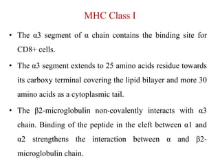
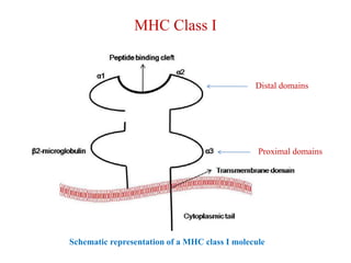
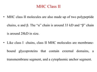
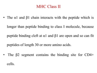
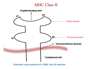
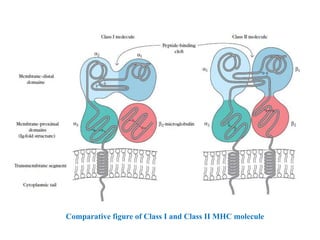
Major Histocompatibility Complex
(Structure)
Dr. Sher Singh Parihar
Introduction
• Every mammalian organism possesses a tightly linked
cluster of genes, the major histocompatibility complex
(MHC), whose products play roles in intercellular
recognition and in discrimination between self and nonself.
• The MHC participates in the development of both humoral
and cell mediated immune responses.
• While antibodies may react with antigens alone, most T cells
recognize antigen only when it is combined with an MHC
molecule.
Introduction
• The concept that the rejection of foreign tissue is the
result of an immune response to cell-surface molecules,
originated from the work of Peter Gorer in the mid-1930s.
• He identified four groups of genes, designated I to IV, that
encoded blood-cell antigens.
• Gorer and George Snell established that antigens encoded
by the genes in the group designated II took part in the
rejection of transplanted tumors and other tissue.
Introduction
• Snell called these genes “histocompatibility genes”; their
current designation as histocompatibility-2 (H-2) genes
was in reference to Gorer’s group II blood-group
antigens. Snell was awarded the Nobel prize in 1980 for
this work.
• Class I and class II MHC molecules are membrane-bound
glycoproteins that are closely related in both structure and
function.
Introduction
• Both class I and class II MHC molecules have been
isolated and purified and the three dimensional structures
of their extracellular domains have been determined by x-
ray crystallography.
• Both types of membrane glycoproteins function as highly
specialized antigen-presenting molecules.
MHC Class I
• The fully formed MHC class I molecule is a heterotrimer
consists of α1, α2, α3 and β2-microglobulin chain.
• MHC class I molecules are made up of two polypeptide
chains, α and β2-microglobulin.
• The “α” chain is around 44 kD and “β2-microglobulin”
chain is around 12kD in size.
• Each α chain is divided into three parts to accommodate
extracellular α1, α2, and α3 domains, transmembrane
domain and a cytoplasmic tail.
MHC Class I
• The α1 and α2 is around 90 amino acids long and binds to
only 8-11 amino acid long peptides (peptide binding cleft).
• Because MHC class I peptide binding cleft is closed and the
larger peptide cannot be accommodated in the designated
space.
• The α1 and α2 contains the polymorphic residues which are
responsible for the variation among the MHC I allele and
their recognition by a specific T cell.
MHC Class I
• The α3 segment of α chain contains the binding site for
CD8+ cells.
• The α3 segment extends to 25 amino acids residue towards
its carboxy terminal covering the lipid bilayer and more 30
amino acids as a cytoplasmic tail.
• The β2-microglobulin non-covalently interacts with α3
chain. Binding of the peptide in the cleft between α1 and
α2 strengthens the interaction between α and β2-
microglobulin chain.
MHC Class I
Distal domains
Proximal domains
Schematic representation of a MHC class I molecule
MHC Class II
• MHC class II molecules are also made up of two polypeptide
chains, α and β. The “α” chain is around 33 kD and “β” chain
is around 28kD in size.
• Like class I chains, class II MHC molecules are membrane-
bound glycoproteins that contain external domains, a
transmembrane segment, and a cytoplasmic anchor segment.
MHC Class II
• The α1 and β1 chain interacts with the peptide which is
longer than peptide binding to class I molecule, because
peptide binding cleft at α1 and β1 are open and so can fit
peptides of length 30 or more amino acids.
• The β2 segment contains the binding site for CD4+
cells.
MHC Class II
• The fully formed MHC class II molecule is a dimer of
dimer consisting of α1, α2, β1 and β2 microglobulin
chain.
• The dimer is oriented so that the two peptide-binding
clefts face in opposite directions.
MHC Class II
• While it has not yet been determined whether this
dimeric form exists in vivo, the presence of CD4
binding sites on opposite sides of the class II
molecule suggests that it does.
• These two sites on the 2 and 2 domains are adjacent
in the dimer form and a CD4 molecule binding to
them may stabilize class II dimers.
MHC Class II
Schematic representation of a MHC class II molecule
Distal domain
Proximal domain
Comparative figure of Class I and Class II MHC molecule
Conclusion
• The major histocompatibility complex (MHC)
comprises a stretch of tightly linked genes that
encode proteins associated with intercellular
recognition and antigen presentation to T
lymphocytes.
• A group of linked MHC genes is generally inherited
as a unit from parents; these linked groups are called
haplotypes.
Conclusion
• Class I MHC molecules consist of a large glycoprotein
chain with 3 extracellular domains and a trans-membrane
segment, and 2-microglobulin, a protein with a single
domain.
• Class II MHC molecules are composed of two
noncovalently associated glycoproteins, the and chain,
encoded by separate MHC genes.
Conclusion
• X-ray crystallographic analyses reveal peptide-
binding clefts in the membrane-distal regions of both
class I and class II MHC molecules.
• Both class I and class II MHC molecules present
antigen to T cells.
• Class I molecules present processed endogenous
antigen to CD8 T cells. Class II molecules present
processed exogenous antigen to CD4 T cells.

Major Histocompatibility Complex.ppt

  • 1.
  • 2.
    Introduction • Every mammalianorganism possesses a tightly linked cluster of genes, the major histocompatibility complex (MHC), whose products play roles in intercellular recognition and in discrimination between self and nonself. • The MHC participates in the development of both humoral and cell mediated immune responses. • While antibodies may react with antigens alone, most T cells recognize antigen only when it is combined with an MHC molecule.
  • 3.
    Introduction • The conceptthat the rejection of foreign tissue is the result of an immune response to cell-surface molecules, originated from the work of Peter Gorer in the mid-1930s. • He identified four groups of genes, designated I to IV, that encoded blood-cell antigens. • Gorer and George Snell established that antigens encoded by the genes in the group designated II took part in the rejection of transplanted tumors and other tissue.
  • 4.
    Introduction • Snell calledthese genes “histocompatibility genes”; their current designation as histocompatibility-2 (H-2) genes was in reference to Gorer’s group II blood-group antigens. Snell was awarded the Nobel prize in 1980 for this work. • Class I and class II MHC molecules are membrane-bound glycoproteins that are closely related in both structure and function.
  • 5.
    Introduction • Both classI and class II MHC molecules have been isolated and purified and the three dimensional structures of their extracellular domains have been determined by x- ray crystallography. • Both types of membrane glycoproteins function as highly specialized antigen-presenting molecules.
  • 6.
    MHC Class I •The fully formed MHC class I molecule is a heterotrimer consists of α1, α2, α3 and β2-microglobulin chain. • MHC class I molecules are made up of two polypeptide chains, α and β2-microglobulin. • The “α” chain is around 44 kD and “β2-microglobulin” chain is around 12kD in size. • Each α chain is divided into three parts to accommodate extracellular α1, α2, and α3 domains, transmembrane domain and a cytoplasmic tail.
  • 7.
    MHC Class I •The α1 and α2 is around 90 amino acids long and binds to only 8-11 amino acid long peptides (peptide binding cleft). • Because MHC class I peptide binding cleft is closed and the larger peptide cannot be accommodated in the designated space. • The α1 and α2 contains the polymorphic residues which are responsible for the variation among the MHC I allele and their recognition by a specific T cell.
  • 8.
    MHC Class I •The α3 segment of α chain contains the binding site for CD8+ cells. • The α3 segment extends to 25 amino acids residue towards its carboxy terminal covering the lipid bilayer and more 30 amino acids as a cytoplasmic tail. • The β2-microglobulin non-covalently interacts with α3 chain. Binding of the peptide in the cleft between α1 and α2 strengthens the interaction between α and β2- microglobulin chain.
  • 9.
    MHC Class I Distaldomains Proximal domains Schematic representation of a MHC class I molecule
  • 10.
    MHC Class II •MHC class II molecules are also made up of two polypeptide chains, α and β. The “α” chain is around 33 kD and “β” chain is around 28kD in size. • Like class I chains, class II MHC molecules are membrane- bound glycoproteins that contain external domains, a transmembrane segment, and a cytoplasmic anchor segment.
  • 11.
    MHC Class II •The α1 and β1 chain interacts with the peptide which is longer than peptide binding to class I molecule, because peptide binding cleft at α1 and β1 are open and so can fit peptides of length 30 or more amino acids. • The β2 segment contains the binding site for CD4+ cells.
  • 12.
    MHC Class II •The fully formed MHC class II molecule is a dimer of dimer consisting of α1, α2, β1 and β2 microglobulin chain. • The dimer is oriented so that the two peptide-binding clefts face in opposite directions.
  • 13.
    MHC Class II •While it has not yet been determined whether this dimeric form exists in vivo, the presence of CD4 binding sites on opposite sides of the class II molecule suggests that it does. • These two sites on the 2 and 2 domains are adjacent in the dimer form and a CD4 molecule binding to them may stabilize class II dimers.
  • 14.
    MHC Class II Schematicrepresentation of a MHC class II molecule Distal domain Proximal domain
  • 15.
    Comparative figure ofClass I and Class II MHC molecule
  • 16.
    Conclusion • The majorhistocompatibility complex (MHC) comprises a stretch of tightly linked genes that encode proteins associated with intercellular recognition and antigen presentation to T lymphocytes. • A group of linked MHC genes is generally inherited as a unit from parents; these linked groups are called haplotypes.
  • 17.
    Conclusion • Class IMHC molecules consist of a large glycoprotein chain with 3 extracellular domains and a trans-membrane segment, and 2-microglobulin, a protein with a single domain. • Class II MHC molecules are composed of two noncovalently associated glycoproteins, the and chain, encoded by separate MHC genes.
  • 18.
    Conclusion • X-ray crystallographicanalyses reveal peptide- binding clefts in the membrane-distal regions of both class I and class II MHC molecules. • Both class I and class II MHC molecules present antigen to T cells. • Class I molecules present processed endogenous antigen to CD8 T cells. Class II molecules present processed exogenous antigen to CD4 T cells.