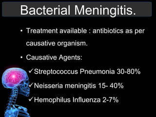
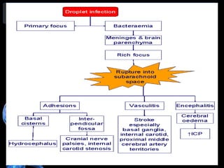
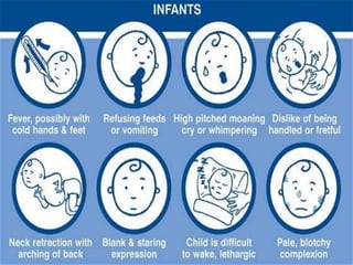
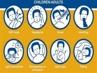
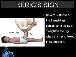
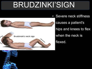
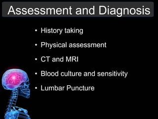
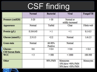
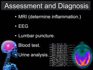
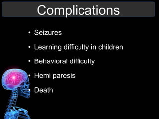
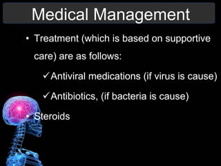
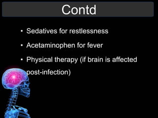
Management of Patient
with meningitis and
encephalitis
Moderator:
Madam Sibi Riju
Lecturer
CON
Mr. Mahesh Kumar
Sharma
M.Sc. Nursing
1st year
Case scenario
• Mr. ‘A’ a 15-year-old male was diagnosed with acute
myeloid leukemia in April ’15. he received two
courses of chemotherapy with partial response.
• Beginning August’15, he complained about an
increasingly disturbed gait due to a weakness. She
also complained of fever and vomiting along with
headache and drowsiness.
• She also complains of nuchal rigidity and
photophobia.
• What is the diagnosis?
Meningitis.
ANATOMY
Introduction.
• Although meningitis is a notifiable disease,
the exact incidence rate is unknown.
• In 2010 – 420, 000 deaths
• In 2013 - 303,000 deaths.
• It can occur as a complication of other
disease and 50% is an opportunistic
infection.
Incidence.
• Of newborns, 20–30% may die from
bacterial meningitis.
• This risk is lower in older children.
• Rises again in adulthood.
• In adults, 66% of all cases emerge
without disability. The main problems
are deafness (14%) and cognitive
impairment (10%)
Definition.
• Meningitis (from Greek méninx,
"membrane”) is
an acute inflammation of the
meninges.
• Caused by either bacteria or virus.
Route of Entry in CNS
• Skull or Back bone Fractures (trauma)
• Medical Procedures
• Along peripheral Nerves
• Blood or Lymphatic system
Etiology.
The causes can be classified into:
• Bacterial Infections
• Viral Infections
• Fungal Infections
• Inflammatory diseases (SLE)
• Cancer
• Trauma to head or spine
Pathophysiology.
Bacteria enters blood stream/ trauma
Enters the mucosal surface/ cavity
Breakdown of normal barriers
Crosses the blood brain barrier
Proliferates in the CSF
Inflammation of the meninges
Increase in ICP
Bacterial Meningitis
• Also known as septic meningitis.
• Extremely serious that requires immediate
care.
• Can lead to permanent damage of brain or
disability and death.
• Spreads by:
-coughing or sneezing
Bacterial Meningitis.
• Treatment available : antibiotics as per
causative organism.
• Causative Agents:
Streptococcus Pneumonia 30-80%
Neisseria meningitis 15- 40%
Hemophilus Influenza 2-7%
Tubercular Meningitis.
• TB meningitis is caused by
Mycobacterium tuberculi.
• Infection with this bacterium begins usually
in the lungs
• 1 – 2% of cases the bacteria travel via the
bloodstream.
• Unlike other types of meningitis its
progresses very slowly and symptoms are
vague
Viral Meningitis
• Also known as aseptic meningitis.
• More common than bacterial form and
usually less serious.
• Less likely to have permanent brain
damage after the infection resolves.
• Treatment: No specific treatment
available.
• Most patients recover completely on their
own
Viral Meningitis.
• Causative agents:
Enterovirus
Adenovirus
Arbovirus
Measles virus
Herpes simplex virus
Varicella
Fungal Meningitis
• It is much less common than the other two
infections.
• It is rare in healthy people but it is more
likely in persons who have impaired
immune system.
Risk factors
• Systemic infections
• Viral RTIs
• Tobacco use
• Impaired Immune system
• Over crowding
Risk Factors
• Vaccinations
• Seasonal:
 Winter
 Spring
Clinical manifestation
Clinical Manifestations.
In Infants:
Contd..
In Adults:
KERIG’S SIGN
• Severe stiffness of
the hamstrings
causes an inability to
straighten the leg
when the hip is flexed
to 90 degrees.
BRUDZINKI’SIGN
• Severe neck stiffness
causes a patient's
hips and knees to flex
when the neck is
flexed.
Assessment and Diagnosis
• History taking
• Physical assessment
• CT and MRI
• Blood culture and sensitivity
• Lumbar Puncture
CSF finding
Complications.
• Sensori-neural hearing loss
• Epilepsy/ seizures
• Memory loss
• Paralysis
• Learning difficulty
Contd.
• Behavioral difficulty
• Decreased intelligence
• Septicemia
• Death
Medical Management
• Bacterial meningitis:
Third-generation cefalosporin such
as cefotaxime or ceftriaxone
Vancomycin is added in the regime in
case of resistance.
Contd..
Dexamethasone
Dehydration and shock can be treated
with fluid therapy.
Phenytoin for seizure management.
Tubercular Meningitis:
ATT medications are started:
Isoniazid; rifampacin; pyrazinamide and
streptomycin.
Second line drugs:
Aminoglycosides; Fluroquinolones
Conventional therapy is given for 6-9
months
 In children BCG vaccine offers (approx
64%) protective effect
Viral Meningitis:
• Treatment is mostly supportive and no
medicines are prescribed.
 Seizure prophylaxis:
Lorazepam or phenytoin or barbiturate.
 Increased ICP:
Inj. Mannitol 1g/kg followed by 0.25-
0.5g/kg Q6H or/and dexamethasone
 Rest is advised
Contd
In case hydrocephalus is present VP or
LP shunt is required.
Adequate hydration is to be maintained
Antipyretics
Anti emetics
Encephalitis.
Definition
• Encephalitis (from Ancient
Greek, enképhalos “brain”) is
an acute inflammation of the brain.
• Encephalitis with meningitis is known
as meningo-encephalitis.
Incidence
• In western countries incidence is 7.4
cases per 100,000 population per year.
• In tropical countries, the incidence is 6.34
per 100,000 per year
• In 2013: 77,000 deaths from encephalitis
from 92,000 in 1990.
Etiology
• Viral cause
HSV encephalitis.
Arthropod borne virus encephalitis.
• Bacterial cause
• Fungal cause
• Auto immune
Herpes Simplex virus
Encephalitis.
• Caused by herpes Simplex virus;
• Its of two types:
1. HSV-I typically affects children
and adults
2. HSV-II common in neonates
• Treatment: Acyclovir or ganciclovir
Arbovirus Encephalitis.
• Arthropod Borne virus belongs to several
family of viruses.
• Most commonly due to mosquito bite.
• Usually increases in summer and autumn
when the mosquitoes increases.
Contd.
• Diagnosed by serology antigen-antibody of
blood or CSF
• Treatment: no specific medication is
prescribed.
Japanese Encephalitis.
• Japanese encephalitis (JE) is a flavivirus
• Spread by mosquitoes.
• There is no cure for the disease.
• Treatment is focused on relieving severe
clinical signs and supporting the patient
to overcome the infection.
Contd
• Safe and effective vaccines are available
to prevent JE.
• WHO recommends JE vaccination in all
regions where the disease is a
recognized public health problem.
Clinical manifestations
Younger children or infants:
• Irritability
• Poor appetite
• Fever
• Drowsy or confused patient.
Contd
In Adults:
• Acute onset of fever
• Headache
• Confusion
• Seizures.
• Malaise
Contd
• Cranial nerve dysfunction
• Hemi paresis
• Dysphasia.
• Change in LOC
• Increased ICP related to
hydrocephalus.
Assessment and Diagnosis
• MRI (determine inflammation.)
• EEG
• Lumbar puncture.
• Blood test.
• Urine analysis.
Complications
• Seizures
• Learning difficulty in children
• Behavioral difficulty
• Hemi paresis
• Death
Medical Management
• Treatment (which is based on supportive
care) are as follows:
Antiviral medications (if virus is cause)
Antibiotics, (if bacteria is cause)
• Steroids
Contd
• Sedatives for restlessness
• Acetaminophen for fever
• Physical therapy (if brain is affected
post-infection)
NURSING
MANAGEMENT
Assessment
• Admission history and physical exam.
• Baseline vital signs.
• Ongoing assessment for disease
progression is critical.
• The patient is monitored for life-
threatening complications e.g, respiratory
failure.
Nursing Diagnosis
• Ineffective gas exchange r/t decreased
tissue perfusion
• Impaired physical mobility r/t paralysis,
fatigue.
• Pain r/t disease condition.
• Altered nutrition less than body requirement
r/t dysphagia ( c. nerve dysfunction).
Contd..
• High risk for injury r/t seizures episodes
• Impaired verbal function r/t cranial nerve
dysfunction.
• Fear and anxiety r/t loss of control and
paralysis.
• Potential for secondary complication
(infections etc)
Maintain Respiratory function.
• Assess respiratory rate and quality
frequently.
• Monitor perfusion with pulse oximetry.
• Monitor the patient for respiratory
insufficiency.
• Ventilator support, oxygen therapy.
Contd
• Chest physiotherapy.
• Elevation of head of bed.
• Monitor vitals
• Suctioning.
Reducing effects of physical
immobility.
• Change position 2hrly
• The paralyzed extremities are supported
in functional positions,
• ROM exercises every 2 hourly
• Use of comfort devices
Contd.
• Adequate nutrition and hydration
• Use of elastic stocking
• Massage
• Hygiene maintenance
Nutritional Support.
• NG tube feeding
• Assess for bowel sounds
• Check the weight of the patient.
• Total parental nutrition if needed
Contd.
• Assesses for the return of the gag reflex
and bowel sounds before resuming oral
nutrition.
• Monitor intake and output
Improving communication.
• Lip reading.
• Use of picture cards.
• Speech therapy.
• Give pen and paper.
Relieving fear and anxiety.
• Providing information about patient’s
condition.
• Positive appraisal
• Encouraging relaxation exercise
• Positive feedback
Contd.
• Clear their doubts.
• Manage pain with analgesic
• Diversion therapy
Any questions?
Question 1
A 10 year old child diagnosed with
meningitis admitted in hospital. According to
nurse child placed which unit in hospital ?
1. In strict isolation
2. With other older infant
3. In respiratory isolation
4. With another child with meningitis
Question 2
during lumber puncture procedure
which position to given to patient ?
1. Prone position
2. Knee chest position
3. Sitting position
4. Side lying position with his neck
flexed
Question 3
• Which are the confirm diagnostic
tests done for bacterial meningitis?
1. Blood test
2. CSF exam.
3. Urine test
4. X ray
Summary
• Introduction
• Definition
• Incidence
• Causes and risk factors
• Patho-physiology
• Clinical manifestations
• Assessment and Diagnosis
• Complications
• Medical and Nursing management.
References.
• Smeltzer. Suzzanne C., et al; Textbook of
Medical Surgical Nursing; Vol. 2;1950-
1952, 1953-1955 12th edition
• Case, Christine.L; Microbiology An
Introduction; 8Th edition
• www.meningitisnow.org
• www.who.int
• www.wikipedia.com
Meningitis

Meningitis

  • 1.
    Management of Patient withmeningitis and encephalitis Moderator: Madam Sibi Riju Lecturer CON Mr. Mahesh Kumar Sharma M.Sc. Nursing 1st year
  • 2.
    Case scenario • Mr.‘A’ a 15-year-old male was diagnosed with acute myeloid leukemia in April ’15. he received two courses of chemotherapy with partial response. • Beginning August’15, he complained about an increasingly disturbed gait due to a weakness. She also complained of fever and vomiting along with headache and drowsiness. • She also complains of nuchal rigidity and photophobia. • What is the diagnosis?
  • 3.
  • 4.
  • 6.
    Introduction. • Although meningitisis a notifiable disease, the exact incidence rate is unknown. • In 2010 – 420, 000 deaths • In 2013 - 303,000 deaths. • It can occur as a complication of other disease and 50% is an opportunistic infection.
  • 7.
    Incidence. • Of newborns,20–30% may die from bacterial meningitis. • This risk is lower in older children. • Rises again in adulthood. • In adults, 66% of all cases emerge without disability. The main problems are deafness (14%) and cognitive impairment (10%)
  • 9.
    Definition. • Meningitis (fromGreek méninx, "membrane”) is an acute inflammation of the meninges. • Caused by either bacteria or virus.
  • 10.
    Route of Entryin CNS • Skull or Back bone Fractures (trauma) • Medical Procedures • Along peripheral Nerves • Blood or Lymphatic system
  • 11.
    Etiology. The causes canbe classified into: • Bacterial Infections • Viral Infections • Fungal Infections • Inflammatory diseases (SLE) • Cancer • Trauma to head or spine
  • 12.
    Pathophysiology. Bacteria enters bloodstream/ trauma Enters the mucosal surface/ cavity Breakdown of normal barriers Crosses the blood brain barrier Proliferates in the CSF Inflammation of the meninges Increase in ICP
  • 13.
    Bacterial Meningitis • Alsoknown as septic meningitis. • Extremely serious that requires immediate care. • Can lead to permanent damage of brain or disability and death. • Spreads by: -coughing or sneezing
  • 14.
    Bacterial Meningitis. • Treatmentavailable : antibiotics as per causative organism. • Causative Agents: Streptococcus Pneumonia 30-80% Neisseria meningitis 15- 40% Hemophilus Influenza 2-7%
  • 15.
    Tubercular Meningitis. • TBmeningitis is caused by Mycobacterium tuberculi. • Infection with this bacterium begins usually in the lungs • 1 – 2% of cases the bacteria travel via the bloodstream. • Unlike other types of meningitis its progresses very slowly and symptoms are vague
  • 17.
    Viral Meningitis • Alsoknown as aseptic meningitis. • More common than bacterial form and usually less serious. • Less likely to have permanent brain damage after the infection resolves. • Treatment: No specific treatment available. • Most patients recover completely on their own
  • 18.
    Viral Meningitis. • Causativeagents: Enterovirus Adenovirus Arbovirus Measles virus Herpes simplex virus Varicella
  • 19.
    Fungal Meningitis • Itis much less common than the other two infections. • It is rare in healthy people but it is more likely in persons who have impaired immune system.
  • 20.
    Risk factors • Systemicinfections • Viral RTIs • Tobacco use • Impaired Immune system • Over crowding
  • 21.
    Risk Factors • Vaccinations •Seasonal:  Winter  Spring
  • 22.
  • 23.
  • 24.
  • 25.
    KERIG’S SIGN • Severestiffness of the hamstrings causes an inability to straighten the leg when the hip is flexed to 90 degrees.
  • 26.
    BRUDZINKI’SIGN • Severe neckstiffness causes a patient's hips and knees to flex when the neck is flexed.
  • 27.
    Assessment and Diagnosis •History taking • Physical assessment • CT and MRI • Blood culture and sensitivity • Lumbar Puncture
  • 28.
  • 29.
    Complications. • Sensori-neural hearingloss • Epilepsy/ seizures • Memory loss • Paralysis • Learning difficulty
  • 30.
    Contd. • Behavioral difficulty •Decreased intelligence • Septicemia • Death
  • 31.
    Medical Management • Bacterialmeningitis: Third-generation cefalosporin such as cefotaxime or ceftriaxone Vancomycin is added in the regime in case of resistance.
  • 32.
    Contd.. Dexamethasone Dehydration and shockcan be treated with fluid therapy. Phenytoin for seizure management.
  • 33.
    Tubercular Meningitis: ATT medicationsare started: Isoniazid; rifampacin; pyrazinamide and streptomycin. Second line drugs: Aminoglycosides; Fluroquinolones Conventional therapy is given for 6-9 months  In children BCG vaccine offers (approx 64%) protective effect
  • 34.
    Viral Meningitis: • Treatmentis mostly supportive and no medicines are prescribed.  Seizure prophylaxis: Lorazepam or phenytoin or barbiturate.  Increased ICP: Inj. Mannitol 1g/kg followed by 0.25- 0.5g/kg Q6H or/and dexamethasone  Rest is advised
  • 35.
    Contd In case hydrocephalusis present VP or LP shunt is required. Adequate hydration is to be maintained Antipyretics Anti emetics
  • 36.
  • 37.
    Definition • Encephalitis (fromAncient Greek, enképhalos “brain”) is an acute inflammation of the brain. • Encephalitis with meningitis is known as meningo-encephalitis.
  • 38.
    Incidence • In westerncountries incidence is 7.4 cases per 100,000 population per year. • In tropical countries, the incidence is 6.34 per 100,000 per year • In 2013: 77,000 deaths from encephalitis from 92,000 in 1990.
  • 39.
    Etiology • Viral cause HSVencephalitis. Arthropod borne virus encephalitis. • Bacterial cause • Fungal cause • Auto immune
  • 40.
    Herpes Simplex virus Encephalitis. •Caused by herpes Simplex virus; • Its of two types: 1. HSV-I typically affects children and adults 2. HSV-II common in neonates • Treatment: Acyclovir or ganciclovir
  • 41.
    Arbovirus Encephalitis. • ArthropodBorne virus belongs to several family of viruses. • Most commonly due to mosquito bite. • Usually increases in summer and autumn when the mosquitoes increases.
  • 42.
    Contd. • Diagnosed byserology antigen-antibody of blood or CSF • Treatment: no specific medication is prescribed.
  • 43.
    Japanese Encephalitis. • Japaneseencephalitis (JE) is a flavivirus • Spread by mosquitoes. • There is no cure for the disease. • Treatment is focused on relieving severe clinical signs and supporting the patient to overcome the infection.
  • 44.
    Contd • Safe andeffective vaccines are available to prevent JE. • WHO recommends JE vaccination in all regions where the disease is a recognized public health problem.
  • 45.
    Clinical manifestations Younger childrenor infants: • Irritability • Poor appetite • Fever • Drowsy or confused patient.
  • 46.
    Contd In Adults: • Acuteonset of fever • Headache • Confusion • Seizures. • Malaise
  • 47.
    Contd • Cranial nervedysfunction • Hemi paresis • Dysphasia. • Change in LOC • Increased ICP related to hydrocephalus.
  • 48.
    Assessment and Diagnosis •MRI (determine inflammation.) • EEG • Lumbar puncture. • Blood test. • Urine analysis.
  • 49.
    Complications • Seizures • Learningdifficulty in children • Behavioral difficulty • Hemi paresis • Death
  • 50.
    Medical Management • Treatment(which is based on supportive care) are as follows: Antiviral medications (if virus is cause) Antibiotics, (if bacteria is cause) • Steroids
  • 51.
    Contd • Sedatives forrestlessness • Acetaminophen for fever • Physical therapy (if brain is affected post-infection)
  • 52.
  • 53.
    Assessment • Admission historyand physical exam. • Baseline vital signs. • Ongoing assessment for disease progression is critical. • The patient is monitored for life- threatening complications e.g, respiratory failure.
  • 54.
    Nursing Diagnosis • Ineffectivegas exchange r/t decreased tissue perfusion • Impaired physical mobility r/t paralysis, fatigue. • Pain r/t disease condition. • Altered nutrition less than body requirement r/t dysphagia ( c. nerve dysfunction).
  • 55.
    Contd.. • High riskfor injury r/t seizures episodes • Impaired verbal function r/t cranial nerve dysfunction. • Fear and anxiety r/t loss of control and paralysis. • Potential for secondary complication (infections etc)
  • 56.
    Maintain Respiratory function. •Assess respiratory rate and quality frequently. • Monitor perfusion with pulse oximetry. • Monitor the patient for respiratory insufficiency. • Ventilator support, oxygen therapy.
  • 57.
    Contd • Chest physiotherapy. •Elevation of head of bed. • Monitor vitals • Suctioning.
  • 58.
    Reducing effects ofphysical immobility. • Change position 2hrly • The paralyzed extremities are supported in functional positions, • ROM exercises every 2 hourly • Use of comfort devices
  • 59.
    Contd. • Adequate nutritionand hydration • Use of elastic stocking • Massage • Hygiene maintenance
  • 60.
    Nutritional Support. • NGtube feeding • Assess for bowel sounds • Check the weight of the patient. • Total parental nutrition if needed
  • 61.
    Contd. • Assesses forthe return of the gag reflex and bowel sounds before resuming oral nutrition. • Monitor intake and output
  • 62.
    Improving communication. • Lipreading. • Use of picture cards. • Speech therapy. • Give pen and paper.
  • 63.
    Relieving fear andanxiety. • Providing information about patient’s condition. • Positive appraisal • Encouraging relaxation exercise • Positive feedback
  • 64.
    Contd. • Clear theirdoubts. • Manage pain with analgesic • Diversion therapy
  • 65.
  • 66.
    Question 1 A 10year old child diagnosed with meningitis admitted in hospital. According to nurse child placed which unit in hospital ? 1. In strict isolation 2. With other older infant 3. In respiratory isolation 4. With another child with meningitis
  • 67.
    Question 2 during lumberpuncture procedure which position to given to patient ? 1. Prone position 2. Knee chest position 3. Sitting position 4. Side lying position with his neck flexed
  • 68.
    Question 3 • Whichare the confirm diagnostic tests done for bacterial meningitis? 1. Blood test 2. CSF exam. 3. Urine test 4. X ray
  • 69.
    Summary • Introduction • Definition •Incidence • Causes and risk factors • Patho-physiology • Clinical manifestations • Assessment and Diagnosis • Complications • Medical and Nursing management.
  • 70.
    References. • Smeltzer. SuzzanneC., et al; Textbook of Medical Surgical Nursing; Vol. 2;1950- 1952, 1953-1955 12th edition • Case, Christine.L; Microbiology An Introduction; 8Th edition • www.meningitisnow.org • www.who.int • www.wikipedia.com

Editor's Notes

  • #28 CSF is sent for glucose, protein, WBC count and culture and sensitivity
  • #32 Third generation : cefotaxim and ceftriaxone; dexa 15-20 min before 1st dose of antibiotic and ever 6hrly for 4 days