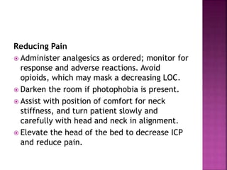
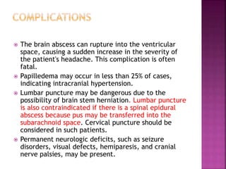
Chapter XII
 Meningitis is an inflammation of the pia
mater and arachnoid membranes that
surround the brain and the spinal cord.
 Viral meningitis is the most common form
and is usually self-limiting.
 Bacterial meningitis.
 Fungal meningitis,
 Parasitic meningitis.
 Hospital-acquired postcraniotomy meningitis,
 Classic symptoms are fever, headache, and nuchal rigidity (a
resistance to flexion of the neck).
 Altered mental status; confusion in older patients.
 Petechial (appears like rug burn) or purpuric rash from
coagulopathy,
 Photophobia.
 Nuchal rigidity, neck tenderness, or a bulging the anterior
fontanelle in infants.
 Children may exhibit behavioral changes, arching of the back and
neck, a blank stare, refusal to feed, and seizures. Viral
meningitis can cause a red, maculopapular rash in children.
 Positive Brudzinski's and Kernig's signs (see figure)
 Neonates may exhibit poor feeding, altered breathing patterns,
or listlessness.
 Onset may be over several hours or several days depending on
the infectious agent, the patient's age, immune status,
comorbidities, and other variables.
 Complete blood count (CBC) with differential
is indicated to detect an elevated leukocyte
count in bacterial and viral meningitis, with
a greater percentage of polymorphonuclear
leukocytes (90%) in bacterial and (less than
50%) in viral meningitis (normal 0% to 15%).
 Blood cultures are obtained to indicate the
organism.
 CSF evaluation for pressure, leukocytes,
protein,
 MRI/CT scan with and without contrast rules
out other disorders. A CT scan with contrast
must be done to detect abscesses.
 Bacterial meningitis, particularly in children,
may result in deafness, learning difficulties,
spasticity, paresis, or cranial nerve disorders.
 Increased ICP in AIDS patients with cryptococcal
meningitis has resulted in severe visual losses.
 Seizures occur in 20% to 30% of patients.
 Increased ICP may result in cerebral edema,
decreased perfusion, and tissue damage.
 Severe brain edema may result in herniation or
compression of the brain stem.
 Purpura may be associated with disseminated
intravascular coagulation.
 Obtain a history of recent infections such as
upper respiratory infection, and exposure to
causative agents.
 Assess neurologic status and vital signs.
 Evaluate for signs of meningeal irritation.
 Assess sensorineural hearing loss (vision and
hearing), cranial nerve damage (eg, facial
nerve palsy), and diminished cognitive
function.
 Hyperthermia related to the infectious
process and cerebral edema
 Risk for Imbalanced Fluid Volume related to
fever and decreased intake
 Ineffective Tissue Perfusion (cerebral)
related to infectious process and cerebral
edema
 Acute Pain related to meningeal irritation
 Impaired Physical Mobility related to
prolonged bed rest
Reducing Fever
 Administer antimicrobial agents on time to
maintain optimal blood levels.
 Monitor temperature frequently or
continuously, and administer antipyretics as
ordered.
 Institute other cooling measures, such as a
hypothermia blanket, as indicated.
Maintaining Fluid Balance
 Prevent I.V. fluid overload, which may
worsen cerebral edema.
 Monitor intake and output closely.
 Monitor CVP frequently.
Enhancing Cerebral Perfusion
 Assess LOC, vital signs, and neurologic
parameters frequently. Observe for signs and
symptoms of ICP (eg, decreased LOC, dilated
pupils, widening pulse pressure).
 Maintain a quiet, calm environment to prevent
agitation, which may cause an increased ICP.
 Prepare patient for a lumbar puncture for CSF
 Notify the health care provider of signs of
deterioration: increasing temperature,
decreasing LOC, seizure activity, or altered
respirations.
Reducing Pain
 Administer analgesics as ordered; monitor for
response and adverse reactions. Avoid
opioids, which may mask a decreasing LOC.
 Darken the room if photophobia is present.
 Assist with position of comfort for neck
stiffness, and turn patient slowly and
carefully with head and neck in alignment.
 Elevate the head of the bed to decrease ICP
and reduce pain.
Promoting Return to Optimal Level of
Functioning
 Implement rehabilitation interventions after
admission (eg, turning, positioning).
 Progress from passive to active exercises
based on the patient's neurologic status.
 Prevent bacterial meningitis by eliminating colonization
and infection with the offending organism.
 Administer vaccines against H. influenzae type B for children;
N. meningitidis serogroups A, C, Y, and W135 for patients at
high risk (especially college students, those without spleens,
immunodeficient); and S. pneumoniae for patients with
chronic illnesses and the elderly.
 Administer vaccines for travelers to countries with a high
incidence of meningococcal disease and household contacts of
someone who has had meningitis.
 Chemoprophylaxis for meningococcal disease, most commonly
with rifampin, may be necessary for health care workers,
household contacts in the community, day care centers, and
other highly susceptible populations.
 If maintenance antifungal prophylaxis is initiated for
patients with low CD4+ counts, as seen in some patients
with AIDS, the patient must understand the importance of
long-term pharmacologic therapy.
 Advise close contacts of the patient with
meningitis that prophylactic treatment may
be indicated; they should check with their
health care providers or the local public
health department.
 Encourage the patient to follow medication
regimen as directed to fully eradicate the
infectious agent.
 Encourage follow-up and prompt attention to
infections in future.
 Afebrile
 Adequate urine output; CVP in normal range
 Alert LOC; normal vital signs
 Pain controlled
 Optimal level of functioning after resolution
 Encephalitis is an inflammation of cerebral
tissue, typically accompanied by meningeal
inflammation. Meningoencephalitis is most
commonly caused by a viral infection. Like
meningitis, encephalitis can be infectious or
noninfectious and acute, subacute, or
chronic.
 In primary encephalitis, the brain or spinal cord is the
predominate foci of the toxin or pathogen. Secondary
encephalitis is a less serious form of encephalitis; it is
caused by an infection that is spread from another part of
the body.
 Acute viral encephalitis,
 Brainstem encephalitis
 Postischemic inflammatory encephalitis occurs due to
brain inflammation following a CVA.
 West Nile Virus (WNV) is an arthropod virus (arbovirus).
The mosquito is the primary vector and birds are the
primary hosts.
 Herpes simplex encephalitis
 Postinfectious encephalomyelitis follows a viral or
bacterial infectious process
 Cytomegalovirus encephalitis
 Toxoplasma encephalitis
 Signs and symptoms may develop hours or weeks
after exposure.
 Classic symptoms include fever, headache, and brain
aberration (eg, disorientation, neurologic deficits,
seizures).
 Increased ICP may result in alteration in
consciousness, nausea, and vomiting.
 Motor weakness, such as hemiparesis, may be
detected.
 Increased deep tendon reflexes and extensor plantar
response are noted.
 Bizarre behavior and personality changes may present
at onset.
 Hypothalamic-pituitary involvement may result in
hypothermia, diabetes insipidus, SIADH
 Neurologic symptoms may include superior quadrant
visual field defects, aphasia, dysphagia, ataxia, and
paresthesias.
 The patient with brainstem encephalitis may present
with nystagmus, decreased extraocular movements,
hearing loss, dysphagia, dysarthria, respiratory
abnormalities, and motor involvement.
 Limbic encephalitis may cause mood and personality
changes that progress to severe memory loss and
delirium.
 In WNV, about two-thirds of symptomatic patients
have encephalitis with signs and symptoms of fever,
vomiting, headache, nuchal rigidity, decreased LOC,
cranial nerve dysfunction, and an erythematous rash.
Seizures are uncommon
 Lumbar puncture
 EEG
 MRI
 Brain tissue biopsy
 Obtain patient history of recent infection,
animal exposure, tick or mosquito bite, recent
travel, exposure to ill contacts.
 Before delivery, women should be questioned
regarding a history of congenital herpes simplex
virus and examined for evidence of this virus; a
cesarean delivery should be explored with the
physician.
 Strict standard precautions should be adhered to
in order to contain drainage from herpetic
lesions.
 Vesicular lesions or rashes on neonates should be
reported immediately because these could
indicate active herpes simplex infection.
 Perform a complete clinical assessment.
 Risk for Injury related to seizures and
cerebral edema
 Ineffective Tissue Perfusion (cerebral)
related to disease process
 Hyperthermia related to infectious process
 Disturbed Thought Processes due to
personality changes
 Risk of Infection related to transmittal
Preventing Injury
 Maintain quiet environment and provide care
gently, avoiding over activity and agitation,
which may cause increased ICP.
 Maintain seizure precautions with side rails
padded, airway, and suction equipment at
bedside.
 Administer medications as ordered; monitor
response and adverse reactions.
Promoting Cerebral Perfusion
 Monitor neurologic status closely. Observe for
subtle changes, such as behavior or personality
changes, weakness, or cranial nerve
involvement. Notify health care provider.
 In arbovirus encephalitis, restrict fluids to
passively dehydrate the brain.
 Reorient patient frequently.
 Provide supportive care if coma develops; may
last several weeks.
 Encourage significant others to interact with
patient, and participate in the patient's
rehabilitation, even while the patient is in a
coma.
Relieving Fever
 Monitor temperature and vital signs
frequently.
 Administer antipyretics and other cooling
measures as indicated.
 Monitor fluid intake and output, and provide
fluid replacement through I.V. lines as
needed. Be alert to signs of other coexisting
infections, such as UTI or pneumonia, and
notify health care provider so cultures can be
obtained and treatment started.
Managing Aberrations in Thought Processes
 Orient to person, place, time.
 Maintain memory book, and provide cues to
perform required activities.
Avoiding Infectious Disease Transmission
 Maintain strict standard precautions.
 Initiate and maintain isolation per your
facility's policy.
 Promote vaccination of patient, family, and
significant others for measles, mumps, and
rubella.
 Pregnant women who have a history of
genital herpes simplex, or their partners,
should inform their physician of this history.
 Contacts of rabies-infected patients should
be offered rabies prophylaxis.
 Explain the effects of the disease process
and the rationale for care.
 Reassure significant others based on patient's
prognosis.
 Encourage follow-up for evaluation of
deficits and rehabilitation progress.
 Educate others about the signs and symptoms
of encephalitis if epidemic is suspected.
 To prevent WNV, advise the use of repellants
when outdoors and removal of standing
water that acts as a breeding ground for
mosquitoes.
 No seizures or signs of increased ICP
 Alert with no neurologic deficits
 Afebrile
 Oriented, memory intact
 No transmission of infection
 A brain abscess is a free or encapsulated
collection of infectious material of brain
parenchyma, between the dura and the
arachnoid linings (subdural abscess) or
between the dura and the skull (epidural
abscess). Spinal abscesses typically occur in
the epidural region.
 Intracranial subdural abscesses
 Intracranial epidural abscesses
 Spinal epidural abscesses
 Intermedullary abscesses
 Fungal brain abscesses
 M. tuberculosis
 Headache is poorly localized with a dull ache.
 Increased ICP may result in nausea, vomiting, decreased
LOC.
 Fever is found in less than 50% of cases.
 Neurologic findings such as hemisensory and paresis
deficits, aphasia, ataxia may be present.
 Seizures are frequently present.
 Dental abscess, sinusitis, and otitis media may be present.
 Signs and symptoms of a cerebral subdural empyema
include severe headache, fever, nuchal rigidity, and
Kernig's sign .
 Patients with intracranial epidural abscess commonly
present with fever, lethargy, and severe headache.
 Spinal epidural abscesses may be evidenced by severe back
pain, fever, headache, lower extremity weakness or
paralysis, nuchal rigidity, Kernig's sign, and local
tenderness
 CT scan, MRI with contrast locate the sites of
abscess, and follow evolution and resolution
of the suppurative process.
 Blood cultures
 Cultures are obtained from the suspected
source of infection
 EEG detects seizure disorders
 The brain abscess can rupture into the ventricular
space, causing a sudden increase in the severity of
the patient's headache. This complication is often
fatal.
 Papilledema may occur in less than 25% of cases,
indicating intracranial hypertension.
 Lumbar puncture may be dangerous due to the
possibility of brain stem herniation. Lumbar puncture
is also contraindicated if there is a spinal epidural
abscess because pus may be transferred into the
subarachnoid space. Cervical puncture should be
considered in such patients.
 Permanent neurologic deficits, such as seizure
disorders, visual defects, hemiparesis, and cranial
nerve palsies, may be present.
 There is greater mortality if the patient has
symptoms of short duration, has severe mental
status changes, and has rapid progression of
neurologic impairment.
 Delayed treatment of a spinal epidural abscess
may result in transaction syndrome, in which
flaccid paraplegia with sensory loss occurs at the
level of the abscess.
 In chronic otitis media, intracranial and
intratemporal complications frequently result
from progressive bony erosion, which may
expose the dura, labyrinth, and facial nerves.
 Obtain history of previous infection,
immunosuppression, headache, and related
symptoms.
 Perform neurologic assessment, including
cranial nerve evaluation, motor, and
cognitive status.
 Acute Pain related to cerebral mass
 Disturbed Thought Processes related to
disease process
 Risk for Injury related to neurologic deficits
 Anxiety related to surgery, prognosis, and
relapse
Relieving Pain
 Administer pain medications as ordered.
 Provide comfort measures, such as quiet
environment, positioning with head slightly
elevated, and assistance with hygiene needs.
 Provide passive relaxation techniques, such
as soft music and backrubs.
Promoting Thought Processes
 Frequently monitor vital signs, LOC,
orientation, and seizure activity.
 Report changes, which can signal increased
ICP, to health care provider.
 Administer medications as ordered, noting
response and adverse reactions.
 Prepare patient for repeated diagnostic tests
to evaluate response to therapy and surgery.
Minimizing Neurologic Deficits
 Maintain a safe environment with side rails
up, call light within reach, and frequent
observation.
 Evaluate other cranial nerve function, and
report changes.
 Refer to occupational therapy, speech
therapist, or other rehabilitation specialist to
provide adjunct to nursing rehabilitation.
Reducing Anxiety
 Prepare patient and family for surgery when
indicated. Encourage discussion with surgeon
to understand risks, benefits of the
procedure.
 Explain postoperative progression and
nursing care
 Patient follow-up is essential for sinusitis,
otitis media, respiratory infections, and
other infectious processes that may result in
a brain abscess.
 Continue with rehabilitation to regain or
compensate for neurologic deficits.
 Continue with pharmacologic regimen in
community setting.
 Observe for recurrence of brain and spinal
abscesses.
 Maintain wellness with vaccinations,
immunizations, and overall health.
 Reinforce need for dental procedure
prophylaxis to avoid dental abscesses.
 Instruct in need for immediate assessment of
head wounds.
 Verbalizes reduced pain
 Oriented to person, place, and time; follows
simple commands
 No injury related to neurologic deficits
 Reduced anxiety regarding disease process
and procedures

10. INFECTIOUS DISORDERS nursing process pptx

  • 1.
  • 2.
     Meningitis isan inflammation of the pia mater and arachnoid membranes that surround the brain and the spinal cord.
  • 3.
     Viral meningitisis the most common form and is usually self-limiting.  Bacterial meningitis.  Fungal meningitis,  Parasitic meningitis.  Hospital-acquired postcraniotomy meningitis,
  • 4.
     Classic symptomsare fever, headache, and nuchal rigidity (a resistance to flexion of the neck).  Altered mental status; confusion in older patients.  Petechial (appears like rug burn) or purpuric rash from coagulopathy,  Photophobia.  Nuchal rigidity, neck tenderness, or a bulging the anterior fontanelle in infants.  Children may exhibit behavioral changes, arching of the back and neck, a blank stare, refusal to feed, and seizures. Viral meningitis can cause a red, maculopapular rash in children.  Positive Brudzinski's and Kernig's signs (see figure)  Neonates may exhibit poor feeding, altered breathing patterns, or listlessness.  Onset may be over several hours or several days depending on the infectious agent, the patient's age, immune status, comorbidities, and other variables.
  • 6.
     Complete bloodcount (CBC) with differential is indicated to detect an elevated leukocyte count in bacterial and viral meningitis, with a greater percentage of polymorphonuclear leukocytes (90%) in bacterial and (less than 50%) in viral meningitis (normal 0% to 15%).  Blood cultures are obtained to indicate the organism.  CSF evaluation for pressure, leukocytes, protein,  MRI/CT scan with and without contrast rules out other disorders. A CT scan with contrast must be done to detect abscesses.
  • 7.
     Bacterial meningitis,particularly in children, may result in deafness, learning difficulties, spasticity, paresis, or cranial nerve disorders.  Increased ICP in AIDS patients with cryptococcal meningitis has resulted in severe visual losses.  Seizures occur in 20% to 30% of patients.  Increased ICP may result in cerebral edema, decreased perfusion, and tissue damage.  Severe brain edema may result in herniation or compression of the brain stem.  Purpura may be associated with disseminated intravascular coagulation.
  • 8.
     Obtain ahistory of recent infections such as upper respiratory infection, and exposure to causative agents.  Assess neurologic status and vital signs.  Evaluate for signs of meningeal irritation.  Assess sensorineural hearing loss (vision and hearing), cranial nerve damage (eg, facial nerve palsy), and diminished cognitive function.
  • 9.
     Hyperthermia relatedto the infectious process and cerebral edema  Risk for Imbalanced Fluid Volume related to fever and decreased intake  Ineffective Tissue Perfusion (cerebral) related to infectious process and cerebral edema  Acute Pain related to meningeal irritation  Impaired Physical Mobility related to prolonged bed rest
  • 10.
    Reducing Fever  Administerantimicrobial agents on time to maintain optimal blood levels.  Monitor temperature frequently or continuously, and administer antipyretics as ordered.  Institute other cooling measures, such as a hypothermia blanket, as indicated.
  • 11.
    Maintaining Fluid Balance Prevent I.V. fluid overload, which may worsen cerebral edema.  Monitor intake and output closely.  Monitor CVP frequently.
  • 12.
    Enhancing Cerebral Perfusion Assess LOC, vital signs, and neurologic parameters frequently. Observe for signs and symptoms of ICP (eg, decreased LOC, dilated pupils, widening pulse pressure).  Maintain a quiet, calm environment to prevent agitation, which may cause an increased ICP.  Prepare patient for a lumbar puncture for CSF  Notify the health care provider of signs of deterioration: increasing temperature, decreasing LOC, seizure activity, or altered respirations.
  • 13.
    Reducing Pain  Administeranalgesics as ordered; monitor for response and adverse reactions. Avoid opioids, which may mask a decreasing LOC.  Darken the room if photophobia is present.  Assist with position of comfort for neck stiffness, and turn patient slowly and carefully with head and neck in alignment.  Elevate the head of the bed to decrease ICP and reduce pain.
  • 14.
    Promoting Return toOptimal Level of Functioning  Implement rehabilitation interventions after admission (eg, turning, positioning).  Progress from passive to active exercises based on the patient's neurologic status.
  • 15.
     Prevent bacterialmeningitis by eliminating colonization and infection with the offending organism.  Administer vaccines against H. influenzae type B for children; N. meningitidis serogroups A, C, Y, and W135 for patients at high risk (especially college students, those without spleens, immunodeficient); and S. pneumoniae for patients with chronic illnesses and the elderly.  Administer vaccines for travelers to countries with a high incidence of meningococcal disease and household contacts of someone who has had meningitis.  Chemoprophylaxis for meningococcal disease, most commonly with rifampin, may be necessary for health care workers, household contacts in the community, day care centers, and other highly susceptible populations.  If maintenance antifungal prophylaxis is initiated for patients with low CD4+ counts, as seen in some patients with AIDS, the patient must understand the importance of long-term pharmacologic therapy.
  • 16.
     Advise closecontacts of the patient with meningitis that prophylactic treatment may be indicated; they should check with their health care providers or the local public health department.  Encourage the patient to follow medication regimen as directed to fully eradicate the infectious agent.  Encourage follow-up and prompt attention to infections in future.
  • 17.
     Afebrile  Adequateurine output; CVP in normal range  Alert LOC; normal vital signs  Pain controlled  Optimal level of functioning after resolution
  • 19.
     Encephalitis isan inflammation of cerebral tissue, typically accompanied by meningeal inflammation. Meningoencephalitis is most commonly caused by a viral infection. Like meningitis, encephalitis can be infectious or noninfectious and acute, subacute, or chronic.
  • 20.
     In primaryencephalitis, the brain or spinal cord is the predominate foci of the toxin or pathogen. Secondary encephalitis is a less serious form of encephalitis; it is caused by an infection that is spread from another part of the body.  Acute viral encephalitis,  Brainstem encephalitis  Postischemic inflammatory encephalitis occurs due to brain inflammation following a CVA.  West Nile Virus (WNV) is an arthropod virus (arbovirus). The mosquito is the primary vector and birds are the primary hosts.  Herpes simplex encephalitis  Postinfectious encephalomyelitis follows a viral or bacterial infectious process  Cytomegalovirus encephalitis  Toxoplasma encephalitis
  • 21.
     Signs andsymptoms may develop hours or weeks after exposure.  Classic symptoms include fever, headache, and brain aberration (eg, disorientation, neurologic deficits, seizures).  Increased ICP may result in alteration in consciousness, nausea, and vomiting.  Motor weakness, such as hemiparesis, may be detected.  Increased deep tendon reflexes and extensor plantar response are noted.  Bizarre behavior and personality changes may present at onset.  Hypothalamic-pituitary involvement may result in hypothermia, diabetes insipidus, SIADH
  • 22.
     Neurologic symptomsmay include superior quadrant visual field defects, aphasia, dysphagia, ataxia, and paresthesias.  The patient with brainstem encephalitis may present with nystagmus, decreased extraocular movements, hearing loss, dysphagia, dysarthria, respiratory abnormalities, and motor involvement.  Limbic encephalitis may cause mood and personality changes that progress to severe memory loss and delirium.  In WNV, about two-thirds of symptomatic patients have encephalitis with signs and symptoms of fever, vomiting, headache, nuchal rigidity, decreased LOC, cranial nerve dysfunction, and an erythematous rash. Seizures are uncommon
  • 23.
     Lumbar puncture EEG  MRI  Brain tissue biopsy
  • 24.
     Obtain patienthistory of recent infection, animal exposure, tick or mosquito bite, recent travel, exposure to ill contacts.  Before delivery, women should be questioned regarding a history of congenital herpes simplex virus and examined for evidence of this virus; a cesarean delivery should be explored with the physician.  Strict standard precautions should be adhered to in order to contain drainage from herpetic lesions.  Vesicular lesions or rashes on neonates should be reported immediately because these could indicate active herpes simplex infection.  Perform a complete clinical assessment.
  • 25.
     Risk forInjury related to seizures and cerebral edema  Ineffective Tissue Perfusion (cerebral) related to disease process  Hyperthermia related to infectious process  Disturbed Thought Processes due to personality changes  Risk of Infection related to transmittal
  • 26.
    Preventing Injury  Maintainquiet environment and provide care gently, avoiding over activity and agitation, which may cause increased ICP.  Maintain seizure precautions with side rails padded, airway, and suction equipment at bedside.  Administer medications as ordered; monitor response and adverse reactions.
  • 27.
    Promoting Cerebral Perfusion Monitor neurologic status closely. Observe for subtle changes, such as behavior or personality changes, weakness, or cranial nerve involvement. Notify health care provider.  In arbovirus encephalitis, restrict fluids to passively dehydrate the brain.  Reorient patient frequently.  Provide supportive care if coma develops; may last several weeks.  Encourage significant others to interact with patient, and participate in the patient's rehabilitation, even while the patient is in a coma.
  • 28.
    Relieving Fever  Monitortemperature and vital signs frequently.  Administer antipyretics and other cooling measures as indicated.  Monitor fluid intake and output, and provide fluid replacement through I.V. lines as needed. Be alert to signs of other coexisting infections, such as UTI or pneumonia, and notify health care provider so cultures can be obtained and treatment started.
  • 29.
    Managing Aberrations inThought Processes  Orient to person, place, time.  Maintain memory book, and provide cues to perform required activities.
  • 30.
    Avoiding Infectious DiseaseTransmission  Maintain strict standard precautions.  Initiate and maintain isolation per your facility's policy.
  • 31.
     Promote vaccinationof patient, family, and significant others for measles, mumps, and rubella.  Pregnant women who have a history of genital herpes simplex, or their partners, should inform their physician of this history.  Contacts of rabies-infected patients should be offered rabies prophylaxis.
  • 32.
     Explain theeffects of the disease process and the rationale for care.  Reassure significant others based on patient's prognosis.  Encourage follow-up for evaluation of deficits and rehabilitation progress.  Educate others about the signs and symptoms of encephalitis if epidemic is suspected.  To prevent WNV, advise the use of repellants when outdoors and removal of standing water that acts as a breeding ground for mosquitoes.
  • 33.
     No seizuresor signs of increased ICP  Alert with no neurologic deficits  Afebrile  Oriented, memory intact  No transmission of infection
  • 35.
     A brainabscess is a free or encapsulated collection of infectious material of brain parenchyma, between the dura and the arachnoid linings (subdural abscess) or between the dura and the skull (epidural abscess). Spinal abscesses typically occur in the epidural region.
  • 36.
     Intracranial subduralabscesses  Intracranial epidural abscesses  Spinal epidural abscesses  Intermedullary abscesses  Fungal brain abscesses  M. tuberculosis
  • 37.
     Headache ispoorly localized with a dull ache.  Increased ICP may result in nausea, vomiting, decreased LOC.  Fever is found in less than 50% of cases.  Neurologic findings such as hemisensory and paresis deficits, aphasia, ataxia may be present.  Seizures are frequently present.  Dental abscess, sinusitis, and otitis media may be present.  Signs and symptoms of a cerebral subdural empyema include severe headache, fever, nuchal rigidity, and Kernig's sign .  Patients with intracranial epidural abscess commonly present with fever, lethargy, and severe headache.  Spinal epidural abscesses may be evidenced by severe back pain, fever, headache, lower extremity weakness or paralysis, nuchal rigidity, Kernig's sign, and local tenderness
  • 38.
     CT scan,MRI with contrast locate the sites of abscess, and follow evolution and resolution of the suppurative process.  Blood cultures  Cultures are obtained from the suspected source of infection  EEG detects seizure disorders
  • 39.
     The brainabscess can rupture into the ventricular space, causing a sudden increase in the severity of the patient's headache. This complication is often fatal.  Papilledema may occur in less than 25% of cases, indicating intracranial hypertension.  Lumbar puncture may be dangerous due to the possibility of brain stem herniation. Lumbar puncture is also contraindicated if there is a spinal epidural abscess because pus may be transferred into the subarachnoid space. Cervical puncture should be considered in such patients.  Permanent neurologic deficits, such as seizure disorders, visual defects, hemiparesis, and cranial nerve palsies, may be present.
  • 40.
     There isgreater mortality if the patient has symptoms of short duration, has severe mental status changes, and has rapid progression of neurologic impairment.  Delayed treatment of a spinal epidural abscess may result in transaction syndrome, in which flaccid paraplegia with sensory loss occurs at the level of the abscess.  In chronic otitis media, intracranial and intratemporal complications frequently result from progressive bony erosion, which may expose the dura, labyrinth, and facial nerves.
  • 41.
     Obtain historyof previous infection, immunosuppression, headache, and related symptoms.  Perform neurologic assessment, including cranial nerve evaluation, motor, and cognitive status.
  • 42.
     Acute Painrelated to cerebral mass  Disturbed Thought Processes related to disease process  Risk for Injury related to neurologic deficits  Anxiety related to surgery, prognosis, and relapse
  • 43.
    Relieving Pain  Administerpain medications as ordered.  Provide comfort measures, such as quiet environment, positioning with head slightly elevated, and assistance with hygiene needs.  Provide passive relaxation techniques, such as soft music and backrubs.
  • 44.
    Promoting Thought Processes Frequently monitor vital signs, LOC, orientation, and seizure activity.  Report changes, which can signal increased ICP, to health care provider.  Administer medications as ordered, noting response and adverse reactions.  Prepare patient for repeated diagnostic tests to evaluate response to therapy and surgery.
  • 45.
    Minimizing Neurologic Deficits Maintain a safe environment with side rails up, call light within reach, and frequent observation.  Evaluate other cranial nerve function, and report changes.  Refer to occupational therapy, speech therapist, or other rehabilitation specialist to provide adjunct to nursing rehabilitation.
  • 46.
    Reducing Anxiety  Preparepatient and family for surgery when indicated. Encourage discussion with surgeon to understand risks, benefits of the procedure.  Explain postoperative progression and nursing care
  • 47.
     Patient follow-upis essential for sinusitis, otitis media, respiratory infections, and other infectious processes that may result in a brain abscess.  Continue with rehabilitation to regain or compensate for neurologic deficits.  Continue with pharmacologic regimen in community setting.  Observe for recurrence of brain and spinal abscesses.
  • 48.
     Maintain wellnesswith vaccinations, immunizations, and overall health.  Reinforce need for dental procedure prophylaxis to avoid dental abscesses.  Instruct in need for immediate assessment of head wounds.
  • 49.
     Verbalizes reducedpain  Oriented to person, place, and time; follows simple commands  No injury related to neurologic deficits  Reduced anxiety regarding disease process and procedures