MENINGITIS
 The inflammation is usually caused by an
infection of the fluid surrounding the
brain and spinal cord.
 Meningitis can be life-threatening
because of the inflammation's proximity
to the brain and spinal cord; therefore the
condition is classified as a medical
emergency.
DEFINITION
Meningitis is the inflammation of
the membranes surrounding the brain
& spinal cord, including the dura,
arachinoid & pia matter.
MENINGES
 The meninges is the system of membranes
which envelops the central nervous system.
 It has 3 layers:
1. Dura mater
2. Arachnoid mater
3. Pia mater
 Subarachnoid space - is
the space which exists
between the arachnoid
and the pia mater, which
is filled with cerebrospinal fluid.
INCIDENCE
 Meningitis can occur at all ages but it is
commonest in infancy.
 While 95% of the cases take place
between 1 month- 5 years of age.
 It is more common in males than females.
ETIOLOGY
 Bacterial
 Viral
 Fungal
 Parasitic/ protozoal
 Physical injury
 Cancer
 Certain drugs ( mainly, NSAID’S)
 Head injury
 Cerebral abscess
 Middle ear infection
ROUTES OF INFECTION
 Nasopharynx
 Blood stream
 Direct spread (skull fracture, meningo
and encephalocele)
 Middle ear infection
 Infected Ventriculoperitoneal shunts.
 Congenital defects
 Sinusitis
TRANSMISSION
 The bacteria are transmitted from person to
person through droplets of respiratory or throat
secretions.
 Close and prolonged contact (e.g. sneezing and
coughing on someone, living in close quarters
or dormitories (military recruits, students),
sharing eating or drinking utensils, etc.)
 The incubation period ranges between 2 -10
days.
RISK FACTORS
 Skipping vaccinations
 Age-Viral meningitis
 occur in children younger than age 5.
Bacterial meningitis is common in those
under age 20.
 Pregnancy.
 Compromised immune system. AIDS,
alcoholism, diabetes, use of
immunosuppressant drugs etc.
TYPES
 Bacterial
 Viral (aseptic)
 Fungal
 Parasitic
 Non-infectious
 Tuberculosis
BACTERIAL MENINGITIS
Pathogenesis:
 Entry of organism through blood brain
barrier
 release of cell wall & membrane products
 Outpouring of polymorphs & fibrin
 cytokines & chemokines
 Inflammatory mediators
 Inflamed meninges covered with exudate
(most marked in pneumoccocal meningitis).
VIRAL MENINGITIS
 Viral meningitis comprises most aseptic
meningitis syndromes. The viral agents for
aseptic meningitis include the following:
 Enterovirus (polio virus, Echovirus,
Coxsackievirus )
 Herpesvirus (Hsv-1,2, Varicella.Z,EBV )
 Paramyxovirus (Mumps, Measles)
 Togavirus (Rubella)
 Rhabdovirus (Rabies)
 Retrovirus (HIV)
FUNGAL MENINGITIS
 It’s rare in healthy people, but is a higher
risk in those who have AIDS, other forms
of immunodeficiency or
immunosuppression.
 The most common agents are
Cryptococcus neoformans, Candida, H
capsulatum.
PARASITIC MENINGITIES
 Infection with free-living amoebas is an
infrequent but often life-threatening human
illness.
 It’s more common in underdeveloped countries
and usually is caused by parasites found in
contaminated water, food, and soil.
 The most common causative agents are:
• Free-living amoebas (ie, Acanthamoeba,
Balamuthia, Naegleria)
• Helminthic eosinophilic meningitis
NON-INFECTIOUS MENINGITIS
 Rarely, meningitis can be caused by exposure
to certain medications, such as the following:
• Immune globulin
• Levamisole
• Metronidazole
• Mumps and rubella vaccines
• Nonsteroidal anti-inflammatory drugs (e.g.,
ibuprofen, diclofenac, naproxen)
TUBERCULOSIS MENINGITIS
 It’s a complication of Childhood
tuberculosis & common cause of
prolonged morbidity, handicap & death.
 Children below 5 years are specially
prone.
PATHOPHYSIOLOGY
Infection from any part of the body like nasopharynx
Organisms invade surrounding blood vessels
Through blood, organisms enter cerebrospinal fluid
Infection spread through subarachnoid space
Inflammatory process begins
Contd……
Increase in CSF exudation in ventricles
CSF flow through ventricular aqueduct
Thrombophlebitis of cerebral vessels
Infection of cerebral cortex, cerebral damage
CLINICAL MANIFESTATIONS
First Phase :
 Vague symptoms.
 Child doesn’t play, is irritable, restless or
drowsy.
 Anorexia & vomiting may be present
 Older child may complain of headache.
 Possibly preceding history of Measles or
another illness with incompletely recovery
Second Phase:
 Child is drowsy with neck stiffness, & rigidity.
 Kernig & Brudzinski sign may become positive,
anterior fontanels bulges
 Twitching of muscles, convulsions, raised
temperature.
 strabismus, nystagmus, and papilloedema may be
present.
 Fundoscopy: Choroidal TB may be seen
BRUDZINSKI’S SIGN
KERNIG’S SIGN
Terminal Phase
 Child is characteristically comatose
with opisthotonus, & multiple focal
paresis.
 Cranial nerve palsies are present.
 High grade fever often occurs
terminally.
COMPLICATIONS
 Hearing loss
 Memory difficulty
 Learning disabilities
 Brain damage
 Gait problems
 Seizures
 Kidney failure
 Shock
 Death
DIAGNOSTIC EVALUATION
 History collection
 Physical examination
 CT and MRI
 Blood culture and sensitivity
 CSF examination
 Lumbar puncture: pressure usually
raised,10-500 PMNs early but later
lymphocytes predominate.
 Tuberculin skin test
MANAGEMENT
MEDICAL MANAGEMENT
A. PHARMACOLOGICAL MANAGEMENT
 Antibiotics- ampicillin, penicillin, amoxycillin
 Antiviral- tenofovir
 Antifungal- fluconazole
 Corticosteroid- dexamethasone
 IV mannitol for diuresis
 IV phenytoin
 Antipyretics- Acetaminophen
B. NON PHARMACOLOGICAL
MANAGEMENT
 Maintenance of fluid-electrolyte balance
by IV fluid therapy
 Nasogastric tube feeding
 Vitamin supplementation
 Head end elevation 30-45degree
 Emotional support and necessary
information for continuation of care at
home, follow-up and rehabilitation.
SURGICAL MANAGEMENT
 Cochlear implantation rehabilitation
due to deafness
NURSING MANAGEMENT
ASSESSMENT
 Obtain a history of recent infections such as
upper respiratory infection, and exposure to
causative agents.
 Assess neurologic status and vital signs.
 Evaluate for signs of meningeal irritation.
 Assess sensorineural hearing loss (vision and
hearing), cranial nerve damage (eg, facial nerve
palsy), and diminished cognitive function.
NURSING DIAGNOSIS
 Elevated body temperature related to positive
bacterial infection as manifested by flushed and
warm to touch skin.
 Acute pain related to meningeal irritation with
spasm of extensor muscles (neck, shoulders and
back) as manifested by positive kernig’s and
brudzinski’s sign.
 Risk for ineffective cerebral tissue perfusion
related to increased intracranial pressure(ICP)
 Risk for infection related to presence of
infective organisms
 Risk for injury related to presence of
infection
 Altered heat regulation related to
compression of hypothalamus
 Altered family processes related to
having a child with a serious illness
GOAL
 To Enhanced Cerebral Tissue Perfusion
 To Reduce Fever
 To Maintain Fluid Balance
 To Reduce Pain
 To Return to Optimal Level
of Functioning/ mobility
INTERVENTIONS
 Enhancing Cerebral Perfusion
 Assess LOC, vital signs, and neurologic parameters
frequently. Observe for signs and symptoms of ICP
(e.g. decreased LOC, dilated pupils, widening pulse
pressure).
 Maintain a quiet, calm environment to prevent
agitation, which may cause an increased ICP.
Contd….
 Prepare patient for a lumbar puncture for
CSF evaluation, and repeat spinal tap, if
indicated. Lumbar puncture typically
precedes neuroimaging
 Notify the health care provider of signs of
deterioration: increasing temperature,
decreasing LOC, seizure activity, or altered
respirations.
 I/V mannitol is administered.
EXPECTED OUTCOMES
 Enhanced Cerebral Tissue Perfusion
 Fluid Balance Maintained
 Reduced Fever
 Reduced Pain
 Return to Optimal Level of Functioning
PREVENTION
 The vaccines against Hib, measles, mumps,
polio, meningococcus, and pneumococcus can
protect against meningitis
 Hib vaccine: all infants should receive at
2,4,6 months of age & booster 1 year later.
 After 1 year 1 dose is given till the age of 5
years.
PREVENTION
 Pneumococcal vaccine: 0.5 ml is given IM (<2
yrs)
 High-risk children should also be immunized
routinely.
 Vaccination before travelling
to an endemic area
PREVENTION
 Chemoprophylaxis for susceptible individuals or
close contacts:
H influenzae type b : Rifampin(20 mg/kg/d) for
4 days
N meningitidis: Rifampin (600 mg PO q12h) for
2 days upto 10weeks
Ceftriaxone (250 mg IM) single dose or
Ciprofloxacin(500-750 mg) single dose.
CONCLUSION
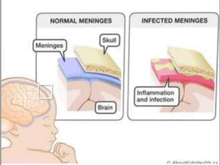
 Meningitis is an inflammation of the
meninges. The meninges are the three
membranes that cover the brain and
spinal cord. Meningitis can occur when
fluid surrounding the meninges becomes
infected
Meningitis

Meningitis

  • 3.
    MENINGITIS  The inflammationis usually caused by an infection of the fluid surrounding the brain and spinal cord.  Meningitis can be life-threatening because of the inflammation's proximity to the brain and spinal cord; therefore the condition is classified as a medical emergency.
  • 4.
    DEFINITION Meningitis is theinflammation of the membranes surrounding the brain & spinal cord, including the dura, arachinoid & pia matter.
  • 5.
    MENINGES  The meningesis the system of membranes which envelops the central nervous system.  It has 3 layers: 1. Dura mater 2. Arachnoid mater 3. Pia mater  Subarachnoid space - is the space which exists between the arachnoid and the pia mater, which is filled with cerebrospinal fluid.
  • 8.
    INCIDENCE  Meningitis canoccur at all ages but it is commonest in infancy.  While 95% of the cases take place between 1 month- 5 years of age.  It is more common in males than females.
  • 9.
    ETIOLOGY  Bacterial  Viral Fungal  Parasitic/ protozoal  Physical injury  Cancer  Certain drugs ( mainly, NSAID’S)  Head injury  Cerebral abscess  Middle ear infection
  • 10.
    ROUTES OF INFECTION Nasopharynx  Blood stream  Direct spread (skull fracture, meningo and encephalocele)  Middle ear infection  Infected Ventriculoperitoneal shunts.  Congenital defects  Sinusitis
  • 11.
    TRANSMISSION  The bacteriaare transmitted from person to person through droplets of respiratory or throat secretions.  Close and prolonged contact (e.g. sneezing and coughing on someone, living in close quarters or dormitories (military recruits, students), sharing eating or drinking utensils, etc.)  The incubation period ranges between 2 -10 days.
  • 12.
    RISK FACTORS  Skippingvaccinations  Age-Viral meningitis  occur in children younger than age 5. Bacterial meningitis is common in those under age 20.  Pregnancy.  Compromised immune system. AIDS, alcoholism, diabetes, use of immunosuppressant drugs etc.
  • 13.
    TYPES  Bacterial  Viral(aseptic)  Fungal  Parasitic  Non-infectious  Tuberculosis
  • 14.
    BACTERIAL MENINGITIS Pathogenesis:  Entryof organism through blood brain barrier  release of cell wall & membrane products  Outpouring of polymorphs & fibrin  cytokines & chemokines  Inflammatory mediators  Inflamed meninges covered with exudate (most marked in pneumoccocal meningitis).
  • 15.
    VIRAL MENINGITIS  Viralmeningitis comprises most aseptic meningitis syndromes. The viral agents for aseptic meningitis include the following:  Enterovirus (polio virus, Echovirus, Coxsackievirus )  Herpesvirus (Hsv-1,2, Varicella.Z,EBV )  Paramyxovirus (Mumps, Measles)  Togavirus (Rubella)  Rhabdovirus (Rabies)  Retrovirus (HIV)
  • 16.
    FUNGAL MENINGITIS  It’srare in healthy people, but is a higher risk in those who have AIDS, other forms of immunodeficiency or immunosuppression.  The most common agents are Cryptococcus neoformans, Candida, H capsulatum.
  • 17.
    PARASITIC MENINGITIES  Infectionwith free-living amoebas is an infrequent but often life-threatening human illness.  It’s more common in underdeveloped countries and usually is caused by parasites found in contaminated water, food, and soil.  The most common causative agents are: • Free-living amoebas (ie, Acanthamoeba, Balamuthia, Naegleria) • Helminthic eosinophilic meningitis
  • 18.
    NON-INFECTIOUS MENINGITIS  Rarely,meningitis can be caused by exposure to certain medications, such as the following: • Immune globulin • Levamisole • Metronidazole • Mumps and rubella vaccines • Nonsteroidal anti-inflammatory drugs (e.g., ibuprofen, diclofenac, naproxen)
  • 19.
    TUBERCULOSIS MENINGITIS  It’sa complication of Childhood tuberculosis & common cause of prolonged morbidity, handicap & death.  Children below 5 years are specially prone.
  • 20.
    PATHOPHYSIOLOGY Infection from anypart of the body like nasopharynx Organisms invade surrounding blood vessels Through blood, organisms enter cerebrospinal fluid Infection spread through subarachnoid space Inflammatory process begins
  • 21.
    Contd…… Increase in CSFexudation in ventricles CSF flow through ventricular aqueduct Thrombophlebitis of cerebral vessels Infection of cerebral cortex, cerebral damage
  • 22.
    CLINICAL MANIFESTATIONS First Phase:  Vague symptoms.  Child doesn’t play, is irritable, restless or drowsy.  Anorexia & vomiting may be present  Older child may complain of headache.  Possibly preceding history of Measles or another illness with incompletely recovery
  • 23.
    Second Phase:  Childis drowsy with neck stiffness, & rigidity.  Kernig & Brudzinski sign may become positive, anterior fontanels bulges  Twitching of muscles, convulsions, raised temperature.  strabismus, nystagmus, and papilloedema may be present.  Fundoscopy: Choroidal TB may be seen
  • 24.
  • 25.
  • 26.
    Terminal Phase  Childis characteristically comatose with opisthotonus, & multiple focal paresis.  Cranial nerve palsies are present.  High grade fever often occurs terminally.
  • 27.
    COMPLICATIONS  Hearing loss Memory difficulty  Learning disabilities  Brain damage  Gait problems  Seizures  Kidney failure  Shock  Death
  • 28.
    DIAGNOSTIC EVALUATION  Historycollection  Physical examination  CT and MRI  Blood culture and sensitivity  CSF examination  Lumbar puncture: pressure usually raised,10-500 PMNs early but later lymphocytes predominate.  Tuberculin skin test
  • 29.
    MANAGEMENT MEDICAL MANAGEMENT A. PHARMACOLOGICALMANAGEMENT  Antibiotics- ampicillin, penicillin, amoxycillin  Antiviral- tenofovir  Antifungal- fluconazole  Corticosteroid- dexamethasone  IV mannitol for diuresis  IV phenytoin  Antipyretics- Acetaminophen
  • 30.
    B. NON PHARMACOLOGICAL MANAGEMENT Maintenance of fluid-electrolyte balance by IV fluid therapy  Nasogastric tube feeding  Vitamin supplementation  Head end elevation 30-45degree  Emotional support and necessary information for continuation of care at home, follow-up and rehabilitation.
  • 31.
    SURGICAL MANAGEMENT  Cochlearimplantation rehabilitation due to deafness
  • 32.
    NURSING MANAGEMENT ASSESSMENT  Obtaina history of recent infections such as upper respiratory infection, and exposure to causative agents.  Assess neurologic status and vital signs.  Evaluate for signs of meningeal irritation.  Assess sensorineural hearing loss (vision and hearing), cranial nerve damage (eg, facial nerve palsy), and diminished cognitive function.
  • 33.
    NURSING DIAGNOSIS  Elevatedbody temperature related to positive bacterial infection as manifested by flushed and warm to touch skin.  Acute pain related to meningeal irritation with spasm of extensor muscles (neck, shoulders and back) as manifested by positive kernig’s and brudzinski’s sign.  Risk for ineffective cerebral tissue perfusion related to increased intracranial pressure(ICP)
  • 34.
     Risk forinfection related to presence of infective organisms  Risk for injury related to presence of infection  Altered heat regulation related to compression of hypothalamus  Altered family processes related to having a child with a serious illness
  • 35.
    GOAL  To EnhancedCerebral Tissue Perfusion  To Reduce Fever  To Maintain Fluid Balance  To Reduce Pain  To Return to Optimal Level of Functioning/ mobility
  • 36.
    INTERVENTIONS  Enhancing CerebralPerfusion  Assess LOC, vital signs, and neurologic parameters frequently. Observe for signs and symptoms of ICP (e.g. decreased LOC, dilated pupils, widening pulse pressure).  Maintain a quiet, calm environment to prevent agitation, which may cause an increased ICP.
  • 37.
    Contd….  Prepare patientfor a lumbar puncture for CSF evaluation, and repeat spinal tap, if indicated. Lumbar puncture typically precedes neuroimaging  Notify the health care provider of signs of deterioration: increasing temperature, decreasing LOC, seizure activity, or altered respirations.  I/V mannitol is administered.
  • 38.
    EXPECTED OUTCOMES  EnhancedCerebral Tissue Perfusion  Fluid Balance Maintained  Reduced Fever  Reduced Pain  Return to Optimal Level of Functioning
  • 39.
    PREVENTION  The vaccinesagainst Hib, measles, mumps, polio, meningococcus, and pneumococcus can protect against meningitis  Hib vaccine: all infants should receive at 2,4,6 months of age & booster 1 year later.  After 1 year 1 dose is given till the age of 5 years.
  • 40.
    PREVENTION  Pneumococcal vaccine:0.5 ml is given IM (<2 yrs)  High-risk children should also be immunized routinely.  Vaccination before travelling to an endemic area
  • 41.
    PREVENTION  Chemoprophylaxis forsusceptible individuals or close contacts: H influenzae type b : Rifampin(20 mg/kg/d) for 4 days N meningitidis: Rifampin (600 mg PO q12h) for 2 days upto 10weeks Ceftriaxone (250 mg IM) single dose or Ciprofloxacin(500-750 mg) single dose.
  • 42.
    CONCLUSION  Meningitis isan inflammation of the meninges. The meninges are the three membranes that cover the brain and spinal cord. Meningitis can occur when fluid surrounding the meninges becomes infected