SlideShare a Scribd company logo
Dr. Avinash Gholave
Department of Botany
K. V. N. Naik Arts, Commerce And Science College Nashik.
agholave@gmail.com
Gnetum is a genus of gymnosperms, the sole genus in the family Gnetaceae
and order Gnetales. They are tropical evergreen threes, shrubs and lianas.
Unlike other gymnosperms they possess vessel element in the xylem.
Systematic position:
Kingdom: Plantae
Division: Gnetophyta
Class: Gnetopsida
Order: Gnetales
Family: Gnetaceae
Genus: Gnetum
Distribution: The genus Gnetum includes about 35 species. The species are
distributed mainly in tropical and humid region of Africa, Asia and south
America. The five species of Gnetum have been reported in India.
Distribution of five Indian species:
1) Gnetum gnemon: A small tree or shrub with small drooping branches. The
species is found in Arunachal Pradesh, Assam, Manipur and Khasi and Jaintia
hiils.
2) Gnetum latifolium: An evergreen shrubs or climber. The species is mainly
distributed in Andaman and Nicobar Island.
3) Gnetum montanum: It is a large woody climbers. The species is confined to
north India. It occurs in tropical Himalayas. Sikkim to East Bengal, Assam,
Manipur and Khasi hiils.
4) Gnetum contractum: A scandent shrubs. It occurs in Travancore, Kerala,
Coonoon and Nilgiri hills.
5) Gnetum ula: It is large woody climber with scaly bark and swollen nodes. The
species is distributed along Eastern and Western Ghats.
Gnetum gnemon
Gnetum latifolium
Gnetum montanum
Gnetum ula
Sporophyte of Gnetum:
Gnetum resembles very much in its characteristics to an
angiosperm than a gymnosperm. Based on the studies of
58 characteristics, Gnetum shares more than 60% of the
characteristics with angiosperm and about 30%
characteristics with gymnosperm. Gnetum is easily
mistaken for a dicot plant unless it is in flowering or
fruiting stage. The plant body is differentiated into root,
stem and leaves.
1. Root of Gnetum:
Gnetum shows a typical tap root system which is
profusely branched. The mature roots show normal
secondary growth.
Internal Structure:
Internally, the root of Gnetum resembles the root of
angiosperm. The root is differentiated into the outermost
layer epidermis, multilayered cortex and diarch vascular
cylinder. The cortex consists of polygonal or oval-
shaped parenchymatous cells containing starch grains.
The thick-walled fibre cells are often present in the
cortex. A single-layered endodermis encircles a
multilayered pericycle.
The primary vascular cylinder is diarch, radial and
exarch. The secondary growth in roots is of normal
type. The arcs of cambium form below the phloem
groups and above the xylem groups which join
together to form a cambium ring. The secondary
xylem consists of tracheids possessing uniseriate
bordered pits with conspicuous Bars of Sanio. The
vessels are also present.
The pits on the vessels are bordered or simple, small
and multiseriate with or without inconspicuous Bars
of Sanio. The xylem ray is composed of thin-walled
parenchymatous cells containing starch grains. The
phloem consists of sieve cells and parenchyma. The
periderm is formed due to the extrastelar secondary
growth.
2. Stem of Gnetum:
External Morphology:
Almost all the species of Gnetum, except the tree type
like G. gnemon, exhibits two types of branches viz. dwarf
shoots or branches of limited growth and long shoots or
branches of unlimited growth. In climbing and scandent
species of Gnetum, the stem is articulated with prominent
joints.
The joint consists of two parts: one just above the node
and the other below the node and these two are separated
by an annular groove. In arboreal species such as G.
gnemon, the stem exhibits uniform type of branching.
The cells of the inner regions are referred to as spicular
cells . Many fibrous cells are often present which have
branched or unbranched pit canals. The cortex is
followed by endodermis and pericycle layers which are
not distinct. Next to pericycle, there is an endarch
siphonostele.
The vascular cylinder is comprised of 20-24 collateral,
open and endarch vascular bundles, arranged in a ring.
The vascular bundles are separated from one other by
wide primary medullary rays. The xylem consists of
tracheids and few vessels . The protoxylem of
tracheids have annular or spiral thickening and the
metaxylem have reticulate thickening with uniseriate
bordered pits. The phloem is composed of sieve cells
and phloem parechyma. Laticiferous elements are
often found in pith as well as in cortex.
Secondary Growth in Thickness:
In tree species like G. gnemon, the secondary growth is of
normal type. The fascicular cambium joins with
interfascicular cambium to form a complete cambium ring
that cuts off a continuous cylinder of secondary xylem
towards the inside and secondary phloem towards the
outside. The secondary xylem is produced much more than
the secondary phloem.
In climbing species (G. ula, G. africanum), the anomalous
secondary growth is noted. Initially, the stem shows
normal secondary growth. Later, several successive
cambium rings are formed in the cortex.
Thus, several extra- stellar rings of xylem and phloem are
formed which are separated into wedge-shaped bundles
because of medullary rays . Some of these accessory rings
remain incomplete and as a result, the vascular bundles
become concentric with regard to pith.
The periderm is also formed by the activity of phellogen
during the third or fourth year of growth of the stem.
The wood of Gnetum is composed of vessels, tracheids
and xylem parenchyma. The vessels are of different sizes
and have a single pore on their end walls . The tracheids
are much longer than the vessels and have bordered pits
on both of their radial and tangential walls. The xylem
parenchyma cells have simple pits. There are many uni- to
multiseriate vascular rays.
The secondary phloem consists of sieve cells and phloem
parenchyma. The companion cells are totally absent in
Gnetum, although some small cells are present near the
sieve tube.
The vascular rays are very large and multi- seriate which
appear boat-shaped in T.L.S. Occasionally, some short
biseriate or uniseriate rays are present. The ray cells are
thick-walled and pitted.
3. Leaf of Gnetum:
External Morphology:
Gnetum exhibits leaf dimorphism bearing both the foliage leaves and scale leaves.
A dwarf shoot bears 9 to 10 foliage leaves, arranged in an opposite decussate
manner. The leaves are large, simple having an oval-shaped broad coriaceous
lamina with unicostate reticulate venation. The leaves are exstipulate, shortly
petiolate with entire margin. The leaf of Gnetum resembles a dicot leaf.
Internal Structure:
Anatomically, the leaves of Gnetum resemble those of
dorsiventral dicot leaves. The epidermis (both upper and
lower) has undulating walls, covered with thick cuticle. The
mesophyll is differentiated into palisade and spongy
parenchyma.
The palisade consists of single-layered compactly arranged
elongated cells. The spongy parenchyma is composed of
loosely arranged lobed cells. In addition, some spicular
cells, sclerotic cells, latex tubes and fibres are also present.
The vascular bundles are arranged in a curved in the midrib
region. Each bundle is of conjoint, collateral and endarch
type with an adaxial xylem and abaxial phloem. There are
distinct patches of stone cells outside the phloem.
The xylem is composed of vessels, tracheids and xylem
parenchyma, while the phloem consists of sieve cells and
phloem parenchyma. Transfusion tissue is present in the
petiole in association with vascular bundles.
Essay # 2. Reproduction:
Gnetum reproduces sexually. Gnetum is dioecious
and both the male and female strobili
(inflorescence) are compound. The inflorescence is
either axillary or terminal in position which occurs
singly or in groups. The inflorescence is composed
of a stout long axis with two opposite decussate,
connate bracts at the base and a series of cup-like
bracts called cupules or collars that are superposed
one above the other.
Male inflorescence
Female inflorescence
The strobilus becomes compact at young stage
because of the suppression of internodes. Thus,
the collars of a young strobilus appear to be
continuous . At maturity, the axis becomes
elongated and the collars get separated. There
are many rings of flowers in the axil of collars.
The collars are developed in acropetal
succession and the flowers are initiated as
mounds of meristematic cells from the lower
surface of a collar.
Male Strobilus:
The male strobilus is compound and has a long slender axis
bearing 10-25 whorl of bracts (collars). About 12-25 male
flowers are arranged in three to six rings above each collar .
A single ring of 7-15 imperfect female flowers or abortive
ovules is present just above the male flowers.
Male Flower:
A male flower consists of two unilocular anthers on a stalk
(antherophore) enclosed in a small sheathing perianth. The
stalk of the anther elongates rapidly at maturity pushing the
anther beyond the collars through a slit in the perianth.
Female Strobilus:
The female strobilus is very much similar to that of the male strobilus
in the young stage. Like male strobilus, the female strobilus consists of
an axis bearing several whorl of collars arranged one above the other. A
ring of 4-10 ovules (female flowers) is present above each collar. The
male flowers are not found in the female strobilus. The upper few
collars are devoid of ovules and are thus sterile.
Female Strobilus
Development of Male Gametophyte before Pollination:
Pearson view: The pollen grain is inapeturate and spherical, bounded by the two concentric
wall layers: the outer thick, spiny exine and the inner thin intine. The pollen nucleus divides
mitotically to produce a small lens-shaped prothallial cell and a large antheridiai initial. The
prothallial cell does not divide further and eventually degenerates. The antheridiail initial
divides to form an antheridiail cell and a tube nucleus. The antheridiail cell directly functions
as spermatogenous cell. The pollen grains are released from the microsporangium at this 3-
celled stage (one prothallial cell, an antheridiai or spermatogenous cell and a tube nucleus).
Thomson view (1961), the prothallial cell is not formed in Gnetum. He proposed
that the pollen nucleus cuts off a tube nucleus and a generative cell. The generative
cell again divides forming a stalk cell and a body cell. Thus, the pollen grains are
released at this 3-celled stage (tube nucleus, stalk cell and body cell).
Negi and madhulata View: According to Negi and Madhulata (1957) a small prothallial cell is
formed. But it never enters in to pollen tube. The other nucleus to form tube nucleus and
generative nucleus. The tube nucleus enters into the pollen tube. The generative nucleus divides
directly to form two male nuclei. According to this authors stalk cell and body cells are not
formed.
Tube nucleus
Development of Female Gametophyte:
At the initial stage, before the gametophyte
formation, the nucellar cells immediately below
the megaspore mother cell divide to form a tissue.
The cells of this tissue are arranged in radiating
rows. This tissue is termed as ‘pavement tissue’
which eventually gets absorbed and seems to be
nutritive in function.
There is a free nuclear division in the
coenomegaspore, as a result a large number of
free nuclei are formed (Fig. 1.77A). The number of
nuclei thus formed varies in different species, viz.
256 in G. gnemon, 512 in G. africanum and 1500 in
G. ula. At this stage, a large central vacuole appears
and the free nuclei lie in a thin film of cytoplasm
around the vacuole towards the periphery (Fig.
1.77B). Later, the nuclei in the peripheral
cytoplasm divide repeatedly.
At this stage, the upper part of the gametophyte surrounding a vacuole widens, while the
lower part of the gametophyte shows accumulation of cytoplasm. Thus, the gametophyte
becomes an inverted flask-shaped structure (Fig. 1.77C). The wall formation starts very
slowly from the chalazal end towards the micropylar end. Thus, the nuclei remain free at
the microphylar end even at the time of fertilisation.
The important characteristic in the female gametophyte of Gnetum is the absence of
archegonia. One to three nuclei of the gametophyte in the micropylar end enlarge several
times and accumulate dense cytoplasm around them. These large and densely cytoplasmic
cells are the eggs (Fig. 1.77B). It is important to note that all the eggs do not mature
simultaneously.
Ovule:
A single ovule represents a female flower. The ovule
is stalked in G. ula, but may be sub- sessile or even
sessile. The ovules are orthotropous, crassinucellate
(with massive nucellar tissue) and are protected by
three envelopes. The outer envelop which becomes
thickened and succulent at maturity is considered to
be the perianth corresponding to the perianth of male
flower.
The middle and the inner envelopes are actually the
integuments. Numerous laticiferous ducts and
sclerides are present in the perianth with some
epidermal stomata. The middle envelop is called the
outer integument which is anatomically similar to the
outer envelop. The inner envelop, i.e., the inner
integument, elongates far beyond the apical cleft of
the perianth and forms a long micropylar tube.
Anatomically, the inner integument is
different from the other two envelopes,
because neither sclerides nor stomata
develop in the inner integument. The
inner integument is free from the nucellus
except at the chalazal end.
Two sets of vascular bundles are formed,
of which the outer set passes to the
perianth and the inner set again divides
and one of its branches passes to the outer
integument and the other to the inner
integument. All the three envelopes of
ovules develop in acropetal manner. A
shallow pollen chamber is present at the
tip of the nucellus.
Pollination:
Gnetum is wind-pollinated. The pollen grains are dispersed from the anther and remain
suspended in the air for some time. At the free nuclear stage of the female gametophyte, the
nucellar beak in the ovule disorganises forming a viscous sugary liquid which comes out
through the microphyle in the form of a pollination drop. The pollen grains are caught in the
pollination drop.
Due to the drying off of the fluid, the pollen grains are sucked into the micropylar canal and
are finally collected in the pollen chamber. The mouth of the micropyle is then sealed from
the outer environment due to the development of flage (a circular rim or an umbrella-
shaped structure develops from the inner integument) and micropylar closing tissue (a tissue
develops by the proliferation of the inner epidermis of integument at the level of flage).
Fertilisation:
The pollen tube enters the female gametophyte and the male gametes
move ahead of tube nucleus (Fig. 1.77B). The pollen tube ruptures to
discharge the male gametes into the egg cell. The cell sheath of male cell
is left outside the egg cell. Usually one of the male nuclei fuses with the
egg nucleus and thus a zygote is formed. Sometimes, two male gametes
may fuse two different eggs if those eggs are in the vicinity of the pollen
tube.
Endosperm:
In gymnosperms, endosperms are cellular and haploid and are formed before fertilisation. However, in
Gnetum the development of endosperms starts before fertilization very slowly from lower part of the
gametophyte which eventually proceeds upward. After fertilization, the wall formation starts in such a
way that the cytoplasm divides into many multinucleate compartments (Fig. 1.78A). Later, the nuclei in
each cell fuse to form a single polyploid nucleus (Fig. 1.78B).
In this stage, the lower part of the gametophyte becomes cellular, while the upper part remains free
nuclear even after fertilization.Thus, the development of endosperm takes place even after fertilization.
There is a great variation in the development of endosperm in Gnetum. In some cases, the wall formation
starts either from the upper part or from the middle part of the gametophyte instead of the lower part and
the whole gametophyte may become cellular. Though some portions of the endosperms are formed after
fertilization, the characteristic triploid endosperm through double fertilization is, however, absent in
Gnetum.
Seeds:
Gnetum seeds are oval in shape and green to brown-red in colour. The seeds remain
covered with a three-layered envelop, of which outer is fleshy, middle is stony and
inner is pepary. The nucellus is used up and the embryo is embedded within the
endosperm. Gnetum shows one-year reproductive cycle where pollination, fertilisation
and development of embryo take place in one year.
The germination of seed is epigeal. The seeds of G. ula germinate after one year’s of
resting phase.
Life cycle of Gnetum.

More Related Content

What's hot

General Characters of Gymnosperms.pptx
General Characters of Gymnosperms.pptxGeneral Characters of Gymnosperms.pptx
General Characters of Gymnosperms.pptx
Aliya Fathima Ilyas
 
All about Gnetum (Gymnosperm)
All about Gnetum (Gymnosperm)All about Gnetum (Gymnosperm)
All about Gnetum (Gymnosperm)
Ratnakar Upadhyay
 
Chara
CharaChara
Anamalous secondary growth
Anamalous secondary growthAnamalous secondary growth
Anamalous secondary growth
University of Allahabad
 
Marsilea structure and reproduction
Marsilea structure and reproductionMarsilea structure and reproduction
Marsilea structure and reproduction
Muhammad ArSlan
 
Pteris ppt
Pteris pptPteris ppt
Pteris ppt
mamtey1
 
Gnetum
 Gnetum Gnetum
Classification of brayophyta
Classification of brayophytaClassification of brayophyta
Classification of brayophyta
vaishalidandge3
 
Classification of Gymnosperms (K.R.Sporne).pptx
Classification of Gymnosperms (K.R.Sporne).pptxClassification of Gymnosperms (K.R.Sporne).pptx
Classification of Gymnosperms (K.R.Sporne).pptx
Aliya Fathima Ilyas
 
Type polytrichum
Type polytrichumType polytrichum
Type polytrichum
Lŭqmãñ Adil
 
Anthoceros
AnthocerosAnthoceros
Anthoceros
Jayakara Bhandary
 
Classification of pteridophyta
Classification of pteridophytaClassification of pteridophyta
Classification of pteridophyta
khadija habib
 
Funaria
FunariaFunaria
Anatomy of leaf and stem of cycas
Anatomy of leaf and stem of cycasAnatomy of leaf and stem of cycas
Anatomy of leaf and stem of cycas
HarshadaShewale5
 
Anatomy of angiosperms: Lenticels and rhytidome
Anatomy of angiosperms: Lenticels and rhytidomeAnatomy of angiosperms: Lenticels and rhytidome
Anatomy of angiosperms: Lenticels and rhytidome
Dr. Harwinder Kaur
 
Anthoceros
AnthocerosAnthoceros
Anthoceros
SyedaFari2
 
Notothylas by p.n
Notothylas by p.nNotothylas by p.n
Notothylas by p.n
priyankanagal
 
Riccia ppt
Riccia ppt Riccia ppt
Riccia ppt
sumitachoudhary
 
1.3.Fossil Gymnosperms B.Sc I Sem II SGBAU
1.3.Fossil Gymnosperms B.Sc I Sem II SGBAU1.3.Fossil Gymnosperms B.Sc I Sem II SGBAU
1.3.Fossil Gymnosperms B.Sc I Sem II SGBAU
Pranav Gadkar
 
Cycas
CycasCycas
Cycas
SyedaFari2
 

What's hot (20)

General Characters of Gymnosperms.pptx
General Characters of Gymnosperms.pptxGeneral Characters of Gymnosperms.pptx
General Characters of Gymnosperms.pptx
 
All about Gnetum (Gymnosperm)
All about Gnetum (Gymnosperm)All about Gnetum (Gymnosperm)
All about Gnetum (Gymnosperm)
 
Chara
CharaChara
Chara
 
Anamalous secondary growth
Anamalous secondary growthAnamalous secondary growth
Anamalous secondary growth
 
Marsilea structure and reproduction
Marsilea structure and reproductionMarsilea structure and reproduction
Marsilea structure and reproduction
 
Pteris ppt
Pteris pptPteris ppt
Pteris ppt
 
Gnetum
 Gnetum Gnetum
Gnetum
 
Classification of brayophyta
Classification of brayophytaClassification of brayophyta
Classification of brayophyta
 
Classification of Gymnosperms (K.R.Sporne).pptx
Classification of Gymnosperms (K.R.Sporne).pptxClassification of Gymnosperms (K.R.Sporne).pptx
Classification of Gymnosperms (K.R.Sporne).pptx
 
Type polytrichum
Type polytrichumType polytrichum
Type polytrichum
 
Anthoceros
AnthocerosAnthoceros
Anthoceros
 
Classification of pteridophyta
Classification of pteridophytaClassification of pteridophyta
Classification of pteridophyta
 
Funaria
FunariaFunaria
Funaria
 
Anatomy of leaf and stem of cycas
Anatomy of leaf and stem of cycasAnatomy of leaf and stem of cycas
Anatomy of leaf and stem of cycas
 
Anatomy of angiosperms: Lenticels and rhytidome
Anatomy of angiosperms: Lenticels and rhytidomeAnatomy of angiosperms: Lenticels and rhytidome
Anatomy of angiosperms: Lenticels and rhytidome
 
Anthoceros
AnthocerosAnthoceros
Anthoceros
 
Notothylas by p.n
Notothylas by p.nNotothylas by p.n
Notothylas by p.n
 
Riccia ppt
Riccia ppt Riccia ppt
Riccia ppt
 
1.3.Fossil Gymnosperms B.Sc I Sem II SGBAU
1.3.Fossil Gymnosperms B.Sc I Sem II SGBAU1.3.Fossil Gymnosperms B.Sc I Sem II SGBAU
1.3.Fossil Gymnosperms B.Sc I Sem II SGBAU
 
Cycas
CycasCycas
Cycas
 

Similar to Life cycle of gnetum

GNETUM................................pptx
GNETUM................................pptxGNETUM................................pptx
GNETUM................................pptx
Cherry
 
Gnetum Botany Characters used for B>Sc and M.Sc.
Gnetum Botany Characters used for B>Sc and M.Sc.Gnetum Botany Characters used for B>Sc and M.Sc.
Gnetum Botany Characters used for B>Sc and M.Sc.
RashmiYadav137850
 
Anatomy of dicot and monocot root,stem and leaf (2).pdf
Anatomy of dicot and monocot root,stem and leaf (2).pdfAnatomy of dicot and monocot root,stem and leaf (2).pdf
Anatomy of dicot and monocot root,stem and leaf (2).pdf
SubhrajitNayak10
 
Different type of plant cell in tissue system of plant
Different type of plant cell in tissue system of plantDifferent type of plant cell in tissue system of plant
Different type of plant cell in tissue system of plant
Zuby Gohar Ansari
 
Pteris : features, anatomy, morphology and lifecycle
Pteris : features, anatomy, morphology and lifecyclePteris : features, anatomy, morphology and lifecycle
Pteris : features, anatomy, morphology and lifecycle
Cherry
 
Anatomy of Dicot and monocot Root.pptx
Anatomy of Dicot and monocot Root.pptxAnatomy of Dicot and monocot Root.pptx
Anatomy of Dicot and monocot Root.pptx
Dilip Gavande
 
Marsilea is a genus of approximately 65 species of aquatic ferns of the famil...
Marsilea is a genus of approximately 65 species of aquatic ferns of the famil...Marsilea is a genus of approximately 65 species of aquatic ferns of the famil...
Marsilea is a genus of approximately 65 species of aquatic ferns of the famil...
Cherry
 
Simple and complex tissue (permanent tissue)
Simple and complex tissue (permanent tissue)Simple and complex tissue (permanent tissue)
Simple and complex tissue (permanent tissue)
Vimal Priya subramanian
 
Vegetative characters of equisetum
Vegetative characters of equisetumVegetative characters of equisetum
Vegetative characters of equisetum
Sijo A
 
Gymnosperms.pptx
Gymnosperms.pptxGymnosperms.pptx
Gymnosperms.pptx
Adnan Tariq
 
Sexual Reproduction in Flowering Plants (Part I)
Sexual Reproduction in Flowering Plants (Part I)Sexual Reproduction in Flowering Plants (Part I)
Sexual Reproduction in Flowering Plants (Part I)
Shashank Tripathi
 
Ginko
GinkoGinko
Ginko
kaslinsas
 
Adiantum .pptx
Adiantum .pptxAdiantum .pptx
Adiantum .pptx
Dr. Reshmi G R
 
Marchantia jb
Marchantia jbMarchantia jb
Marchantia jb
Jayakara Bhandary
 
Marchantia
MarchantiaMarchantia
Marchantia
vaishalidandge3
 
MARSILEA notes in detail for II year Botany.ppt
MARSILEA  notes in detail for II year Botany.pptMARSILEA  notes in detail for II year Botany.ppt
MARSILEA notes in detail for II year Botany.ppt
aigil2
 
Bryopsida – funaria
Bryopsida – funariaBryopsida – funaria
Bryopsida – funaria
vaishalidandge3
 

Similar to Life cycle of gnetum (20)

GNETUM................................pptx
GNETUM................................pptxGNETUM................................pptx
GNETUM................................pptx
 
Gnetum Botany Characters used for B>Sc and M.Sc.
Gnetum Botany Characters used for B>Sc and M.Sc.Gnetum Botany Characters used for B>Sc and M.Sc.
Gnetum Botany Characters used for B>Sc and M.Sc.
 
Anatomy of dicot and monocot root,stem and leaf (2).pdf
Anatomy of dicot and monocot root,stem and leaf (2).pdfAnatomy of dicot and monocot root,stem and leaf (2).pdf
Anatomy of dicot and monocot root,stem and leaf (2).pdf
 
Different type of plant cell in tissue system of plant
Different type of plant cell in tissue system of plantDifferent type of plant cell in tissue system of plant
Different type of plant cell in tissue system of plant
 
Pteris : features, anatomy, morphology and lifecycle
Pteris : features, anatomy, morphology and lifecyclePteris : features, anatomy, morphology and lifecycle
Pteris : features, anatomy, morphology and lifecycle
 
Anatomy of Dicot and monocot Root.pptx
Anatomy of Dicot and monocot Root.pptxAnatomy of Dicot and monocot Root.pptx
Anatomy of Dicot and monocot Root.pptx
 
Marsilea is a genus of approximately 65 species of aquatic ferns of the famil...
Marsilea is a genus of approximately 65 species of aquatic ferns of the famil...Marsilea is a genus of approximately 65 species of aquatic ferns of the famil...
Marsilea is a genus of approximately 65 species of aquatic ferns of the famil...
 
Life cycle of_funaria
Life cycle of_funariaLife cycle of_funaria
Life cycle of_funaria
 
Life cycle of_funaria-jb
Life cycle of_funaria-jbLife cycle of_funaria-jb
Life cycle of_funaria-jb
 
Simple and complex tissue (permanent tissue)
Simple and complex tissue (permanent tissue)Simple and complex tissue (permanent tissue)
Simple and complex tissue (permanent tissue)
 
Vegetative characters of equisetum
Vegetative characters of equisetumVegetative characters of equisetum
Vegetative characters of equisetum
 
Gymnosperms.pptx
Gymnosperms.pptxGymnosperms.pptx
Gymnosperms.pptx
 
Sexual Reproduction in Flowering Plants (Part I)
Sexual Reproduction in Flowering Plants (Part I)Sexual Reproduction in Flowering Plants (Part I)
Sexual Reproduction in Flowering Plants (Part I)
 
Ginko
GinkoGinko
Ginko
 
Adiantum .pptx
Adiantum .pptxAdiantum .pptx
Adiantum .pptx
 
Marchantia jb
Marchantia jbMarchantia jb
Marchantia jb
 
Marchantia
MarchantiaMarchantia
Marchantia
 
MARSILEA notes in detail for II year Botany.ppt
MARSILEA  notes in detail for II year Botany.pptMARSILEA  notes in detail for II year Botany.ppt
MARSILEA notes in detail for II year Botany.ppt
 
Plant anatomy
Plant anatomyPlant anatomy
Plant anatomy
 
Bryopsida – funaria
Bryopsida – funariaBryopsida – funaria
Bryopsida – funaria
 

Recently uploaded

Synthetic Fiber Construction in lab .pptx
Synthetic Fiber Construction in lab .pptxSynthetic Fiber Construction in lab .pptx
Synthetic Fiber Construction in lab .pptx
Pavel ( NSTU)
 
Guidance_and_Counselling.pdf B.Ed. 4th Semester
Guidance_and_Counselling.pdf B.Ed. 4th SemesterGuidance_and_Counselling.pdf B.Ed. 4th Semester
Guidance_and_Counselling.pdf B.Ed. 4th Semester
Atul Kumar Singh
 
Polish students' mobility in the Czech Republic
Polish students' mobility in the Czech RepublicPolish students' mobility in the Czech Republic
Polish students' mobility in the Czech Republic
Anna Sz.
 
678020731-Sumas-y-Restas-Para-Colorear.pdf
678020731-Sumas-y-Restas-Para-Colorear.pdf678020731-Sumas-y-Restas-Para-Colorear.pdf
678020731-Sumas-y-Restas-Para-Colorear.pdf
CarlosHernanMontoyab2
 
Biological Screening of Herbal Drugs in detailed.
Biological Screening of Herbal Drugs in detailed.Biological Screening of Herbal Drugs in detailed.
Biological Screening of Herbal Drugs in detailed.
Ashokrao Mane college of Pharmacy Peth-Vadgaon
 
Language Across the Curriculm LAC B.Ed.
Language Across the  Curriculm LAC B.Ed.Language Across the  Curriculm LAC B.Ed.
Language Across the Curriculm LAC B.Ed.
Atul Kumar Singh
 
Welcome to TechSoup New Member Orientation and Q&A (May 2024).pdf
Welcome to TechSoup   New Member Orientation and Q&A (May 2024).pdfWelcome to TechSoup   New Member Orientation and Q&A (May 2024).pdf
Welcome to TechSoup New Member Orientation and Q&A (May 2024).pdf
TechSoup
 
Overview on Edible Vaccine: Pros & Cons with Mechanism
Overview on Edible Vaccine: Pros & Cons with MechanismOverview on Edible Vaccine: Pros & Cons with Mechanism
Overview on Edible Vaccine: Pros & Cons with Mechanism
DeeptiGupta154
 
CLASS 11 CBSE B.St Project AIDS TO TRADE - INSURANCE
CLASS 11 CBSE B.St Project AIDS TO TRADE - INSURANCECLASS 11 CBSE B.St Project AIDS TO TRADE - INSURANCE
CLASS 11 CBSE B.St Project AIDS TO TRADE - INSURANCE
BhavyaRajput3
 
Palestine last event orientationfvgnh .pptx
Palestine last event orientationfvgnh .pptxPalestine last event orientationfvgnh .pptx
Palestine last event orientationfvgnh .pptx
RaedMohamed3
 
Instructions for Submissions thorugh G- Classroom.pptx
Instructions for Submissions thorugh G- Classroom.pptxInstructions for Submissions thorugh G- Classroom.pptx
Instructions for Submissions thorugh G- Classroom.pptx
Jheel Barad
 
Supporting (UKRI) OA monographs at Salford.pptx
Supporting (UKRI) OA monographs at Salford.pptxSupporting (UKRI) OA monographs at Salford.pptx
Supporting (UKRI) OA monographs at Salford.pptx
Jisc
 
Honest Reviews of Tim Han LMA Course Program.pptx
Honest Reviews of Tim Han LMA Course Program.pptxHonest Reviews of Tim Han LMA Course Program.pptx
Honest Reviews of Tim Han LMA Course Program.pptx
timhan337
 
Acetabularia Information For Class 9 .docx
Acetabularia Information For Class 9  .docxAcetabularia Information For Class 9  .docx
Acetabularia Information For Class 9 .docx
vaibhavrinwa19
 
Mule 4.6 & Java 17 Upgrade | MuleSoft Mysore Meetup #46
Mule 4.6 & Java 17 Upgrade | MuleSoft Mysore Meetup #46Mule 4.6 & Java 17 Upgrade | MuleSoft Mysore Meetup #46
Mule 4.6 & Java 17 Upgrade | MuleSoft Mysore Meetup #46
MysoreMuleSoftMeetup
 
The approach at University of Liverpool.pptx
The approach at University of Liverpool.pptxThe approach at University of Liverpool.pptx
The approach at University of Liverpool.pptx
Jisc
 
Unit 2- Research Aptitude (UGC NET Paper I).pdf
Unit 2- Research Aptitude (UGC NET Paper I).pdfUnit 2- Research Aptitude (UGC NET Paper I).pdf
Unit 2- Research Aptitude (UGC NET Paper I).pdf
Thiyagu K
 
Introduction to AI for Nonprofits with Tapp Network
Introduction to AI for Nonprofits with Tapp NetworkIntroduction to AI for Nonprofits with Tapp Network
Introduction to AI for Nonprofits with Tapp Network
TechSoup
 
June 3, 2024 Anti-Semitism Letter Sent to MIT President Kornbluth and MIT Cor...
June 3, 2024 Anti-Semitism Letter Sent to MIT President Kornbluth and MIT Cor...June 3, 2024 Anti-Semitism Letter Sent to MIT President Kornbluth and MIT Cor...
June 3, 2024 Anti-Semitism Letter Sent to MIT President Kornbluth and MIT Cor...
Levi Shapiro
 
The Roman Empire A Historical Colossus.pdf
The Roman Empire A Historical Colossus.pdfThe Roman Empire A Historical Colossus.pdf
The Roman Empire A Historical Colossus.pdf
kaushalkr1407
 

Recently uploaded (20)

Synthetic Fiber Construction in lab .pptx
Synthetic Fiber Construction in lab .pptxSynthetic Fiber Construction in lab .pptx
Synthetic Fiber Construction in lab .pptx
 
Guidance_and_Counselling.pdf B.Ed. 4th Semester
Guidance_and_Counselling.pdf B.Ed. 4th SemesterGuidance_and_Counselling.pdf B.Ed. 4th Semester
Guidance_and_Counselling.pdf B.Ed. 4th Semester
 
Polish students' mobility in the Czech Republic
Polish students' mobility in the Czech RepublicPolish students' mobility in the Czech Republic
Polish students' mobility in the Czech Republic
 
678020731-Sumas-y-Restas-Para-Colorear.pdf
678020731-Sumas-y-Restas-Para-Colorear.pdf678020731-Sumas-y-Restas-Para-Colorear.pdf
678020731-Sumas-y-Restas-Para-Colorear.pdf
 
Biological Screening of Herbal Drugs in detailed.
Biological Screening of Herbal Drugs in detailed.Biological Screening of Herbal Drugs in detailed.
Biological Screening of Herbal Drugs in detailed.
 
Language Across the Curriculm LAC B.Ed.
Language Across the  Curriculm LAC B.Ed.Language Across the  Curriculm LAC B.Ed.
Language Across the Curriculm LAC B.Ed.
 
Welcome to TechSoup New Member Orientation and Q&A (May 2024).pdf
Welcome to TechSoup   New Member Orientation and Q&A (May 2024).pdfWelcome to TechSoup   New Member Orientation and Q&A (May 2024).pdf
Welcome to TechSoup New Member Orientation and Q&A (May 2024).pdf
 
Overview on Edible Vaccine: Pros & Cons with Mechanism
Overview on Edible Vaccine: Pros & Cons with MechanismOverview on Edible Vaccine: Pros & Cons with Mechanism
Overview on Edible Vaccine: Pros & Cons with Mechanism
 
CLASS 11 CBSE B.St Project AIDS TO TRADE - INSURANCE
CLASS 11 CBSE B.St Project AIDS TO TRADE - INSURANCECLASS 11 CBSE B.St Project AIDS TO TRADE - INSURANCE
CLASS 11 CBSE B.St Project AIDS TO TRADE - INSURANCE
 
Palestine last event orientationfvgnh .pptx
Palestine last event orientationfvgnh .pptxPalestine last event orientationfvgnh .pptx
Palestine last event orientationfvgnh .pptx
 
Instructions for Submissions thorugh G- Classroom.pptx
Instructions for Submissions thorugh G- Classroom.pptxInstructions for Submissions thorugh G- Classroom.pptx
Instructions for Submissions thorugh G- Classroom.pptx
 
Supporting (UKRI) OA monographs at Salford.pptx
Supporting (UKRI) OA monographs at Salford.pptxSupporting (UKRI) OA monographs at Salford.pptx
Supporting (UKRI) OA monographs at Salford.pptx
 
Honest Reviews of Tim Han LMA Course Program.pptx
Honest Reviews of Tim Han LMA Course Program.pptxHonest Reviews of Tim Han LMA Course Program.pptx
Honest Reviews of Tim Han LMA Course Program.pptx
 
Acetabularia Information For Class 9 .docx
Acetabularia Information For Class 9  .docxAcetabularia Information For Class 9  .docx
Acetabularia Information For Class 9 .docx
 
Mule 4.6 & Java 17 Upgrade | MuleSoft Mysore Meetup #46
Mule 4.6 & Java 17 Upgrade | MuleSoft Mysore Meetup #46Mule 4.6 & Java 17 Upgrade | MuleSoft Mysore Meetup #46
Mule 4.6 & Java 17 Upgrade | MuleSoft Mysore Meetup #46
 
The approach at University of Liverpool.pptx
The approach at University of Liverpool.pptxThe approach at University of Liverpool.pptx
The approach at University of Liverpool.pptx
 
Unit 2- Research Aptitude (UGC NET Paper I).pdf
Unit 2- Research Aptitude (UGC NET Paper I).pdfUnit 2- Research Aptitude (UGC NET Paper I).pdf
Unit 2- Research Aptitude (UGC NET Paper I).pdf
 
Introduction to AI for Nonprofits with Tapp Network
Introduction to AI for Nonprofits with Tapp NetworkIntroduction to AI for Nonprofits with Tapp Network
Introduction to AI for Nonprofits with Tapp Network
 
June 3, 2024 Anti-Semitism Letter Sent to MIT President Kornbluth and MIT Cor...
June 3, 2024 Anti-Semitism Letter Sent to MIT President Kornbluth and MIT Cor...June 3, 2024 Anti-Semitism Letter Sent to MIT President Kornbluth and MIT Cor...
June 3, 2024 Anti-Semitism Letter Sent to MIT President Kornbluth and MIT Cor...
 
The Roman Empire A Historical Colossus.pdf
The Roman Empire A Historical Colossus.pdfThe Roman Empire A Historical Colossus.pdf
The Roman Empire A Historical Colossus.pdf
 

Life cycle of gnetum

  • 1. Dr. Avinash Gholave Department of Botany K. V. N. Naik Arts, Commerce And Science College Nashik. agholave@gmail.com
  • 2. Gnetum is a genus of gymnosperms, the sole genus in the family Gnetaceae and order Gnetales. They are tropical evergreen threes, shrubs and lianas. Unlike other gymnosperms they possess vessel element in the xylem. Systematic position: Kingdom: Plantae Division: Gnetophyta Class: Gnetopsida Order: Gnetales Family: Gnetaceae Genus: Gnetum Distribution: The genus Gnetum includes about 35 species. The species are distributed mainly in tropical and humid region of Africa, Asia and south America. The five species of Gnetum have been reported in India.
  • 3. Distribution of five Indian species: 1) Gnetum gnemon: A small tree or shrub with small drooping branches. The species is found in Arunachal Pradesh, Assam, Manipur and Khasi and Jaintia hiils. 2) Gnetum latifolium: An evergreen shrubs or climber. The species is mainly distributed in Andaman and Nicobar Island. 3) Gnetum montanum: It is a large woody climbers. The species is confined to north India. It occurs in tropical Himalayas. Sikkim to East Bengal, Assam, Manipur and Khasi hiils. 4) Gnetum contractum: A scandent shrubs. It occurs in Travancore, Kerala, Coonoon and Nilgiri hills. 5) Gnetum ula: It is large woody climber with scaly bark and swollen nodes. The species is distributed along Eastern and Western Ghats. Gnetum gnemon Gnetum latifolium Gnetum montanum Gnetum ula
  • 4. Sporophyte of Gnetum: Gnetum resembles very much in its characteristics to an angiosperm than a gymnosperm. Based on the studies of 58 characteristics, Gnetum shares more than 60% of the characteristics with angiosperm and about 30% characteristics with gymnosperm. Gnetum is easily mistaken for a dicot plant unless it is in flowering or fruiting stage. The plant body is differentiated into root, stem and leaves.
  • 5. 1. Root of Gnetum: Gnetum shows a typical tap root system which is profusely branched. The mature roots show normal secondary growth. Internal Structure: Internally, the root of Gnetum resembles the root of angiosperm. The root is differentiated into the outermost layer epidermis, multilayered cortex and diarch vascular cylinder. The cortex consists of polygonal or oval- shaped parenchymatous cells containing starch grains. The thick-walled fibre cells are often present in the cortex. A single-layered endodermis encircles a multilayered pericycle.
  • 6. The primary vascular cylinder is diarch, radial and exarch. The secondary growth in roots is of normal type. The arcs of cambium form below the phloem groups and above the xylem groups which join together to form a cambium ring. The secondary xylem consists of tracheids possessing uniseriate bordered pits with conspicuous Bars of Sanio. The vessels are also present. The pits on the vessels are bordered or simple, small and multiseriate with or without inconspicuous Bars of Sanio. The xylem ray is composed of thin-walled parenchymatous cells containing starch grains. The phloem consists of sieve cells and parenchyma. The periderm is formed due to the extrastelar secondary growth.
  • 7. 2. Stem of Gnetum: External Morphology: Almost all the species of Gnetum, except the tree type like G. gnemon, exhibits two types of branches viz. dwarf shoots or branches of limited growth and long shoots or branches of unlimited growth. In climbing and scandent species of Gnetum, the stem is articulated with prominent joints. The joint consists of two parts: one just above the node and the other below the node and these two are separated by an annular groove. In arboreal species such as G. gnemon, the stem exhibits uniform type of branching.
  • 8. The cells of the inner regions are referred to as spicular cells . Many fibrous cells are often present which have branched or unbranched pit canals. The cortex is followed by endodermis and pericycle layers which are not distinct. Next to pericycle, there is an endarch siphonostele. The vascular cylinder is comprised of 20-24 collateral, open and endarch vascular bundles, arranged in a ring. The vascular bundles are separated from one other by wide primary medullary rays. The xylem consists of tracheids and few vessels . The protoxylem of tracheids have annular or spiral thickening and the metaxylem have reticulate thickening with uniseriate bordered pits. The phloem is composed of sieve cells and phloem parechyma. Laticiferous elements are often found in pith as well as in cortex.
  • 9. Secondary Growth in Thickness: In tree species like G. gnemon, the secondary growth is of normal type. The fascicular cambium joins with interfascicular cambium to form a complete cambium ring that cuts off a continuous cylinder of secondary xylem towards the inside and secondary phloem towards the outside. The secondary xylem is produced much more than the secondary phloem. In climbing species (G. ula, G. africanum), the anomalous secondary growth is noted. Initially, the stem shows normal secondary growth. Later, several successive cambium rings are formed in the cortex. Thus, several extra- stellar rings of xylem and phloem are formed which are separated into wedge-shaped bundles because of medullary rays . Some of these accessory rings remain incomplete and as a result, the vascular bundles become concentric with regard to pith.
  • 10. The periderm is also formed by the activity of phellogen during the third or fourth year of growth of the stem. The wood of Gnetum is composed of vessels, tracheids and xylem parenchyma. The vessels are of different sizes and have a single pore on their end walls . The tracheids are much longer than the vessels and have bordered pits on both of their radial and tangential walls. The xylem parenchyma cells have simple pits. There are many uni- to multiseriate vascular rays. The secondary phloem consists of sieve cells and phloem parenchyma. The companion cells are totally absent in Gnetum, although some small cells are present near the sieve tube. The vascular rays are very large and multi- seriate which appear boat-shaped in T.L.S. Occasionally, some short biseriate or uniseriate rays are present. The ray cells are thick-walled and pitted.
  • 11. 3. Leaf of Gnetum: External Morphology: Gnetum exhibits leaf dimorphism bearing both the foliage leaves and scale leaves. A dwarf shoot bears 9 to 10 foliage leaves, arranged in an opposite decussate manner. The leaves are large, simple having an oval-shaped broad coriaceous lamina with unicostate reticulate venation. The leaves are exstipulate, shortly petiolate with entire margin. The leaf of Gnetum resembles a dicot leaf.
  • 12. Internal Structure: Anatomically, the leaves of Gnetum resemble those of dorsiventral dicot leaves. The epidermis (both upper and lower) has undulating walls, covered with thick cuticle. The mesophyll is differentiated into palisade and spongy parenchyma. The palisade consists of single-layered compactly arranged elongated cells. The spongy parenchyma is composed of loosely arranged lobed cells. In addition, some spicular cells, sclerotic cells, latex tubes and fibres are also present. The vascular bundles are arranged in a curved in the midrib region. Each bundle is of conjoint, collateral and endarch type with an adaxial xylem and abaxial phloem. There are distinct patches of stone cells outside the phloem. The xylem is composed of vessels, tracheids and xylem parenchyma, while the phloem consists of sieve cells and phloem parenchyma. Transfusion tissue is present in the petiole in association with vascular bundles.
  • 13. Essay # 2. Reproduction: Gnetum reproduces sexually. Gnetum is dioecious and both the male and female strobili (inflorescence) are compound. The inflorescence is either axillary or terminal in position which occurs singly or in groups. The inflorescence is composed of a stout long axis with two opposite decussate, connate bracts at the base and a series of cup-like bracts called cupules or collars that are superposed one above the other. Male inflorescence Female inflorescence
  • 14. The strobilus becomes compact at young stage because of the suppression of internodes. Thus, the collars of a young strobilus appear to be continuous . At maturity, the axis becomes elongated and the collars get separated. There are many rings of flowers in the axil of collars. The collars are developed in acropetal succession and the flowers are initiated as mounds of meristematic cells from the lower surface of a collar.
  • 15. Male Strobilus: The male strobilus is compound and has a long slender axis bearing 10-25 whorl of bracts (collars). About 12-25 male flowers are arranged in three to six rings above each collar . A single ring of 7-15 imperfect female flowers or abortive ovules is present just above the male flowers. Male Flower: A male flower consists of two unilocular anthers on a stalk (antherophore) enclosed in a small sheathing perianth. The stalk of the anther elongates rapidly at maturity pushing the anther beyond the collars through a slit in the perianth.
  • 16. Female Strobilus: The female strobilus is very much similar to that of the male strobilus in the young stage. Like male strobilus, the female strobilus consists of an axis bearing several whorl of collars arranged one above the other. A ring of 4-10 ovules (female flowers) is present above each collar. The male flowers are not found in the female strobilus. The upper few collars are devoid of ovules and are thus sterile. Female Strobilus
  • 17. Development of Male Gametophyte before Pollination: Pearson view: The pollen grain is inapeturate and spherical, bounded by the two concentric wall layers: the outer thick, spiny exine and the inner thin intine. The pollen nucleus divides mitotically to produce a small lens-shaped prothallial cell and a large antheridiai initial. The prothallial cell does not divide further and eventually degenerates. The antheridiail initial divides to form an antheridiail cell and a tube nucleus. The antheridiail cell directly functions as spermatogenous cell. The pollen grains are released from the microsporangium at this 3- celled stage (one prothallial cell, an antheridiai or spermatogenous cell and a tube nucleus).
  • 18. Thomson view (1961), the prothallial cell is not formed in Gnetum. He proposed that the pollen nucleus cuts off a tube nucleus and a generative cell. The generative cell again divides forming a stalk cell and a body cell. Thus, the pollen grains are released at this 3-celled stage (tube nucleus, stalk cell and body cell).
  • 19. Negi and madhulata View: According to Negi and Madhulata (1957) a small prothallial cell is formed. But it never enters in to pollen tube. The other nucleus to form tube nucleus and generative nucleus. The tube nucleus enters into the pollen tube. The generative nucleus divides directly to form two male nuclei. According to this authors stalk cell and body cells are not formed. Tube nucleus
  • 20. Development of Female Gametophyte: At the initial stage, before the gametophyte formation, the nucellar cells immediately below the megaspore mother cell divide to form a tissue. The cells of this tissue are arranged in radiating rows. This tissue is termed as ‘pavement tissue’ which eventually gets absorbed and seems to be nutritive in function. There is a free nuclear division in the coenomegaspore, as a result a large number of free nuclei are formed (Fig. 1.77A). The number of nuclei thus formed varies in different species, viz. 256 in G. gnemon, 512 in G. africanum and 1500 in G. ula. At this stage, a large central vacuole appears and the free nuclei lie in a thin film of cytoplasm around the vacuole towards the periphery (Fig. 1.77B). Later, the nuclei in the peripheral cytoplasm divide repeatedly.
  • 21. At this stage, the upper part of the gametophyte surrounding a vacuole widens, while the lower part of the gametophyte shows accumulation of cytoplasm. Thus, the gametophyte becomes an inverted flask-shaped structure (Fig. 1.77C). The wall formation starts very slowly from the chalazal end towards the micropylar end. Thus, the nuclei remain free at the microphylar end even at the time of fertilisation. The important characteristic in the female gametophyte of Gnetum is the absence of archegonia. One to three nuclei of the gametophyte in the micropylar end enlarge several times and accumulate dense cytoplasm around them. These large and densely cytoplasmic cells are the eggs (Fig. 1.77B). It is important to note that all the eggs do not mature simultaneously.
  • 22. Ovule: A single ovule represents a female flower. The ovule is stalked in G. ula, but may be sub- sessile or even sessile. The ovules are orthotropous, crassinucellate (with massive nucellar tissue) and are protected by three envelopes. The outer envelop which becomes thickened and succulent at maturity is considered to be the perianth corresponding to the perianth of male flower. The middle and the inner envelopes are actually the integuments. Numerous laticiferous ducts and sclerides are present in the perianth with some epidermal stomata. The middle envelop is called the outer integument which is anatomically similar to the outer envelop. The inner envelop, i.e., the inner integument, elongates far beyond the apical cleft of the perianth and forms a long micropylar tube.
  • 23. Anatomically, the inner integument is different from the other two envelopes, because neither sclerides nor stomata develop in the inner integument. The inner integument is free from the nucellus except at the chalazal end. Two sets of vascular bundles are formed, of which the outer set passes to the perianth and the inner set again divides and one of its branches passes to the outer integument and the other to the inner integument. All the three envelopes of ovules develop in acropetal manner. A shallow pollen chamber is present at the tip of the nucellus.
  • 24. Pollination: Gnetum is wind-pollinated. The pollen grains are dispersed from the anther and remain suspended in the air for some time. At the free nuclear stage of the female gametophyte, the nucellar beak in the ovule disorganises forming a viscous sugary liquid which comes out through the microphyle in the form of a pollination drop. The pollen grains are caught in the pollination drop. Due to the drying off of the fluid, the pollen grains are sucked into the micropylar canal and are finally collected in the pollen chamber. The mouth of the micropyle is then sealed from the outer environment due to the development of flage (a circular rim or an umbrella- shaped structure develops from the inner integument) and micropylar closing tissue (a tissue develops by the proliferation of the inner epidermis of integument at the level of flage).
  • 25. Fertilisation: The pollen tube enters the female gametophyte and the male gametes move ahead of tube nucleus (Fig. 1.77B). The pollen tube ruptures to discharge the male gametes into the egg cell. The cell sheath of male cell is left outside the egg cell. Usually one of the male nuclei fuses with the egg nucleus and thus a zygote is formed. Sometimes, two male gametes may fuse two different eggs if those eggs are in the vicinity of the pollen tube.
  • 26. Endosperm: In gymnosperms, endosperms are cellular and haploid and are formed before fertilisation. However, in Gnetum the development of endosperms starts before fertilization very slowly from lower part of the gametophyte which eventually proceeds upward. After fertilization, the wall formation starts in such a way that the cytoplasm divides into many multinucleate compartments (Fig. 1.78A). Later, the nuclei in each cell fuse to form a single polyploid nucleus (Fig. 1.78B). In this stage, the lower part of the gametophyte becomes cellular, while the upper part remains free nuclear even after fertilization.Thus, the development of endosperm takes place even after fertilization. There is a great variation in the development of endosperm in Gnetum. In some cases, the wall formation starts either from the upper part or from the middle part of the gametophyte instead of the lower part and the whole gametophyte may become cellular. Though some portions of the endosperms are formed after fertilization, the characteristic triploid endosperm through double fertilization is, however, absent in Gnetum.
  • 27. Seeds: Gnetum seeds are oval in shape and green to brown-red in colour. The seeds remain covered with a three-layered envelop, of which outer is fleshy, middle is stony and inner is pepary. The nucellus is used up and the embryo is embedded within the endosperm. Gnetum shows one-year reproductive cycle where pollination, fertilisation and development of embryo take place in one year. The germination of seed is epigeal. The seeds of G. ula germinate after one year’s of resting phase.
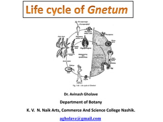
  • 28. Life cycle of Gnetum.