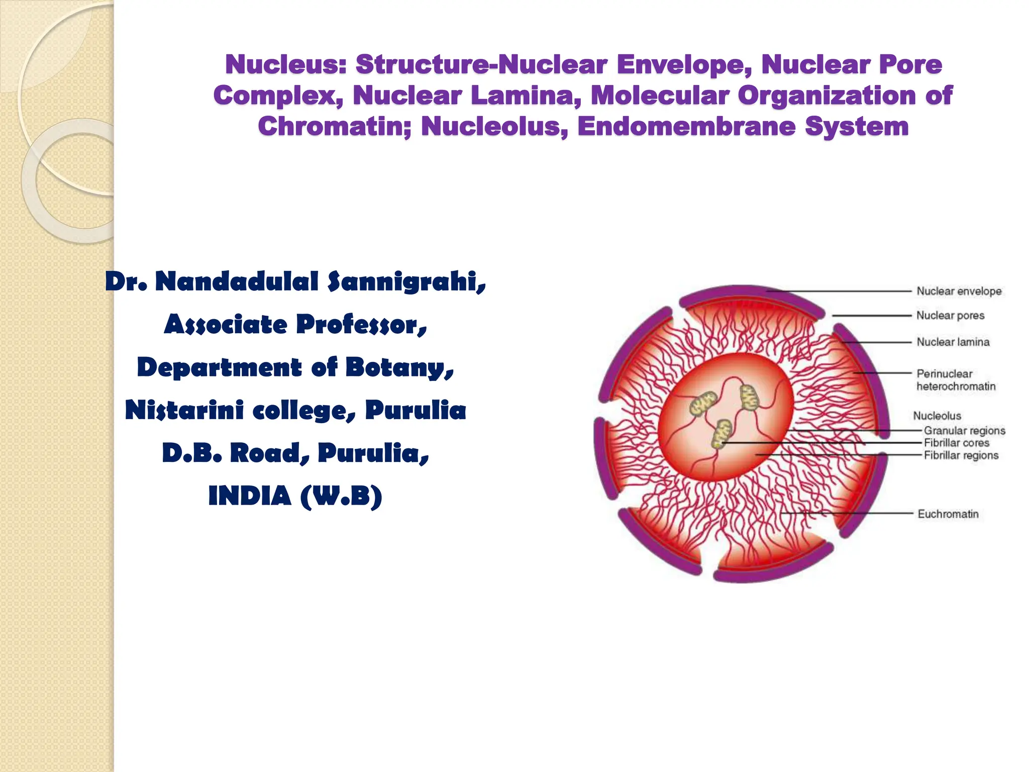
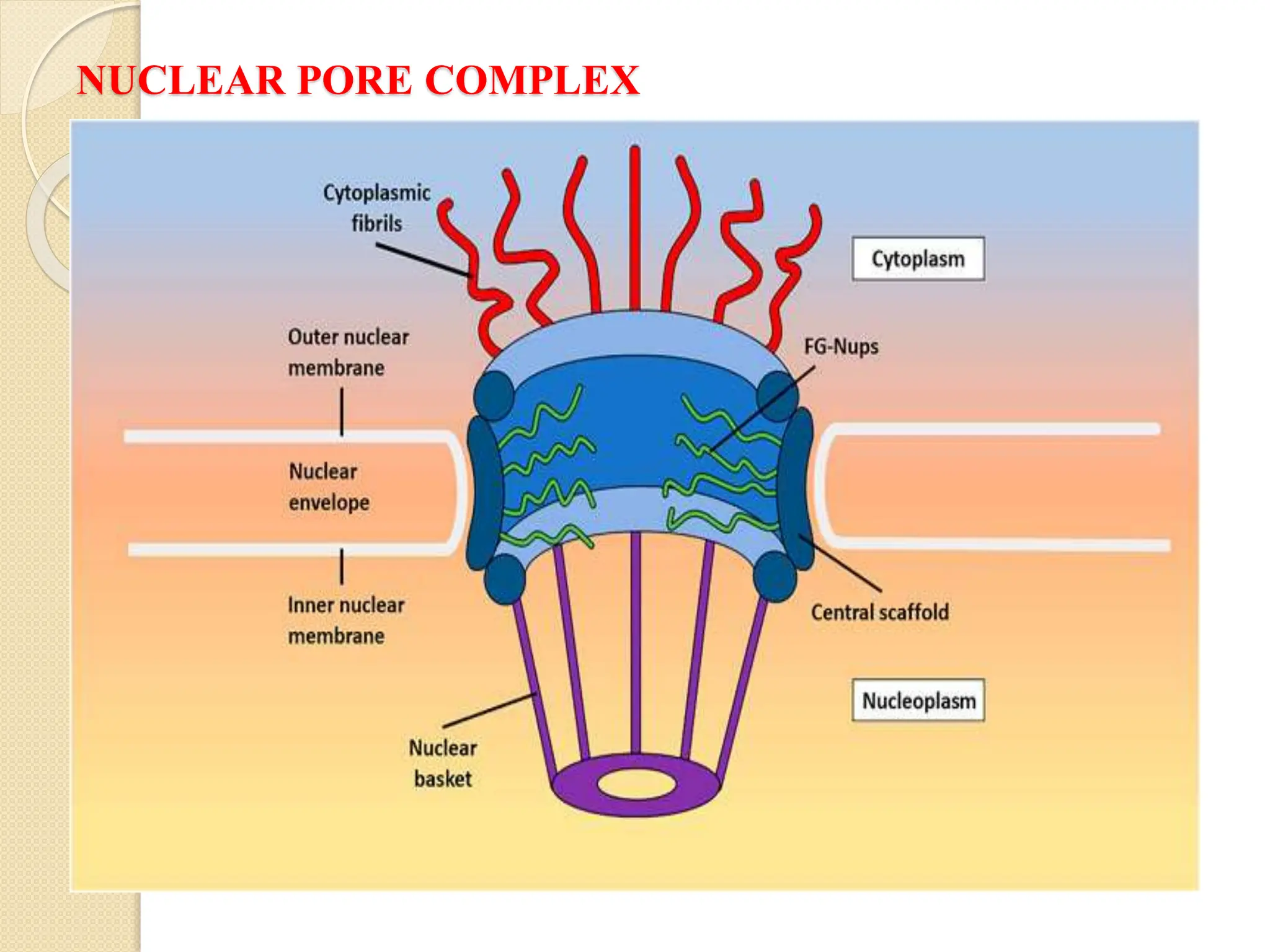
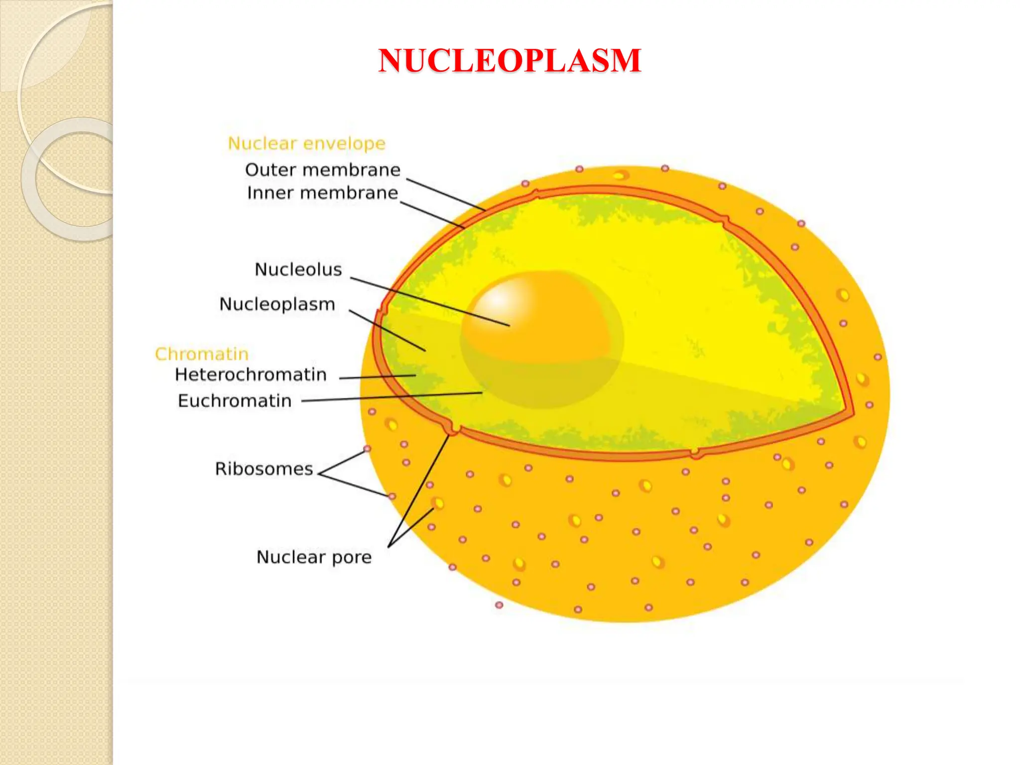
Nucleus: Structure-Nuclear Envelope, Nuclear Pore
Complex, Nuclear Lamina, Molecular Organization of
Chromatin; Nucleolus, Endomembrane System
Dr. Nandadulal Sannigrahi,
Associate Professor,
Department of Botany,
Nistarini college, Purulia
D.B. Road, Purulia,
INDIA (W.B)
CELL- THE BASIC UNIT OF LIFE
 A cell is the smallest living thing in the human organism, and all living
structures in the human body are made of cells. There are hundreds of
different types of cells in the human body, which vary in shape (e.g.
round, flat, long and thin, short and thick) and size (e.g. small granule
cells of the cerebellum in the brain (4 micrometers), up to the huge
oocytes (eggs) produced in the female reproductive organs (100
micrometers) and function.
 However, all cells have three main parts, the plasma membrane, the
cytoplasm and the nucleus. The plasma membrane (often called the
cell membrane) is a thin flexible barrier that separates the inside of the
cell from the environment outside the cell and regulates what can pass
in and out of the cell. Internally, the cell is divided into the cytoplasm
and the nucleus. The cytoplasm (cyto- = cell; -plasm = “something
molded”) is where most functions of the cell are carried out.
 It looks a bit-like mixed fruit jelly, where the watery jelly is called the
cytosol; and the different fruits in it are called organelles. The cytosol
also contains many molecules and ions involved in cell functions.
Different organelles also perform different cell functions and many are
also separated from the cytosol by membranes.
CELL- THE BASIC UNIT OF LIFE
 The largest organelle, the nucleus is separated from the cytoplasm by a
nuclear envelope (membrane). It contains the DNA (genes) that code
for proteins necessary for the cell to function.
 Generally speaking, the inside environment of a cell is called the
intracellular fluid (ICF), (intra- = within; referred to all fluid contained
in cytosol, organelles and nucleus) while the environment outside a
cell is called the extracellular fluid (ECF) (extra- = outside of; referred
to all fluid outside cells). Plasma, the fluid part of blood, is the only
ECF compartment that links all cells in the body.
 The integration of the cell is maintained by the cytoskeleton and the
cell wall conifers protection and cell membrane maintain a selective
permeability to design the cell as an osmotic system.
 In addition to these, different membrane and non-membrane bound
organelles along with other substances make the cell as the structural
& functional unit of life.
NUCLEUS
 Cell is the basic unit of life,
 The cell nucleus is the most noticeable organelle within
the eukaryotic cell, and perhaps the most important and defining
feature of the eukaryotic cells. Most of the genetic material (DNA) is
contained in the nucleus, while a small amount of it is found in
mitochondria.
 The majority of human cells have a single nucleus, although there are
several cell types that have multiple nuclei (e.g.osteoclasts) or don't
have a nucleus at all (erythrocytes).
 Robert Brown (1883) was the discoverer of the nucleus.
 J. Hammerling ( 1934) , a German scientist demonstrated that nucleus
determines the character of cell as well as the character of individual,
 It usually placed centrally but in some plant cells and epithelial cells
of animals, it is peripheral in position,
 It may be spherical, oval, C-shaped, pyriform or may be lobulated.
CELL STRUCTURE-PLANT
NUCLEUS
 The nucleus is normally around 5-10 μm in diameter in many
multicellular organisms, and the largest organelle in the cell. The
smallest nuclei are approximately 1 μm in diameter and are found in
yeast cells.
 Mostly the shape of the nucleus is spherical or oblong. Usually cells
have one nucleus but many at times there are multinucleated cells.
Multi-nucleation in cells may be due to karyokinesis (when cell
undergoes nuclear division) or when cells fuse to form syncytium,
like in mature muscle cells.
 The nucleus has very important roles to play. As it contains genetic
material, it coordinates cell activities like protein synthesis and cell
division.
 Anatomically the nucleus is made up of several components: nuclear
envelope, nuclear lamina, nucleolus, chromosomes, nucleoplasm are
some of these components.
NUCLEUS
NUCLEUS
 All of these components work together in order for the nucleus to
accomplish all of its functions. Namely, these functions are:
 control of the genetically information of the cell and thus the heredity
characteristics of an organism,
 control of the protein and enzyme synthesis
 control of cell division and cell growth
 storage of DNA, RNA and ribosome
 regulation of the transcription of the mRNA to protein
 production of ribosome
 When a cell is histological stained, the nucleus normally appears as a
large, dark organelle, mostly at or near the centre of a cell.
NUCLEAR ENVELOPE
 As its name suggests, the nuclear envelope surrounds the nucleus,
separating it from the cell's cytoplasm. It is a double membrane.
 Each membrane is a phospholipids bilayer associated with proteins, and
the two membranes are divided by 20 to 40 nm of space.
 The two membranes of the nuclear envelope are often referred to as the
inner and outer nuclear membranes.
 The outer membrane is continuous with the cell’s endoplasmic reticulum,
and therefore the space between the inner and outer nuclear membranes
links to the lumen of the endoplasmic reticulum.
 Like the endoplasmic reticulum, the outer nuclear membrane has
ribosome attached to it. Contrastingly, the inner membrane of the nuclear
envelope is attached to proteins that are specific to the nucleus, and
therefore found nowhere else.
 The nuclear envelope is perforated with tiny nuclear pores with diameters
of around 100 nm. The inner and outer membranes of the envelope are
continuous around the pores. Each pore is lined with a structure of 50 to
100 different proteins known as the nuclear pore complex.
NUCLEAR ENVELOPE
 The nucleopores are circular or octagonal in surface view
 It is believed due to the result of distortion during processing and because
by the presence of eight symmetrically arranged annular subunits,
 The pores are arranged in rows in plant cells or in clusters in lymphocytes
but usually are randomly distributed,
 ‘Basket-like structure’ has been found to the inner side of nuclear pore
 ( Don glass. J),
 The pore complex is 125 million Dalton in size with 130 nm diameter
and 50mm in thickness,
 Consists of four separate elements- scaffold, transporter, thick filaments
and a basket,
 Scaffold composed of three closely apposed rings- cytoplasmic ring ,
nucleoplasmic ring and a central ring having eight fold symmetry in each;
the spokes of the central ring are attached to the transporter of the inner
side and to the nucleocytoplasmic and cytoplasmic rings on the outer
side.
 The spikes are interspaced by 9 mm wide aqueous channels through
which proteins and metabolites can be diffused between the nucleus and
cytoplasm,
NUCLEAR ENVELOPE
NUCLEAR ENVELOPE
 Transporters are 36-38 mm wide protenaceous rings consists of two
irises of eight arms in each,
 On the cytoplasmic side of pore, thick fibers of 3.3 mm diameter are
projected into the cytoplasm,
 FUNCTION OF NUCLEAR PORE COMPLEX
 These pore complexes regulate the movements of macromolecules,
RNAs and proteins into and out of the nucleus by three methods-
signals for transport across the pore, import of nuclear proteins ,
export of RNA from the nucleus and Export and reimport of RNA
 This movement of molecules is known as nuclear transport. Small
molecules can move passively through the pores, but larger
molecules, including RNAs and many proteins, are too large for this
and must move actively.
 During this active process, they are selectively recognized and
transported in one specific direction.
 The traffic of RNAs and proteins through the nuclear pore complex is
particularly important, as they play a role in gene expression.
NUCLEAR PORE COMPLEX
NUCLEAR PORE COMPLEX-FUNCTION
 Signal for transport across the membrane- import of proteins and RNPs,
some signal sequences as Nuclear Localization Sequences (NLS) ,
 NLS used for export are nuclear export signal (NES) like HIV Res
protein, Phosphokinase A inhibitor (PKI) and TF III A used for
transcribing 5S RNA and tRNA genes; M9 domain of hnRNA protein in
the only known signal sequence that directs both export and import
processes,
 Import of nuclear proteins- NLS protein receptor complex with the help
of hydrolysis of GTP and Ran GTPase play significant role in this regard,
 Export of RNA from the nucleus- Rev response element (RRE) and its
activation play a significant role here,
 Export and reimport of RNA-Export followed by reimport of 5s rRNA
and Usn RNA occurs through nuclear pore; TF IIIA and ribosomal
protein 1.5 probably act as mediators in export of 5S rRNA . It seems
contain Rev-NES like sequence,
 Thus, the very complex biochemical phenomenon is associated here in
the import, export and reimport of different molecules via nuclear pore
and this pore acts as connecting network between cytoplasm and nucleus.
NUCLEAR LAMINA
 The inner nuclear membrane is internally lined by protein filaments
meshwork organized in a net-like fashion, called nuclear lamina.
 The proteins that make up the nuclear lamina are known as lamins,
which are intermediate filament proteins.
 These support the nuclear envelope, ensuring that the overall shape
and structure of the nucleus is maintained.
 In addition to lamins there is another set of membrane proteins called
lamina associated proteins, which help to mediate the interaction
between the lamina and inner nuclear membrane.
 The nuclear lamina, along with protein fibers called the nuclear
matrix, is also thought to aid in the organization of genetic material,
allowing it to function more efficiently.
NUCLEOLUS
 When a nucleus is not dividing, a structure called a nucleolus becomes
visible. In fact, it is the most prominent structure within the nucleus.
Usually there is only a single nucleolus present, but some nuclei have
multiple nucleoli. It is a mass of granules and fibers attached to
chromatin. It is absent in yeast, bacteria, algae , one in most cases but
two in liver cells, multiple in polyploidy cells,
 First discovered by Fontana in 1874 located in close association with
nucleolar organizer region (NOR) of two or more chromosomes,
 Nucleoli of two types- true nucleoli or plasmasomes stained with
acidic dye and disappear during mitosis and false nucleoli,
karyosomes stained with basic dye, actually flakes of chromatins,
 Under light microscope. Continuous coiled filament called
nucleonemma ,
 Four zones- amorphous matrix, nuclear associated chromatin fibrils
and granules, nuclear associated chromatin consists if DNA with
perinuclear chromatin ,
 Fibrils are small with RNA,
 The granules contain vesicles with central core and dense peripheral
NUCLEOLUS
NUCLEOLOGENESIS
 The process of the formation of nucleolus is called nucleologenesis,
 Conspicuous in interphase but disappear during cell division, The
process involves the following sub stages after Hadjiolov (1985)
 From the pre-metaphase to telophase, a number of non-ribosomal
nucleolar proteins such as B23, fibrillarin, nucleolin,, p52 and U3
snoRNA are found in the peripheral regions of chromosomes,
 The number of nucleolus derived foci (NDF) can reach up to 100 per
cell at late metaphase,
 As NDF decreases, the perinuclear bodies (PNB) gradually appears
and nucleolus formation start,
 The PNB and NDF are the precursors of newly formed nucleoli,
 Actinomycin D blocks the formation of nucleoli from PNB.
 Thus, it is clearly suggested that nucleolus formation requires active
transcription of tRNA from ribosomal genes by RNA pol-1.
FUNCTION OF NUCLEOLUS
 The nucleolus is important because it is the site of ribosomal RNA
(rRNA) production. Inside the nucleolus, rRNA molecules are combined
with proteins to form ribosome. The biogenesis process i.e. initiation,
production, and maturation of ribosome takes place in the nucleolus
region from the centre to the periphery in the three distinct regions,
 In the fibrillar centre (FC) , the rDNAs of NOR are located which
transcribes rRNA. The pre rRNA transcription actually occurs at the edge
of the transcription zone of FC and according to Miller, the structure is
described as Christmas tree.
 The nucleolus is involved in rRNA transcription, pre-rRNA processing
and ribosome subunit assembly.
 The nucleolus is not surrounded by a membrane, but it has a unique
density, separating it from the surrounding nucleoplasm, and allowing it to
be visualized under a microscope.
 As well as being involved in ribosomal biogenesis, the nucleolus is
thought to have other roles, as it contains a number of proteins unrelated
to rRNA and ribosome synthesis. It is thought be play a role in activities
such as DNA damage repair, cell cycle regulation and RNA editing.
FUNCTION OF NUCLEOLUS
 snoRNA and rRNA processing:
 Small Nucleolar RNAs can be grouped into two categories- those
derived from box C (5′RUAUGA3′ ) and box D ( 5′ GUCA3′) present
at the 5′ and 3′ end respectively,
 It is believed that snoRNA of box C/D class are encoded within the
introns of other genes,
 U3snoRNA associates with pre rRNA and passes through different
regions of nucleolus and undergoes passing into mature rRNA which
is then exported ,
 The U3snRNA recycles from GC to DFC again for association with
nascent pre rRNA.
 Seven snoRNAs are now known to be required for processing of pre
rRNA of these U3 and U14 are bound to external terminal
spacer(ETS) in the processing of pre rRNA.
 Probably, these snoRNAs act as chaperones to help correct folding or
they are involved directly in cleavage reaction.
CHROMOSOMES
 The DNA of a cell is found within the nucleus. It is organized into units
known as chromosomes, each containing a long DNA molecule which is
associated with various proteins.
 The DNA coils around protein complexes called nucleosomes, formed of
proteins called histone, making it easier for the chromosome to fit inside
the nucleus.
 The mass of DNA and proteins inside a chromosome is referred to as
chromatin.
 When a cell is not dividing, it is difficult to see the chromosomes within a
cell, even when it is stained. However, when DNA prepares and begins to
divide, the chromosomes can be visualized more clearly.
 During the metaphase of mitosis, the chromosomes become visible as
they prepare to divide by aligning with one another. The chromosomes
are copied, forming sister chromosomes known a chromatids.
 Human cell nuclei contain 46 chromosomes, although gamete nuclei
contain 23. The whole of the nucleus is not filled by chromatin material,
in fact, there are chromatin free regions called interchromosomal
domains containing poly RNAs.
CHROMATIN
 In the interphase stage, the nuclear sap contains twisted filaments of
chromatin which represents the chromosome,
 Chromatin fibers appeared as arrays of spherical particles about 10 nm in
diameter, connected by filaments which are about 2 nm in diameter.
These bodies are called nu bodies which later on confirmed as
nucleosome,
 In the interphase nucleus, the chromatin filaments may show both
centromeric and telomeric attachment,
 The chromatin fibers of interphase nuclei are attached to the that may
function as initiation point of DNA synthesis,
 When chromatin bodies are stained with acetocarmine or Feulgen at
prophase, a linear differentiation into regions having dense stain and that
have light stain s become conspicuous,
 The dark stained regions are heterochromatin and light stained regions
are euchromatin,
 Certain heterochromatin regions particularly proximal to the centromere
are called constitutive heterochromatin serve as chromosome markers
and other heterochromatic regions are called facultatative
heterochromatin.
CHROMATIN
NUCLEOPLASM
 Nucleoplasm is similar to the cytoplasm of a cell, in that it is semi-
liquid, and fills the empty space in the nucleus.
 It is a form of protoplasm and surrounds the chromosomes and
nucleoli inside the nucleus. It also has various proteins and enzymes
dissolved within it.
 Nuclear bodies can be found in the nucleoplasm, and these include
structures such as Cajal bodies, Gemini bodies, and Polycomb bodies.
 Cajal bodies are between 0.3-1.0 µm in diameter, and can be found in
proliferating cells such as embryonic and cancerous cells, as well as
in cells which have a high metabolic rate, such as neurons.
 Sometimes referred to as coiled bodies, Cajal bodies are bound to
nucleoli by specialized proteins called coilin proteins.
 Having these proteins concentrated within Cajal bodies improves the
efficiency of nuclear processes such as the modification and assembly
of UsnRNPs, which can become spliceosome.
NUCLEOPLASM
ENDOMEMBRANE SYSTEM
 The endomembrane system (endo- = “within”) is a group of
membranes and organelles in eukaryotic cells that works together to
modify, package, and transport lipids and proteins. It includes a
variety of organelles, such as the nuclear envelope and lysosomes,
which you may already know, and the endoplasmic reticulum and
Golgi apparatus, which we will cover shortly.
 Although it's not technically inside the cell, the plasma membrane is
also part of the endomembrane system. As we'll see, the plasma
membrane interacts with the other endo-membrane organelles, and it's
the site where secreted proteins (like the pancreatic enzymes in the
intro) are exported. Important note: the endomembrane system does
not include mitochondria, chloroplasts, or peroxisomes.
 The network among the different organelles of the cell is important to
coordinate the different diverse functions of the cell as it needs for the
cell structure and function along with their integrity,
 The ER of two types, ribosome, lysosomes, Golgi bodies etc play an
extensive network by the virtue of the endomembrane system.
THANK YOU FOR YOUR VISIT
 ACKNOWLEDGEMENTS:
a. Google for images,
b. Different websites for enriching the course content,
c. Science Direct pages,
d. A textbook of Botany- Vol III – Hait, Bhattacharya & Ghosh.
e. A Text Book of Cell and Molecular Biology- Ajay Paul,
f. Cell and Molecular Biology- Kar and Halder,
g. Concept of Genetics- Klug. Cummings, Spencer, Palladino,
DISCLAIMER:
This presentation has been designed to address the academic
fraternity without any financial interest. This is absolutely free to
use . The author does not claim any kind of financial benefits from
this content.

Nucleus- Different Parts, structure and Function

  • 1.
    Nucleus: Structure-Nuclear Envelope,Nuclear Pore Complex, Nuclear Lamina, Molecular Organization of Chromatin; Nucleolus, Endomembrane System Dr. Nandadulal Sannigrahi, Associate Professor, Department of Botany, Nistarini college, Purulia D.B. Road, Purulia, INDIA (W.B)
  • 2.
    CELL- THE BASICUNIT OF LIFE  A cell is the smallest living thing in the human organism, and all living structures in the human body are made of cells. There are hundreds of different types of cells in the human body, which vary in shape (e.g. round, flat, long and thin, short and thick) and size (e.g. small granule cells of the cerebellum in the brain (4 micrometers), up to the huge oocytes (eggs) produced in the female reproductive organs (100 micrometers) and function.  However, all cells have three main parts, the plasma membrane, the cytoplasm and the nucleus. The plasma membrane (often called the cell membrane) is a thin flexible barrier that separates the inside of the cell from the environment outside the cell and regulates what can pass in and out of the cell. Internally, the cell is divided into the cytoplasm and the nucleus. The cytoplasm (cyto- = cell; -plasm = “something molded”) is where most functions of the cell are carried out.  It looks a bit-like mixed fruit jelly, where the watery jelly is called the cytosol; and the different fruits in it are called organelles. The cytosol also contains many molecules and ions involved in cell functions. Different organelles also perform different cell functions and many are also separated from the cytosol by membranes.
  • 3.
    CELL- THE BASICUNIT OF LIFE  The largest organelle, the nucleus is separated from the cytoplasm by a nuclear envelope (membrane). It contains the DNA (genes) that code for proteins necessary for the cell to function.  Generally speaking, the inside environment of a cell is called the intracellular fluid (ICF), (intra- = within; referred to all fluid contained in cytosol, organelles and nucleus) while the environment outside a cell is called the extracellular fluid (ECF) (extra- = outside of; referred to all fluid outside cells). Plasma, the fluid part of blood, is the only ECF compartment that links all cells in the body.  The integration of the cell is maintained by the cytoskeleton and the cell wall conifers protection and cell membrane maintain a selective permeability to design the cell as an osmotic system.  In addition to these, different membrane and non-membrane bound organelles along with other substances make the cell as the structural & functional unit of life.
  • 4.
    NUCLEUS  Cell isthe basic unit of life,  The cell nucleus is the most noticeable organelle within the eukaryotic cell, and perhaps the most important and defining feature of the eukaryotic cells. Most of the genetic material (DNA) is contained in the nucleus, while a small amount of it is found in mitochondria.  The majority of human cells have a single nucleus, although there are several cell types that have multiple nuclei (e.g.osteoclasts) or don't have a nucleus at all (erythrocytes).  Robert Brown (1883) was the discoverer of the nucleus.  J. Hammerling ( 1934) , a German scientist demonstrated that nucleus determines the character of cell as well as the character of individual,  It usually placed centrally but in some plant cells and epithelial cells of animals, it is peripheral in position,  It may be spherical, oval, C-shaped, pyriform or may be lobulated.
  • 5.
  • 6.
    NUCLEUS  The nucleusis normally around 5-10 μm in diameter in many multicellular organisms, and the largest organelle in the cell. The smallest nuclei are approximately 1 μm in diameter and are found in yeast cells.  Mostly the shape of the nucleus is spherical or oblong. Usually cells have one nucleus but many at times there are multinucleated cells. Multi-nucleation in cells may be due to karyokinesis (when cell undergoes nuclear division) or when cells fuse to form syncytium, like in mature muscle cells.  The nucleus has very important roles to play. As it contains genetic material, it coordinates cell activities like protein synthesis and cell division.  Anatomically the nucleus is made up of several components: nuclear envelope, nuclear lamina, nucleolus, chromosomes, nucleoplasm are some of these components.
  • 7.
  • 8.
    NUCLEUS  All ofthese components work together in order for the nucleus to accomplish all of its functions. Namely, these functions are:  control of the genetically information of the cell and thus the heredity characteristics of an organism,  control of the protein and enzyme synthesis  control of cell division and cell growth  storage of DNA, RNA and ribosome  regulation of the transcription of the mRNA to protein  production of ribosome  When a cell is histological stained, the nucleus normally appears as a large, dark organelle, mostly at or near the centre of a cell.
  • 9.
    NUCLEAR ENVELOPE  Asits name suggests, the nuclear envelope surrounds the nucleus, separating it from the cell's cytoplasm. It is a double membrane.  Each membrane is a phospholipids bilayer associated with proteins, and the two membranes are divided by 20 to 40 nm of space.  The two membranes of the nuclear envelope are often referred to as the inner and outer nuclear membranes.  The outer membrane is continuous with the cell’s endoplasmic reticulum, and therefore the space between the inner and outer nuclear membranes links to the lumen of the endoplasmic reticulum.  Like the endoplasmic reticulum, the outer nuclear membrane has ribosome attached to it. Contrastingly, the inner membrane of the nuclear envelope is attached to proteins that are specific to the nucleus, and therefore found nowhere else.  The nuclear envelope is perforated with tiny nuclear pores with diameters of around 100 nm. The inner and outer membranes of the envelope are continuous around the pores. Each pore is lined with a structure of 50 to 100 different proteins known as the nuclear pore complex.
  • 10.
    NUCLEAR ENVELOPE  Thenucleopores are circular or octagonal in surface view  It is believed due to the result of distortion during processing and because by the presence of eight symmetrically arranged annular subunits,  The pores are arranged in rows in plant cells or in clusters in lymphocytes but usually are randomly distributed,  ‘Basket-like structure’ has been found to the inner side of nuclear pore  ( Don glass. J),  The pore complex is 125 million Dalton in size with 130 nm diameter and 50mm in thickness,  Consists of four separate elements- scaffold, transporter, thick filaments and a basket,  Scaffold composed of three closely apposed rings- cytoplasmic ring , nucleoplasmic ring and a central ring having eight fold symmetry in each; the spokes of the central ring are attached to the transporter of the inner side and to the nucleocytoplasmic and cytoplasmic rings on the outer side.  The spikes are interspaced by 9 mm wide aqueous channels through which proteins and metabolites can be diffused between the nucleus and cytoplasm,
  • 11.
  • 12.
    NUCLEAR ENVELOPE  Transportersare 36-38 mm wide protenaceous rings consists of two irises of eight arms in each,  On the cytoplasmic side of pore, thick fibers of 3.3 mm diameter are projected into the cytoplasm,  FUNCTION OF NUCLEAR PORE COMPLEX  These pore complexes regulate the movements of macromolecules, RNAs and proteins into and out of the nucleus by three methods- signals for transport across the pore, import of nuclear proteins , export of RNA from the nucleus and Export and reimport of RNA  This movement of molecules is known as nuclear transport. Small molecules can move passively through the pores, but larger molecules, including RNAs and many proteins, are too large for this and must move actively.  During this active process, they are selectively recognized and transported in one specific direction.  The traffic of RNAs and proteins through the nuclear pore complex is particularly important, as they play a role in gene expression.
  • 13.
  • 14.
    NUCLEAR PORE COMPLEX-FUNCTION Signal for transport across the membrane- import of proteins and RNPs, some signal sequences as Nuclear Localization Sequences (NLS) ,  NLS used for export are nuclear export signal (NES) like HIV Res protein, Phosphokinase A inhibitor (PKI) and TF III A used for transcribing 5S RNA and tRNA genes; M9 domain of hnRNA protein in the only known signal sequence that directs both export and import processes,  Import of nuclear proteins- NLS protein receptor complex with the help of hydrolysis of GTP and Ran GTPase play significant role in this regard,  Export of RNA from the nucleus- Rev response element (RRE) and its activation play a significant role here,  Export and reimport of RNA-Export followed by reimport of 5s rRNA and Usn RNA occurs through nuclear pore; TF IIIA and ribosomal protein 1.5 probably act as mediators in export of 5S rRNA . It seems contain Rev-NES like sequence,  Thus, the very complex biochemical phenomenon is associated here in the import, export and reimport of different molecules via nuclear pore and this pore acts as connecting network between cytoplasm and nucleus.
  • 15.
    NUCLEAR LAMINA  Theinner nuclear membrane is internally lined by protein filaments meshwork organized in a net-like fashion, called nuclear lamina.  The proteins that make up the nuclear lamina are known as lamins, which are intermediate filament proteins.  These support the nuclear envelope, ensuring that the overall shape and structure of the nucleus is maintained.  In addition to lamins there is another set of membrane proteins called lamina associated proteins, which help to mediate the interaction between the lamina and inner nuclear membrane.  The nuclear lamina, along with protein fibers called the nuclear matrix, is also thought to aid in the organization of genetic material, allowing it to function more efficiently.
  • 16.
    NUCLEOLUS  When anucleus is not dividing, a structure called a nucleolus becomes visible. In fact, it is the most prominent structure within the nucleus. Usually there is only a single nucleolus present, but some nuclei have multiple nucleoli. It is a mass of granules and fibers attached to chromatin. It is absent in yeast, bacteria, algae , one in most cases but two in liver cells, multiple in polyploidy cells,  First discovered by Fontana in 1874 located in close association with nucleolar organizer region (NOR) of two or more chromosomes,  Nucleoli of two types- true nucleoli or plasmasomes stained with acidic dye and disappear during mitosis and false nucleoli, karyosomes stained with basic dye, actually flakes of chromatins,  Under light microscope. Continuous coiled filament called nucleonemma ,  Four zones- amorphous matrix, nuclear associated chromatin fibrils and granules, nuclear associated chromatin consists if DNA with perinuclear chromatin ,  Fibrils are small with RNA,  The granules contain vesicles with central core and dense peripheral
  • 17.
  • 18.
    NUCLEOLOGENESIS  The processof the formation of nucleolus is called nucleologenesis,  Conspicuous in interphase but disappear during cell division, The process involves the following sub stages after Hadjiolov (1985)  From the pre-metaphase to telophase, a number of non-ribosomal nucleolar proteins such as B23, fibrillarin, nucleolin,, p52 and U3 snoRNA are found in the peripheral regions of chromosomes,  The number of nucleolus derived foci (NDF) can reach up to 100 per cell at late metaphase,  As NDF decreases, the perinuclear bodies (PNB) gradually appears and nucleolus formation start,  The PNB and NDF are the precursors of newly formed nucleoli,  Actinomycin D blocks the formation of nucleoli from PNB.  Thus, it is clearly suggested that nucleolus formation requires active transcription of tRNA from ribosomal genes by RNA pol-1.
  • 19.
    FUNCTION OF NUCLEOLUS The nucleolus is important because it is the site of ribosomal RNA (rRNA) production. Inside the nucleolus, rRNA molecules are combined with proteins to form ribosome. The biogenesis process i.e. initiation, production, and maturation of ribosome takes place in the nucleolus region from the centre to the periphery in the three distinct regions,  In the fibrillar centre (FC) , the rDNAs of NOR are located which transcribes rRNA. The pre rRNA transcription actually occurs at the edge of the transcription zone of FC and according to Miller, the structure is described as Christmas tree.  The nucleolus is involved in rRNA transcription, pre-rRNA processing and ribosome subunit assembly.  The nucleolus is not surrounded by a membrane, but it has a unique density, separating it from the surrounding nucleoplasm, and allowing it to be visualized under a microscope.  As well as being involved in ribosomal biogenesis, the nucleolus is thought to have other roles, as it contains a number of proteins unrelated to rRNA and ribosome synthesis. It is thought be play a role in activities such as DNA damage repair, cell cycle regulation and RNA editing.
  • 20.
    FUNCTION OF NUCLEOLUS snoRNA and rRNA processing:  Small Nucleolar RNAs can be grouped into two categories- those derived from box C (5′RUAUGA3′ ) and box D ( 5′ GUCA3′) present at the 5′ and 3′ end respectively,  It is believed that snoRNA of box C/D class are encoded within the introns of other genes,  U3snoRNA associates with pre rRNA and passes through different regions of nucleolus and undergoes passing into mature rRNA which is then exported ,  The U3snRNA recycles from GC to DFC again for association with nascent pre rRNA.  Seven snoRNAs are now known to be required for processing of pre rRNA of these U3 and U14 are bound to external terminal spacer(ETS) in the processing of pre rRNA.  Probably, these snoRNAs act as chaperones to help correct folding or they are involved directly in cleavage reaction.
  • 21.
    CHROMOSOMES  The DNAof a cell is found within the nucleus. It is organized into units known as chromosomes, each containing a long DNA molecule which is associated with various proteins.  The DNA coils around protein complexes called nucleosomes, formed of proteins called histone, making it easier for the chromosome to fit inside the nucleus.  The mass of DNA and proteins inside a chromosome is referred to as chromatin.  When a cell is not dividing, it is difficult to see the chromosomes within a cell, even when it is stained. However, when DNA prepares and begins to divide, the chromosomes can be visualized more clearly.  During the metaphase of mitosis, the chromosomes become visible as they prepare to divide by aligning with one another. The chromosomes are copied, forming sister chromosomes known a chromatids.  Human cell nuclei contain 46 chromosomes, although gamete nuclei contain 23. The whole of the nucleus is not filled by chromatin material, in fact, there are chromatin free regions called interchromosomal domains containing poly RNAs.
  • 22.
    CHROMATIN  In theinterphase stage, the nuclear sap contains twisted filaments of chromatin which represents the chromosome,  Chromatin fibers appeared as arrays of spherical particles about 10 nm in diameter, connected by filaments which are about 2 nm in diameter. These bodies are called nu bodies which later on confirmed as nucleosome,  In the interphase nucleus, the chromatin filaments may show both centromeric and telomeric attachment,  The chromatin fibers of interphase nuclei are attached to the that may function as initiation point of DNA synthesis,  When chromatin bodies are stained with acetocarmine or Feulgen at prophase, a linear differentiation into regions having dense stain and that have light stain s become conspicuous,  The dark stained regions are heterochromatin and light stained regions are euchromatin,  Certain heterochromatin regions particularly proximal to the centromere are called constitutive heterochromatin serve as chromosome markers and other heterochromatic regions are called facultatative heterochromatin.
  • 23.
  • 24.
    NUCLEOPLASM  Nucleoplasm issimilar to the cytoplasm of a cell, in that it is semi- liquid, and fills the empty space in the nucleus.  It is a form of protoplasm and surrounds the chromosomes and nucleoli inside the nucleus. It also has various proteins and enzymes dissolved within it.  Nuclear bodies can be found in the nucleoplasm, and these include structures such as Cajal bodies, Gemini bodies, and Polycomb bodies.  Cajal bodies are between 0.3-1.0 µm in diameter, and can be found in proliferating cells such as embryonic and cancerous cells, as well as in cells which have a high metabolic rate, such as neurons.  Sometimes referred to as coiled bodies, Cajal bodies are bound to nucleoli by specialized proteins called coilin proteins.  Having these proteins concentrated within Cajal bodies improves the efficiency of nuclear processes such as the modification and assembly of UsnRNPs, which can become spliceosome.
  • 25.
  • 26.
    ENDOMEMBRANE SYSTEM  Theendomembrane system (endo- = “within”) is a group of membranes and organelles in eukaryotic cells that works together to modify, package, and transport lipids and proteins. It includes a variety of organelles, such as the nuclear envelope and lysosomes, which you may already know, and the endoplasmic reticulum and Golgi apparatus, which we will cover shortly.  Although it's not technically inside the cell, the plasma membrane is also part of the endomembrane system. As we'll see, the plasma membrane interacts with the other endo-membrane organelles, and it's the site where secreted proteins (like the pancreatic enzymes in the intro) are exported. Important note: the endomembrane system does not include mitochondria, chloroplasts, or peroxisomes.  The network among the different organelles of the cell is important to coordinate the different diverse functions of the cell as it needs for the cell structure and function along with their integrity,  The ER of two types, ribosome, lysosomes, Golgi bodies etc play an extensive network by the virtue of the endomembrane system.
  • 27.
    THANK YOU FORYOUR VISIT  ACKNOWLEDGEMENTS: a. Google for images, b. Different websites for enriching the course content, c. Science Direct pages, d. A textbook of Botany- Vol III – Hait, Bhattacharya & Ghosh. e. A Text Book of Cell and Molecular Biology- Ajay Paul, f. Cell and Molecular Biology- Kar and Halder, g. Concept of Genetics- Klug. Cummings, Spencer, Palladino, DISCLAIMER: This presentation has been designed to address the academic fraternity without any financial interest. This is absolutely free to use . The author does not claim any kind of financial benefits from this content.