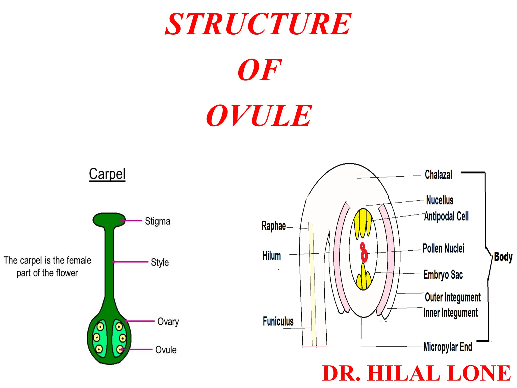
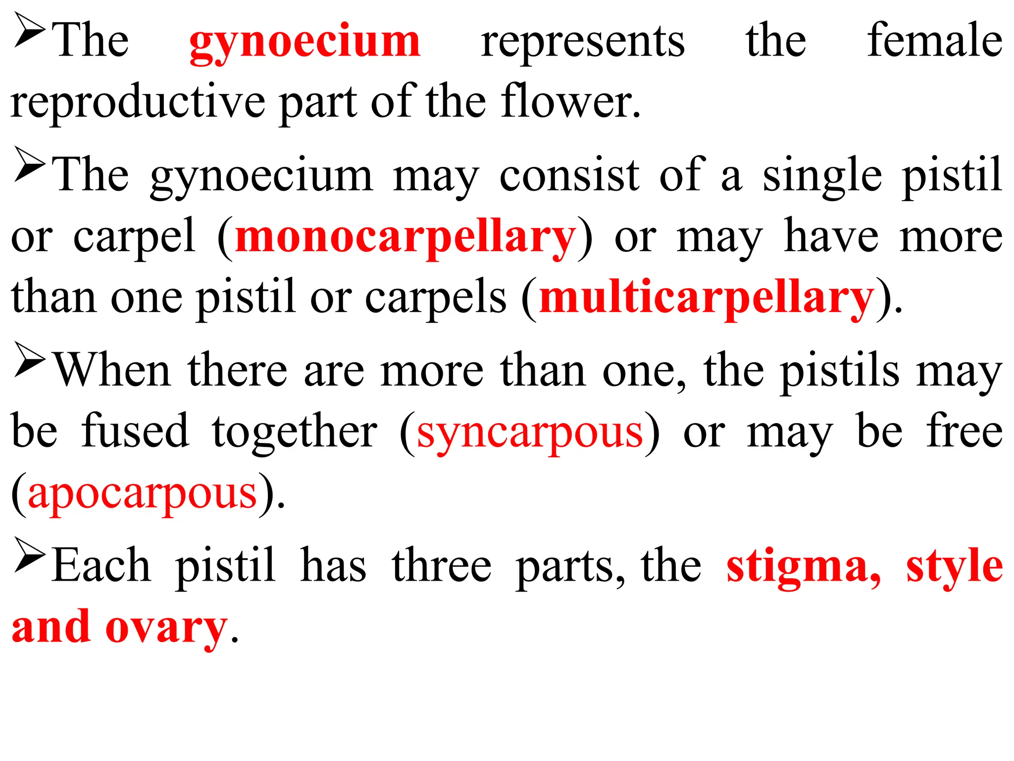
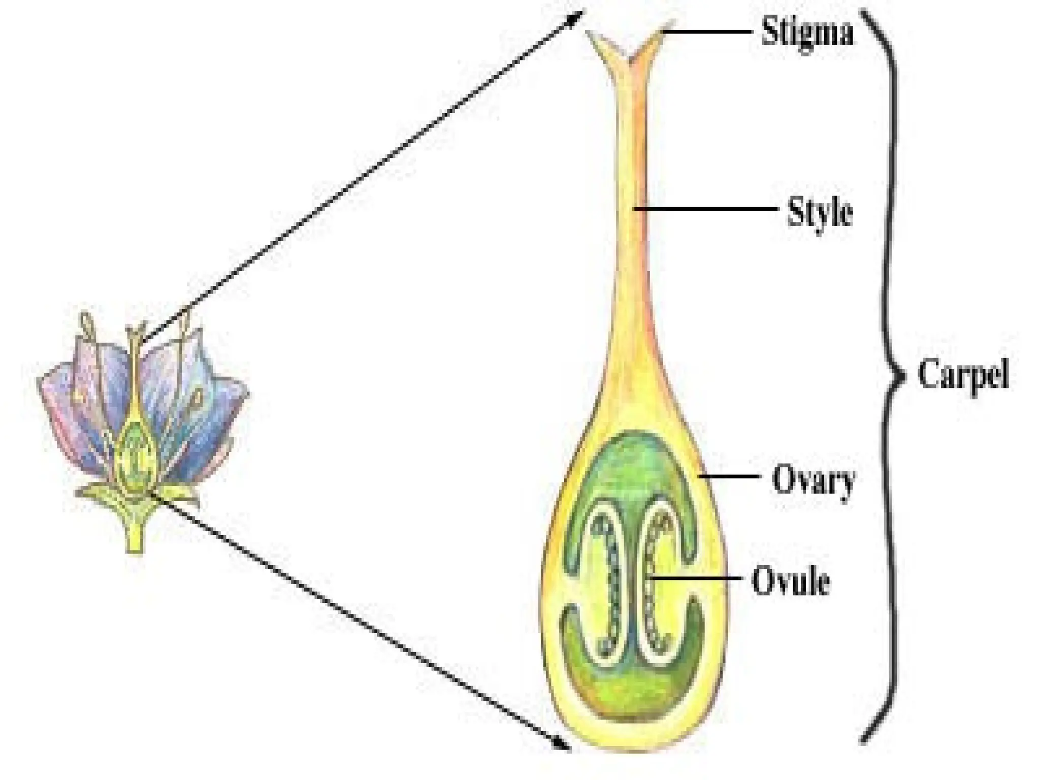
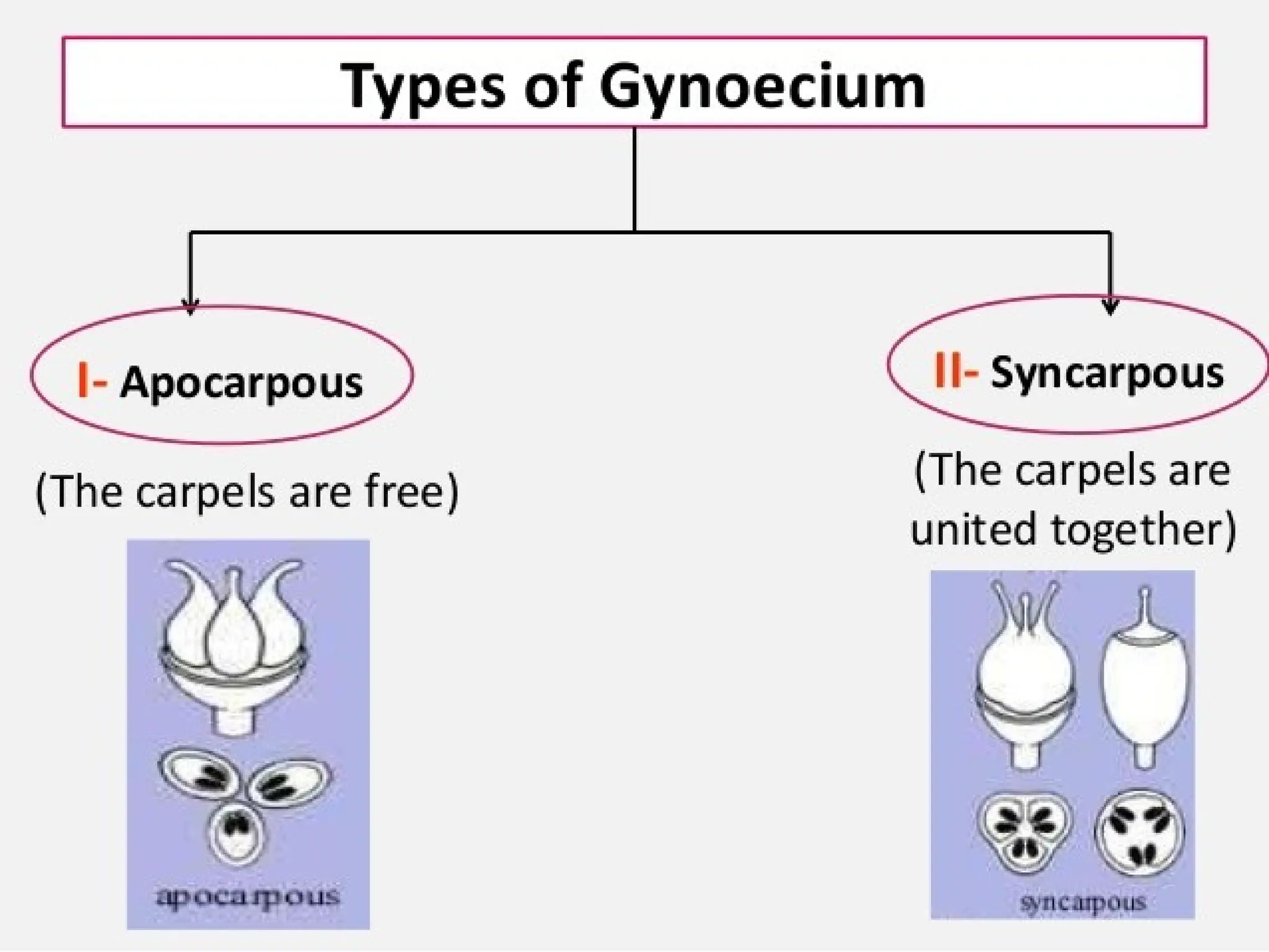
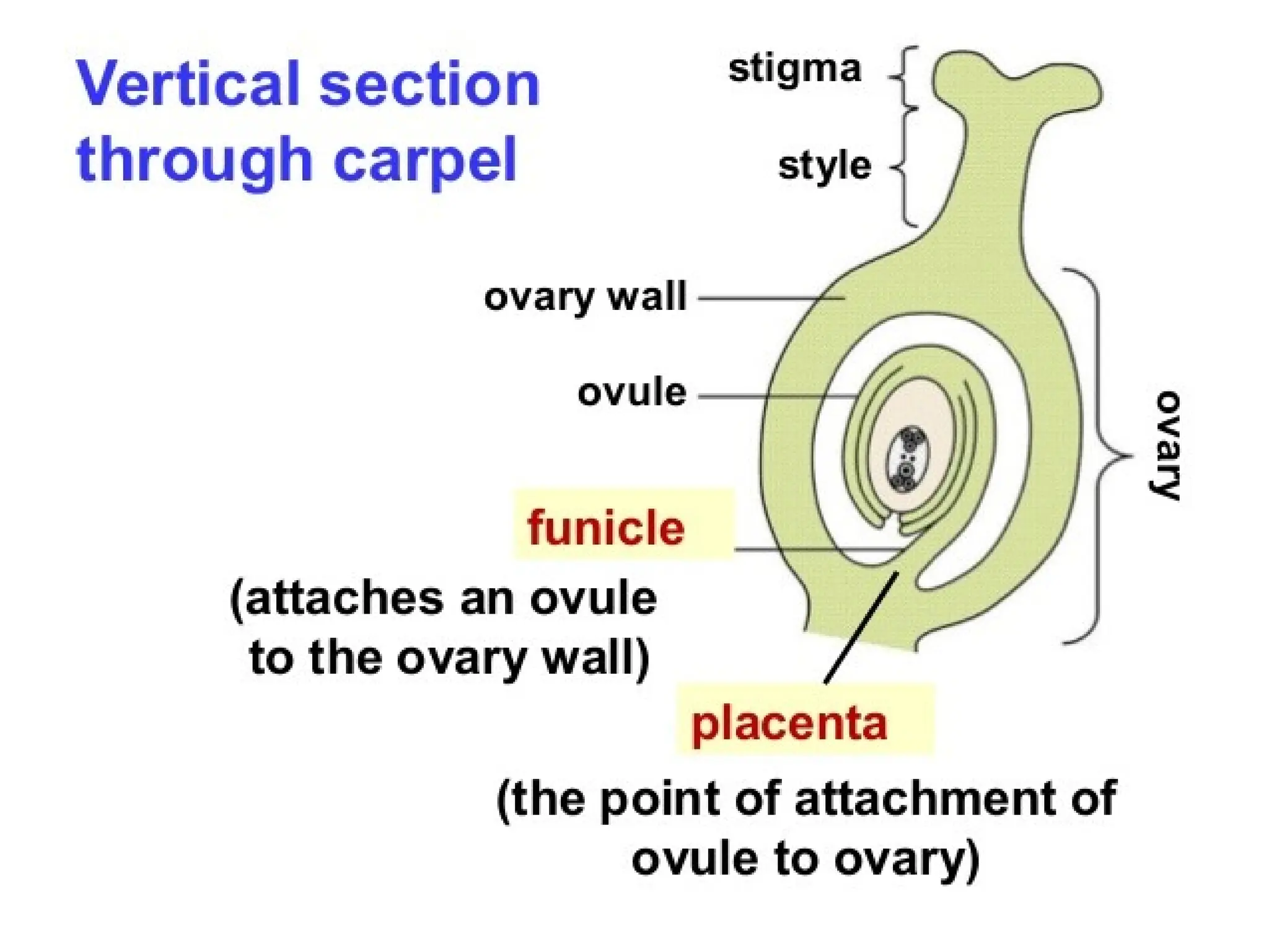
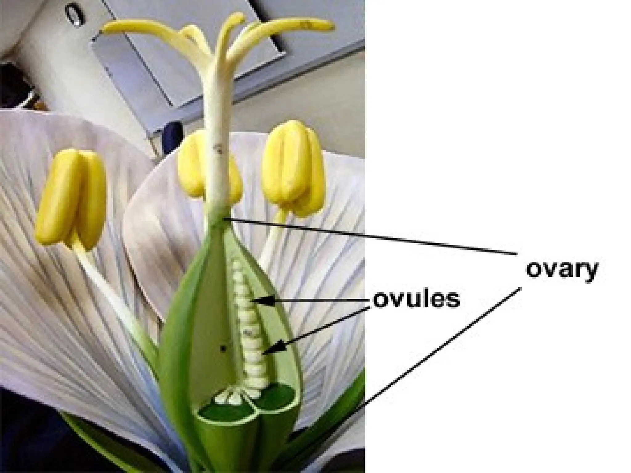
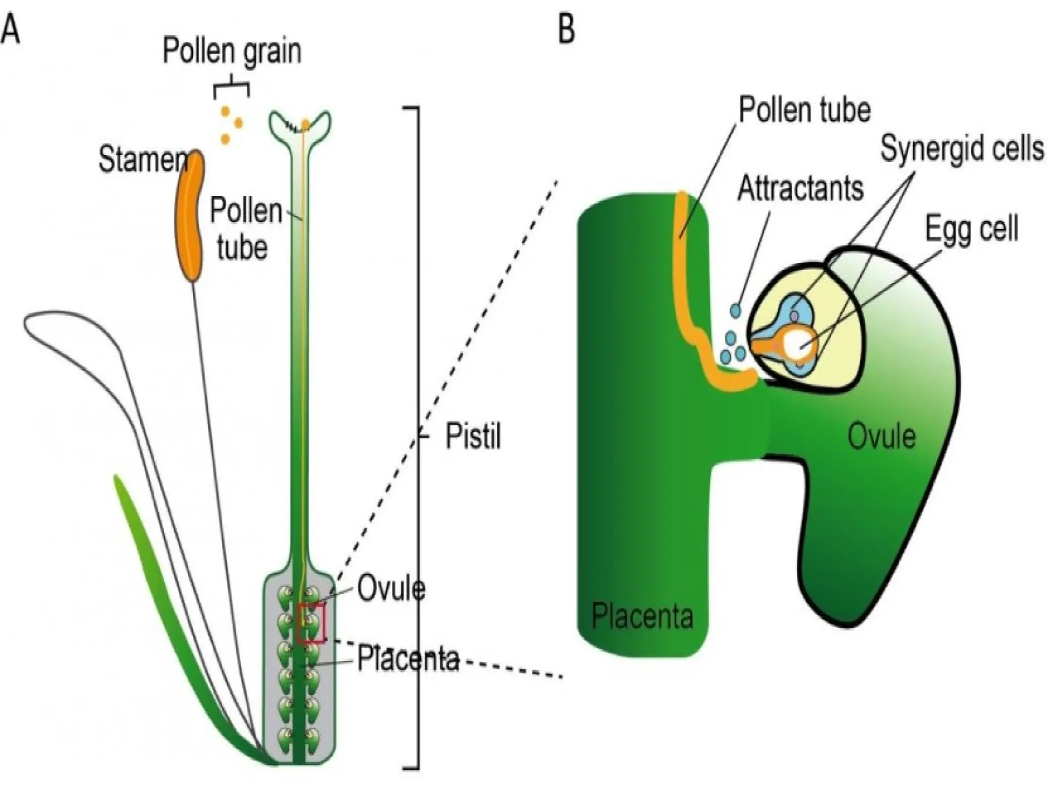
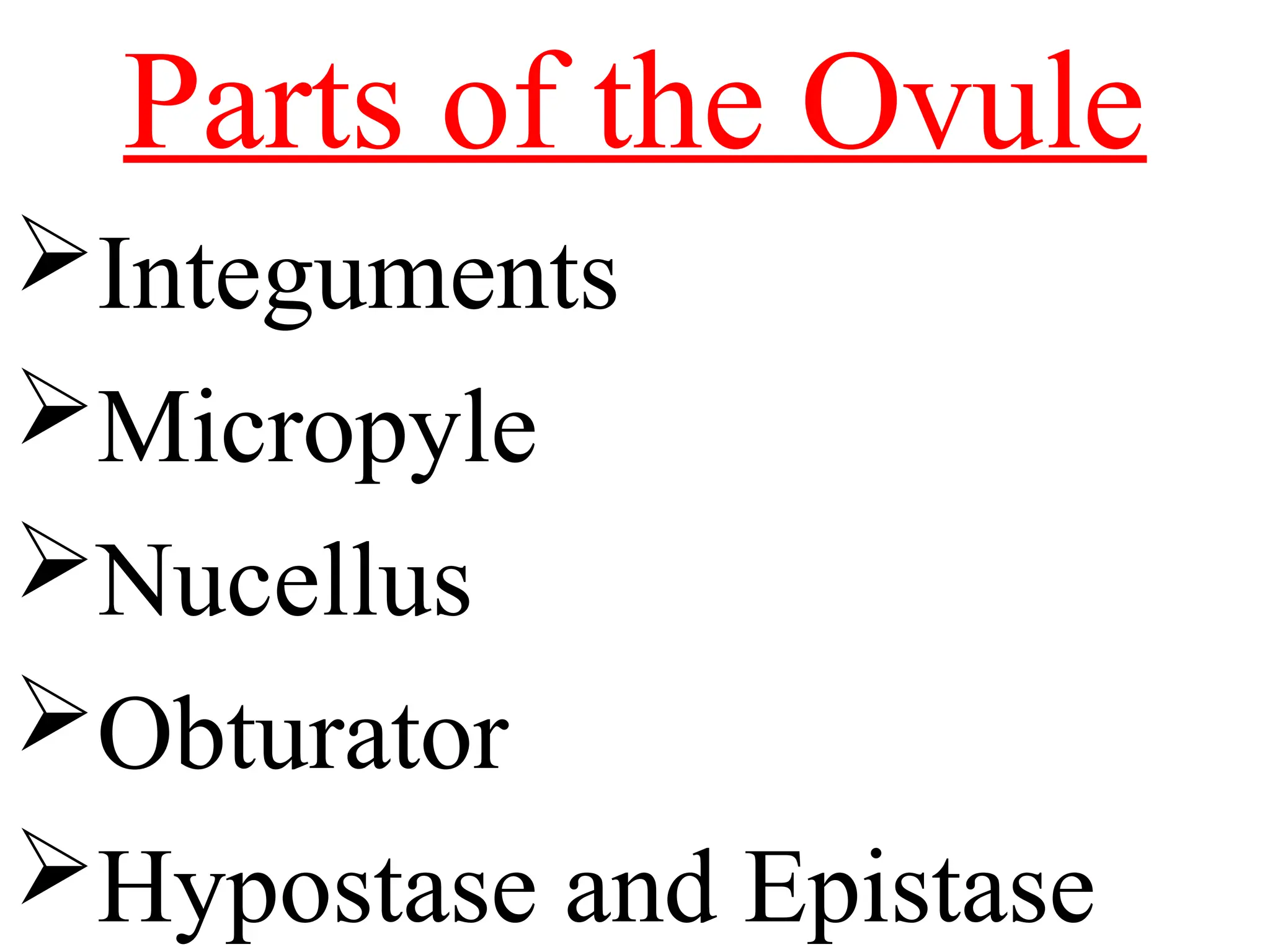
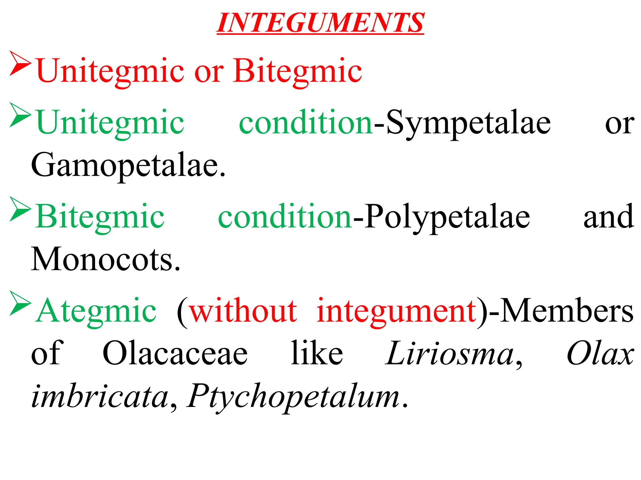
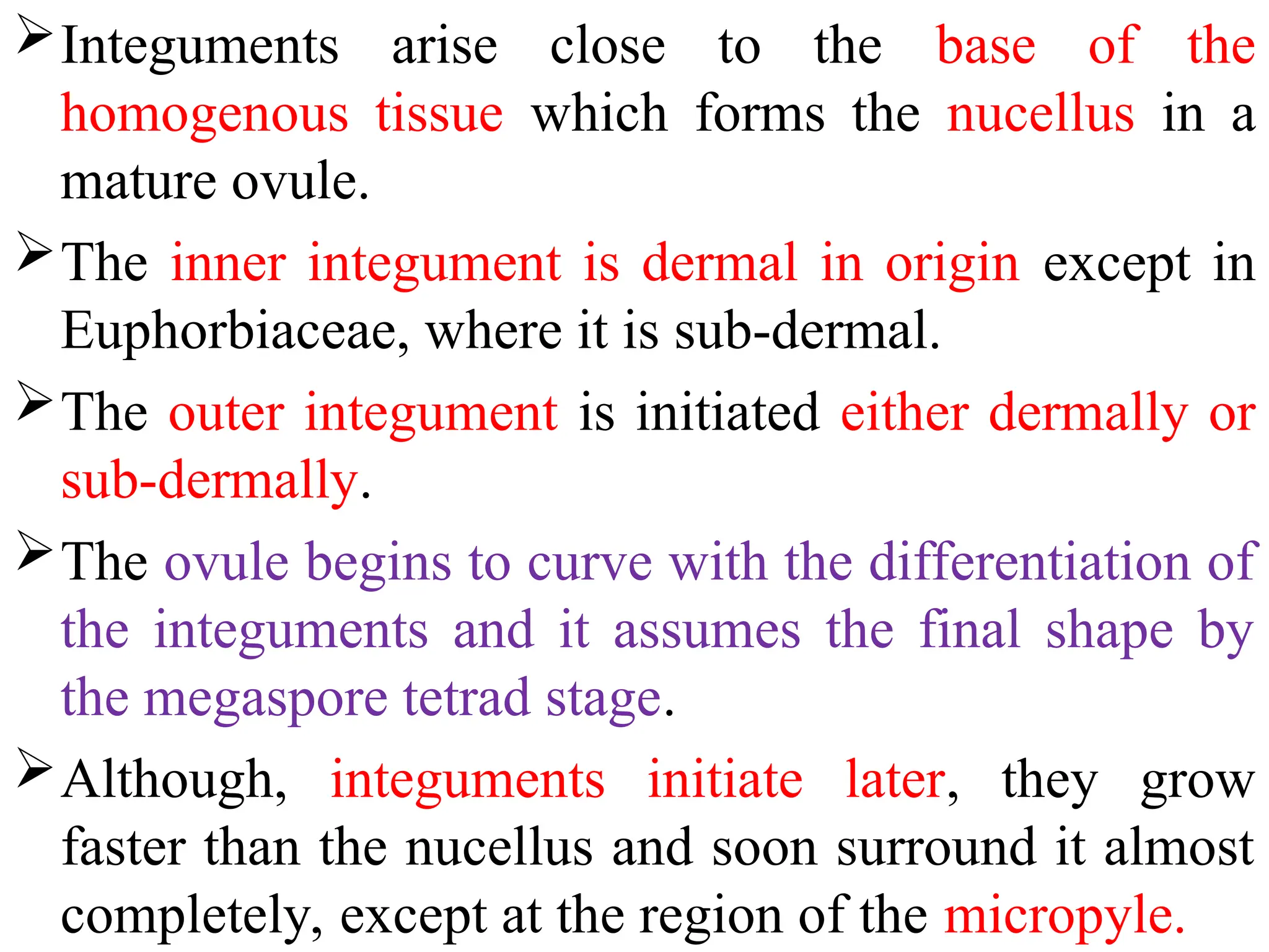
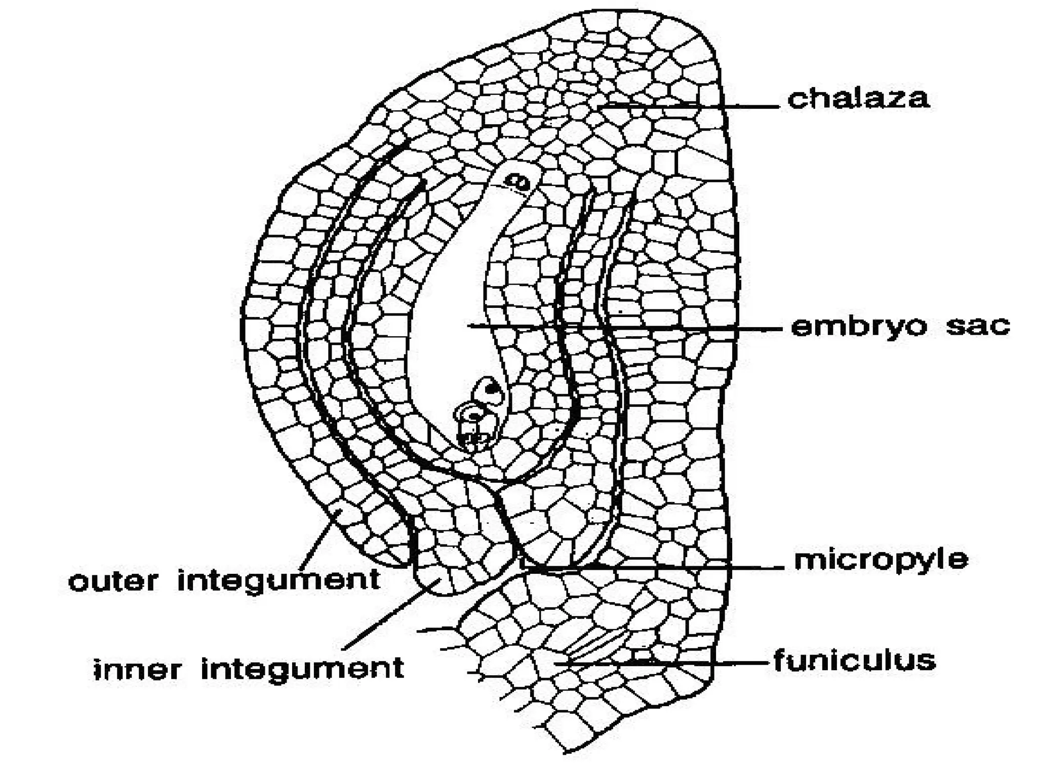
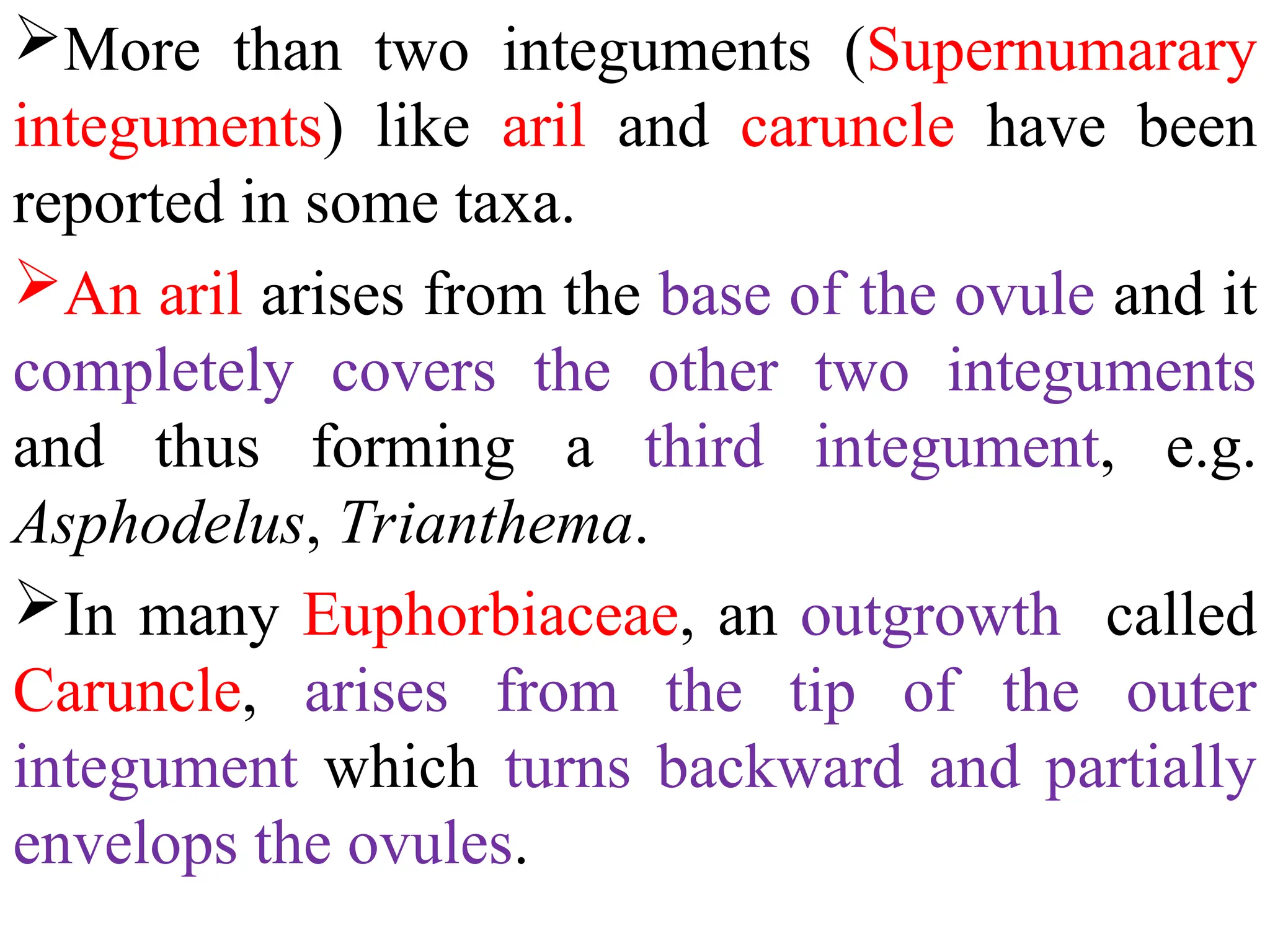
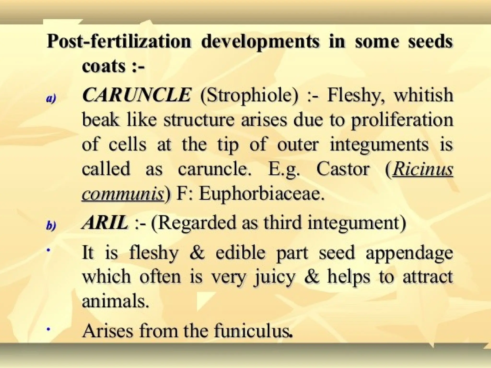
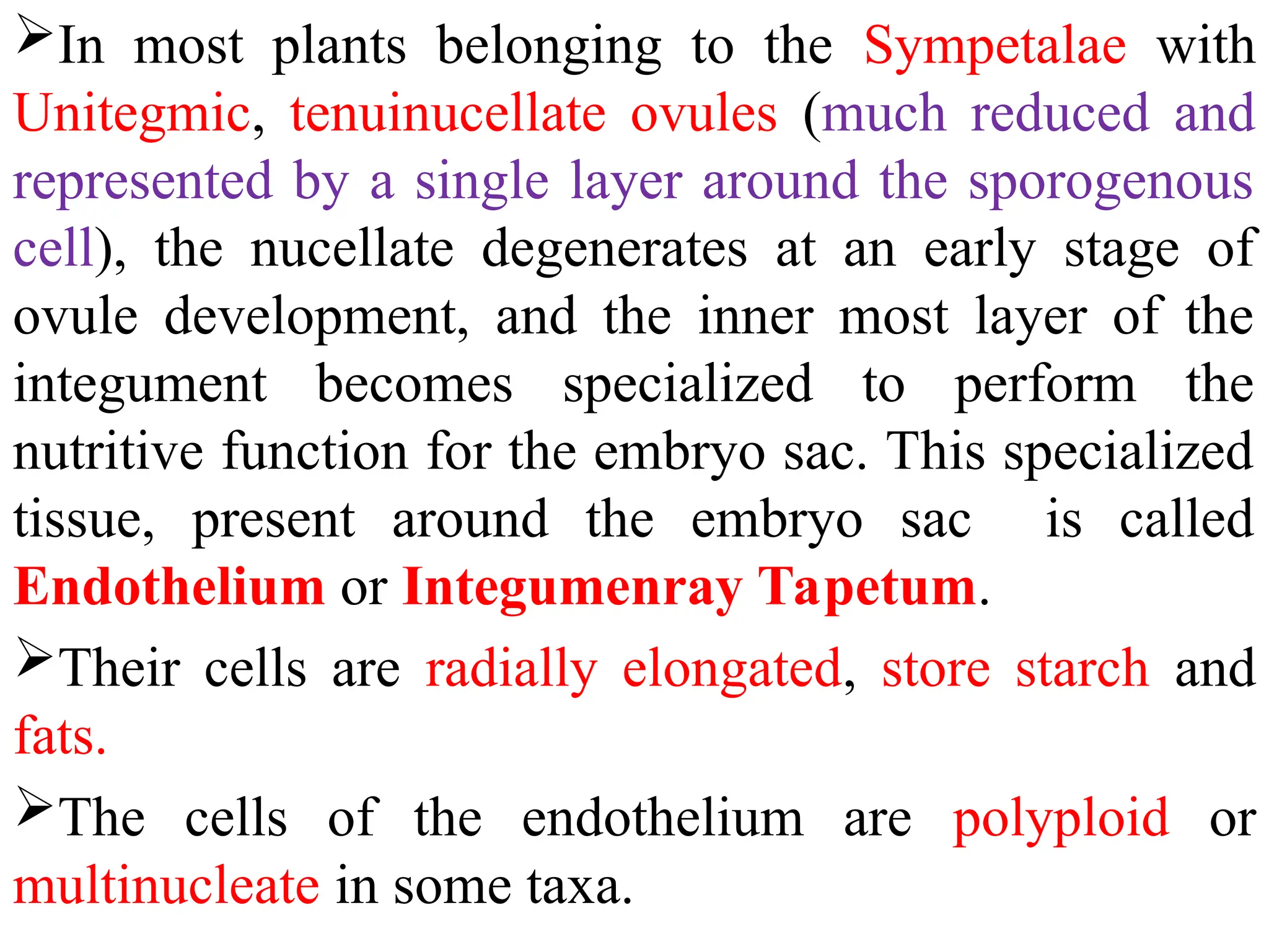
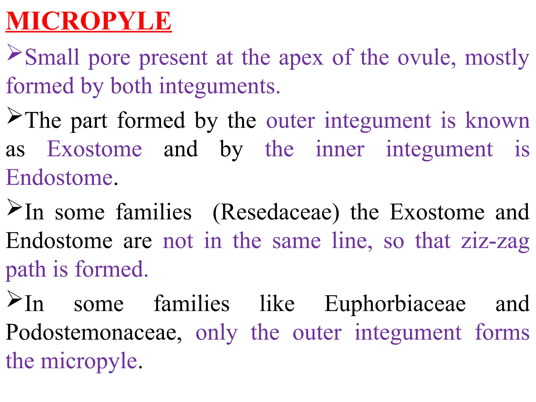
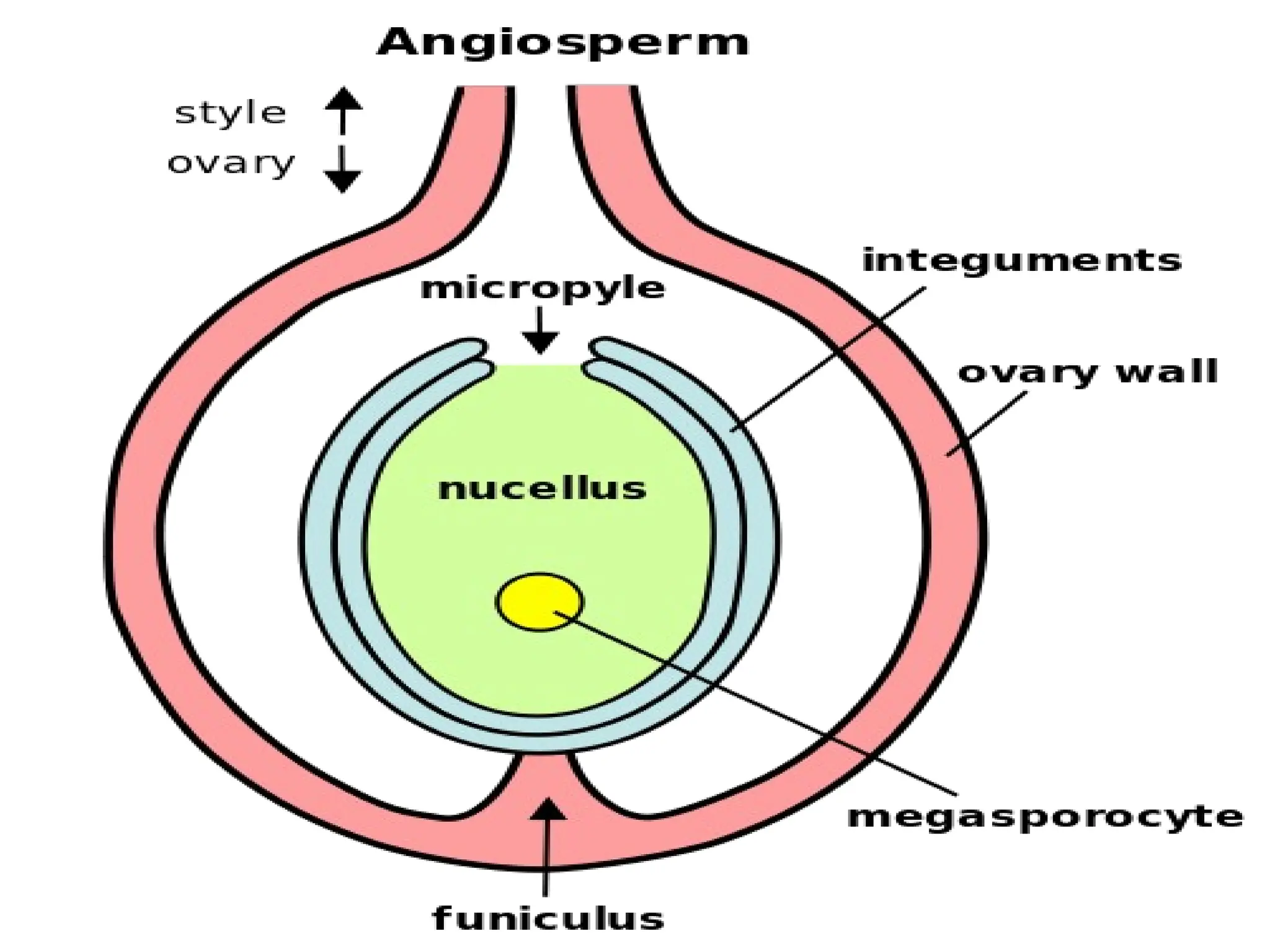
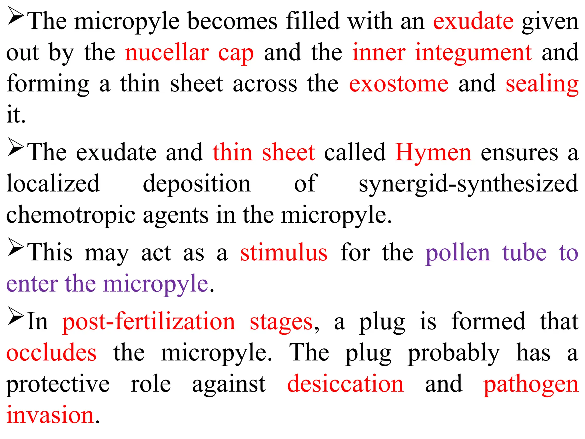
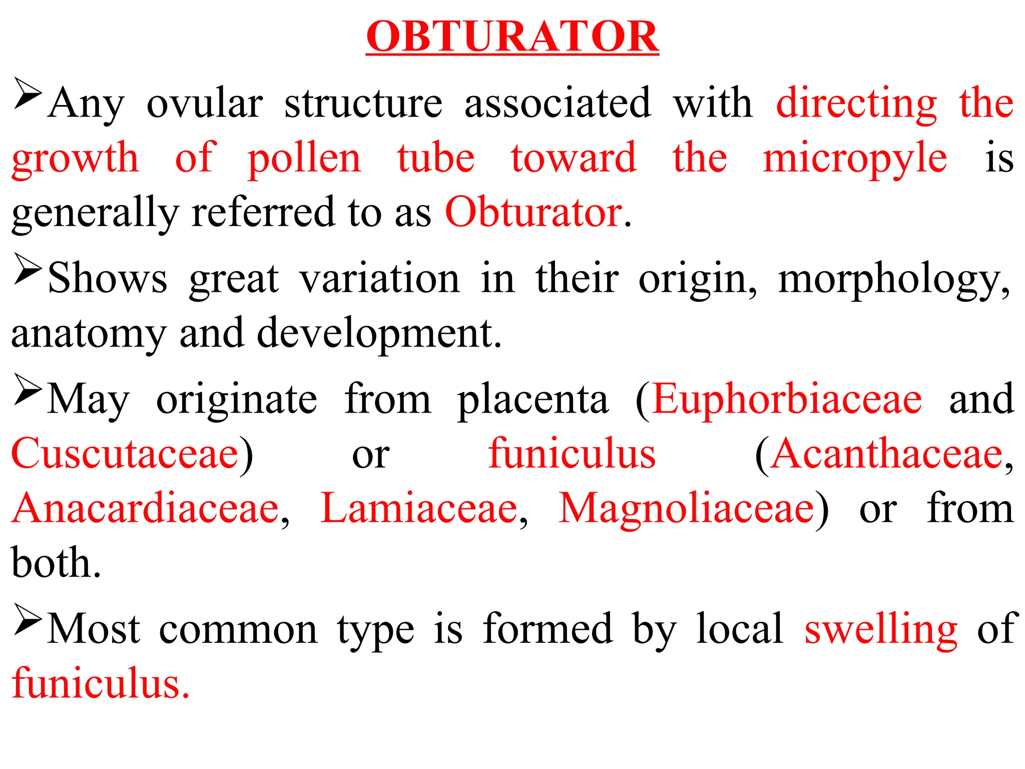
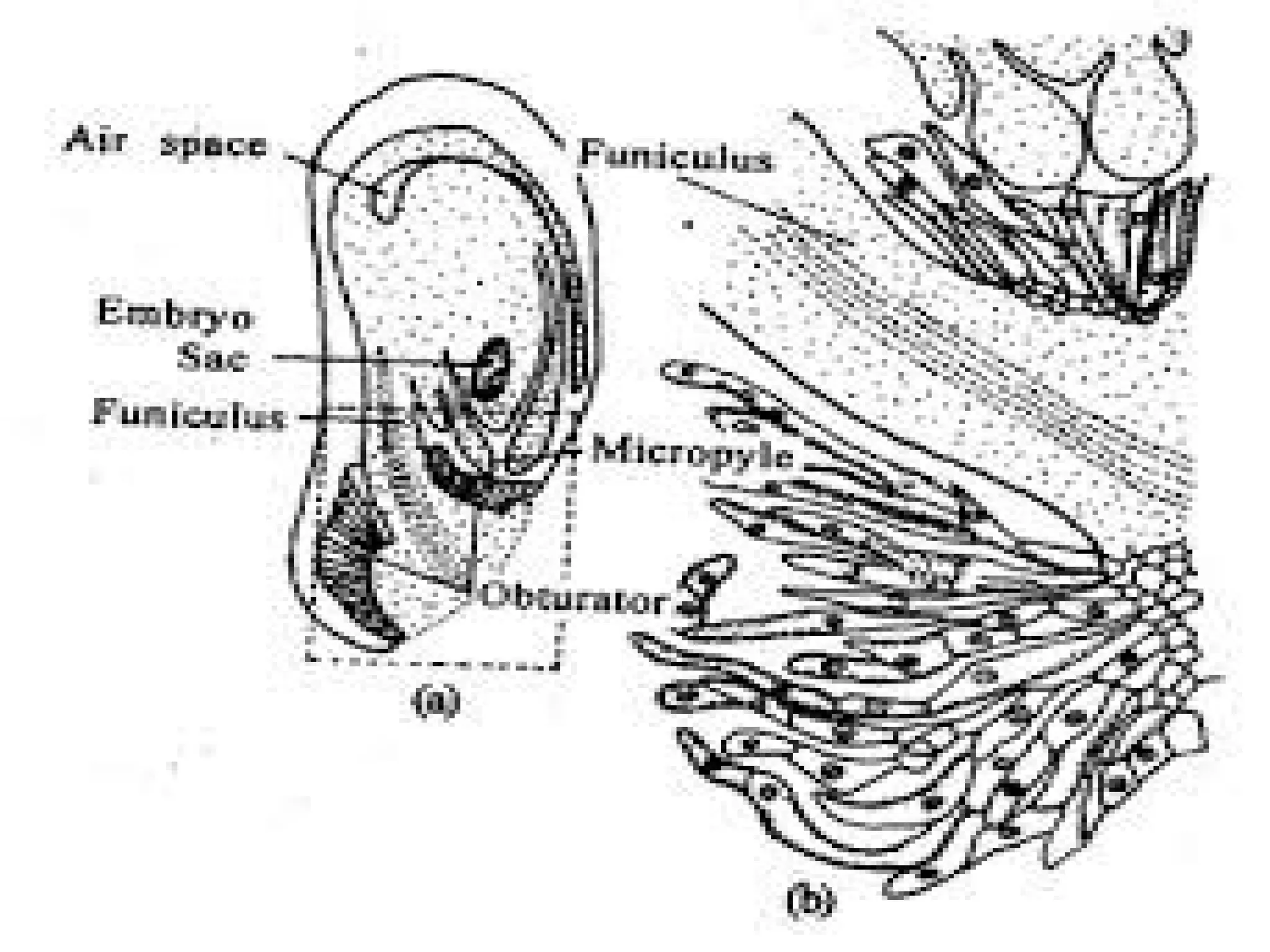
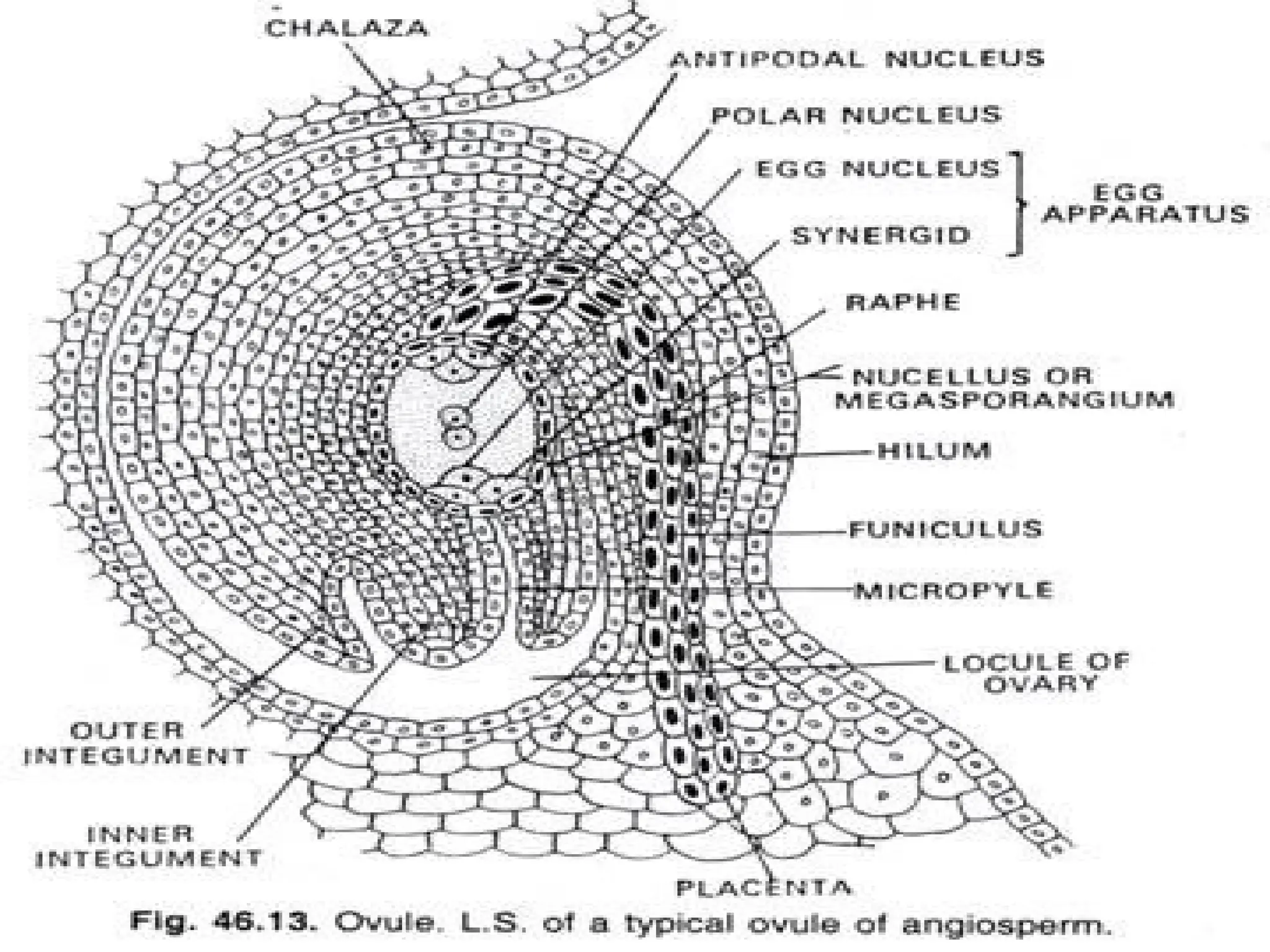
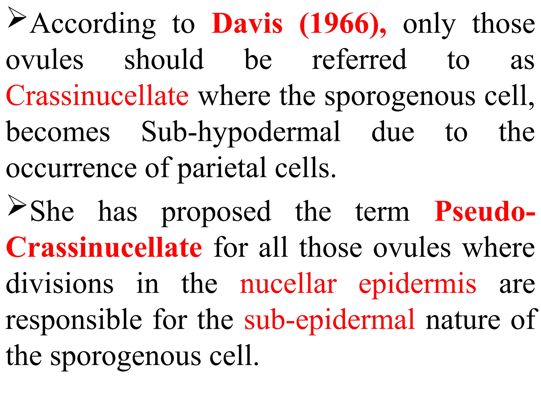
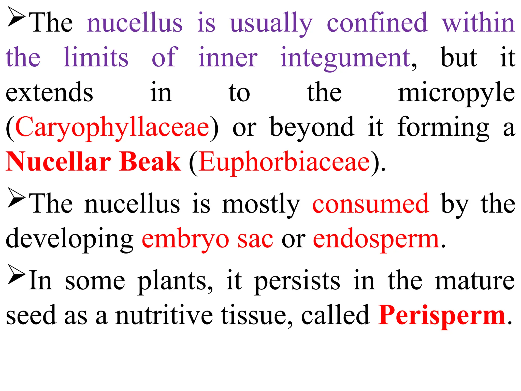
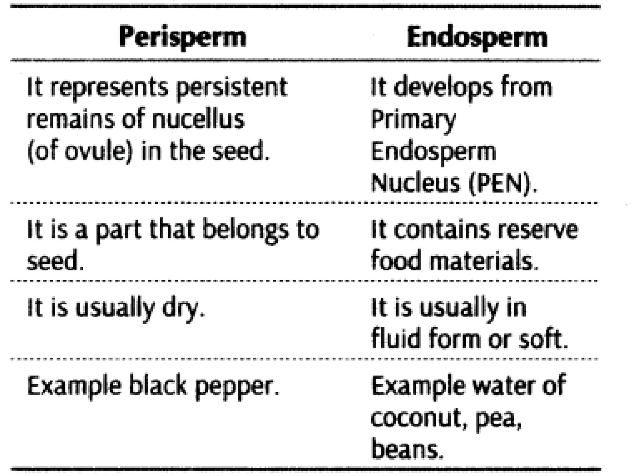
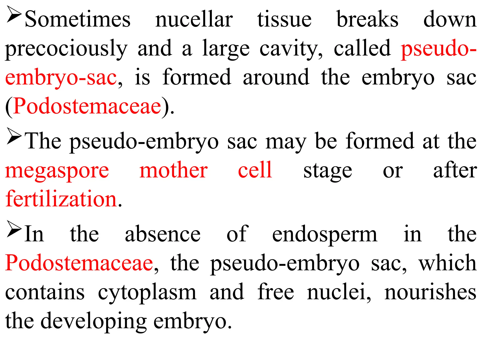
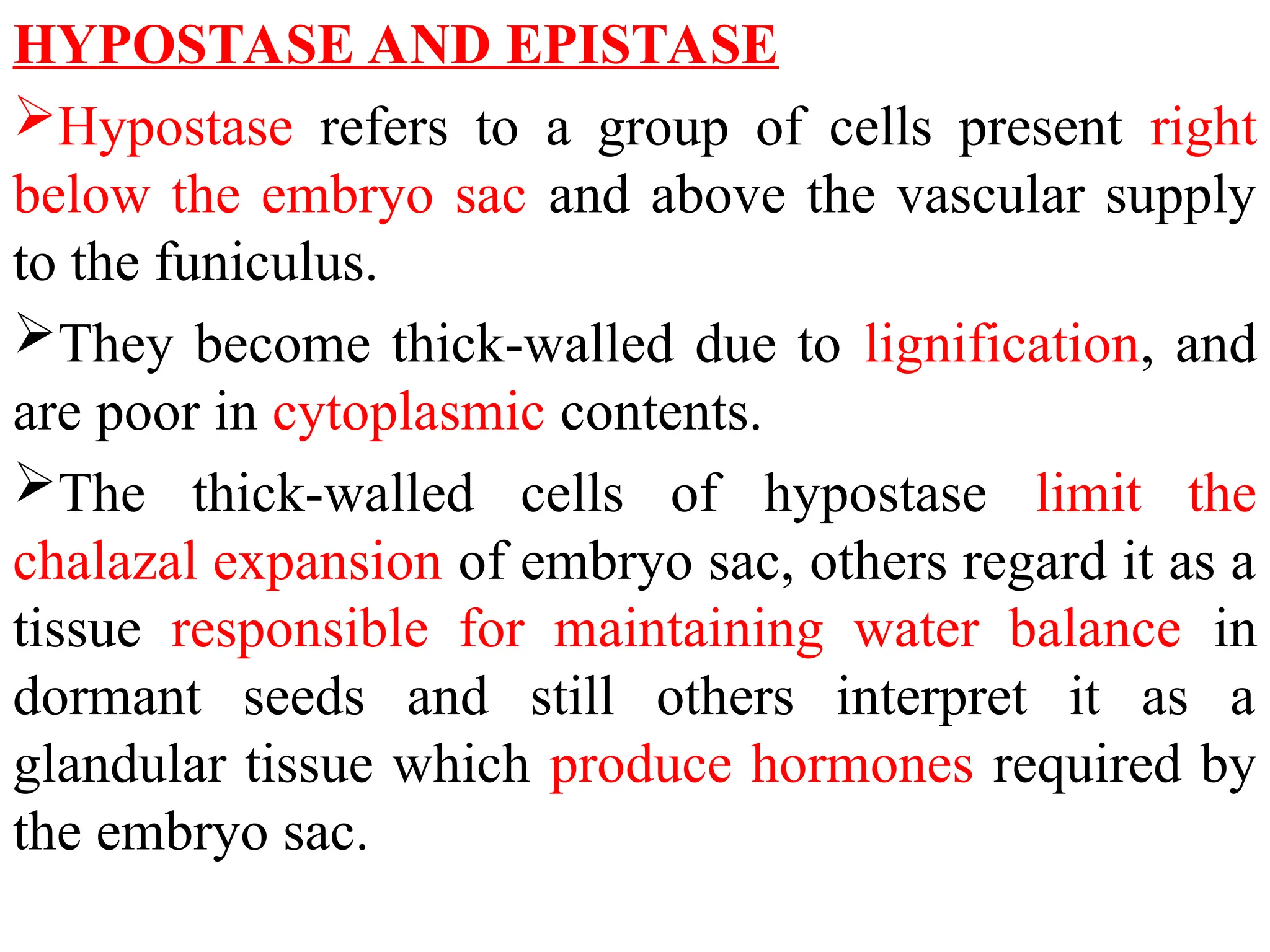
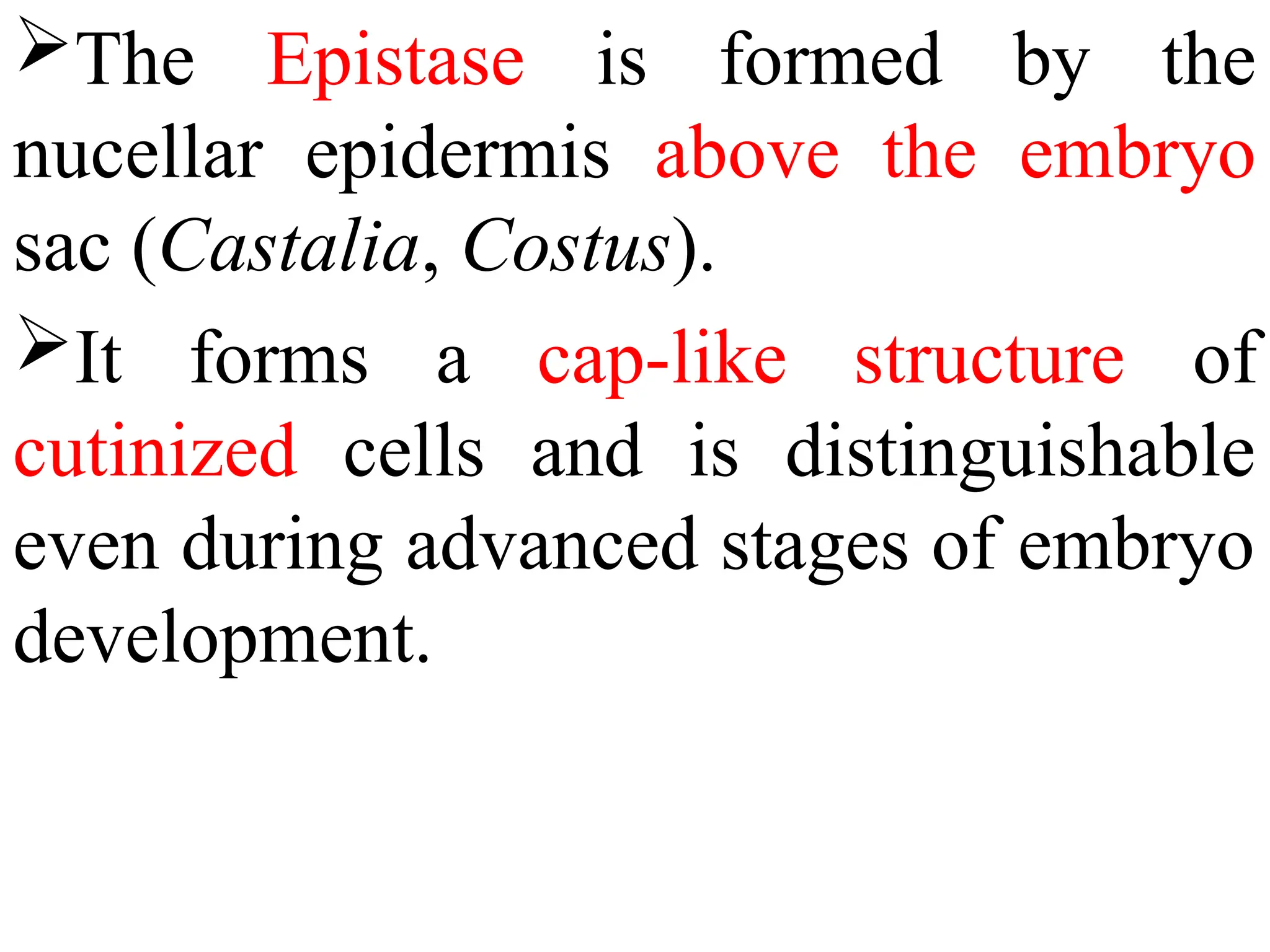
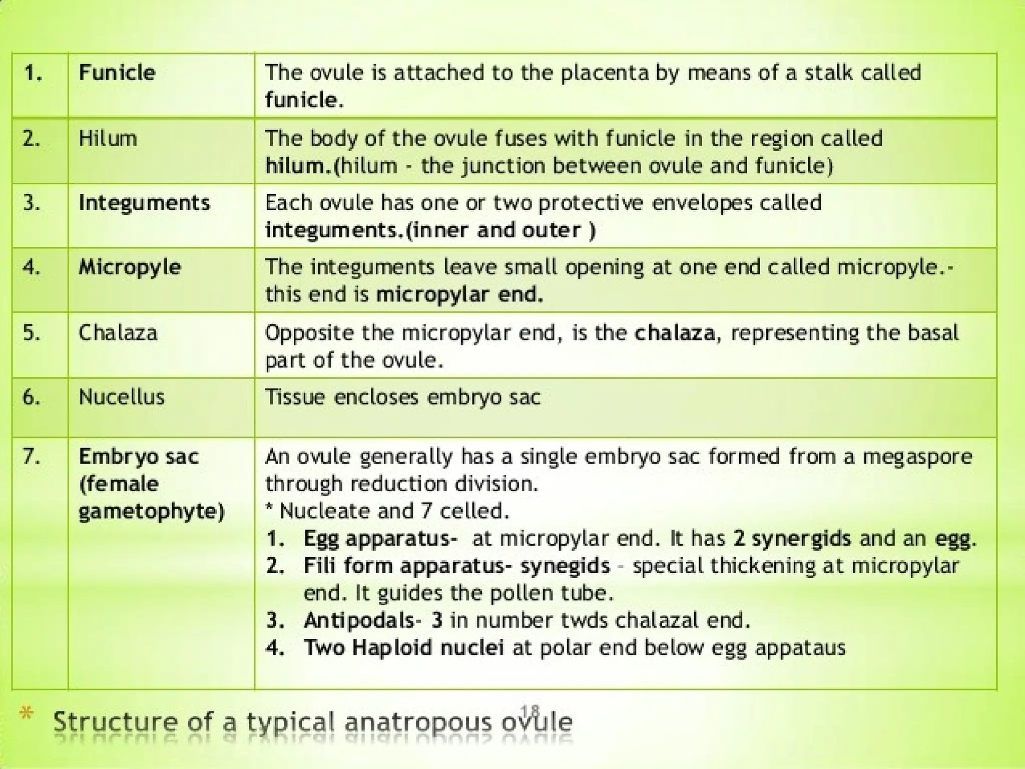
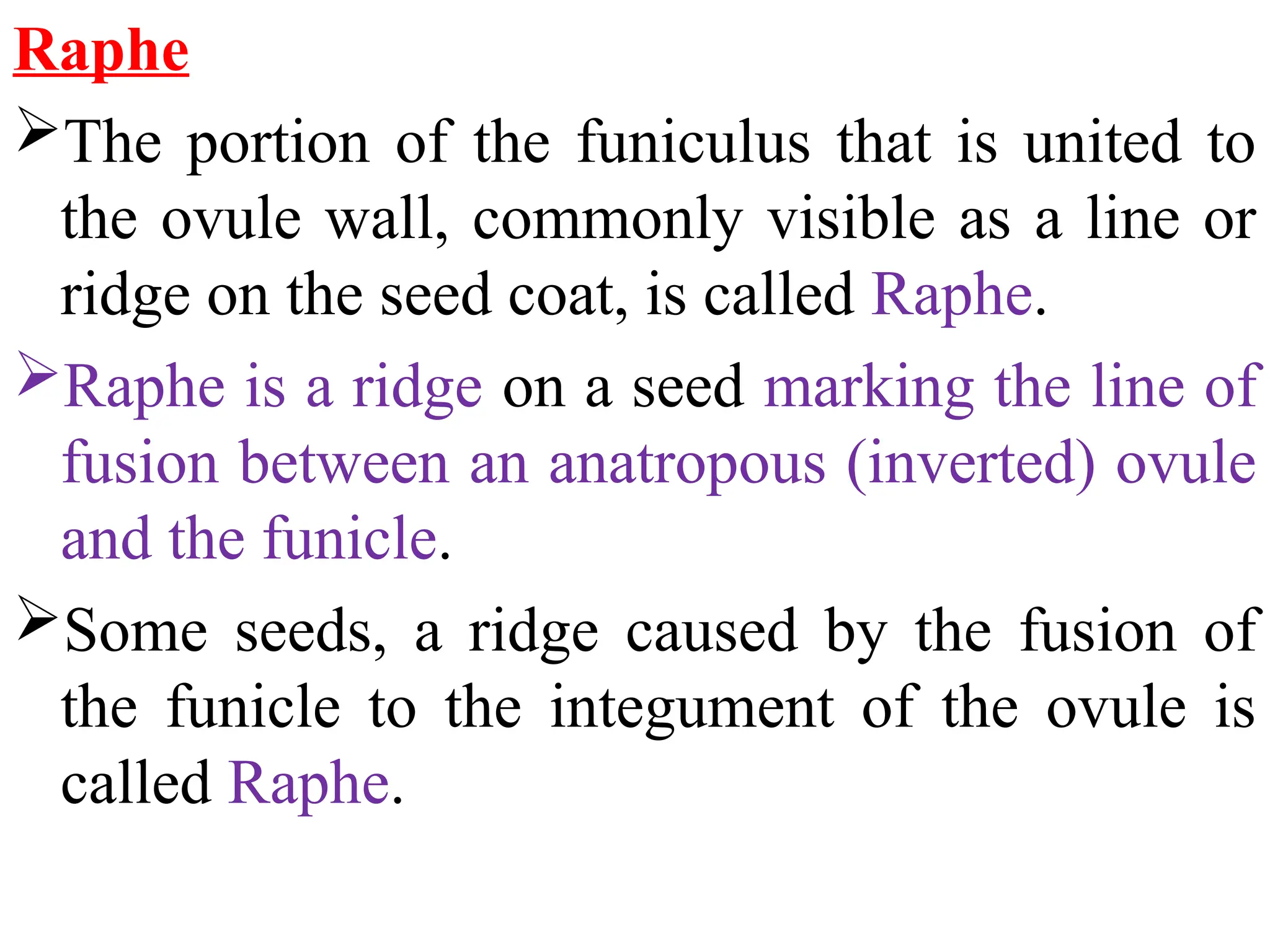
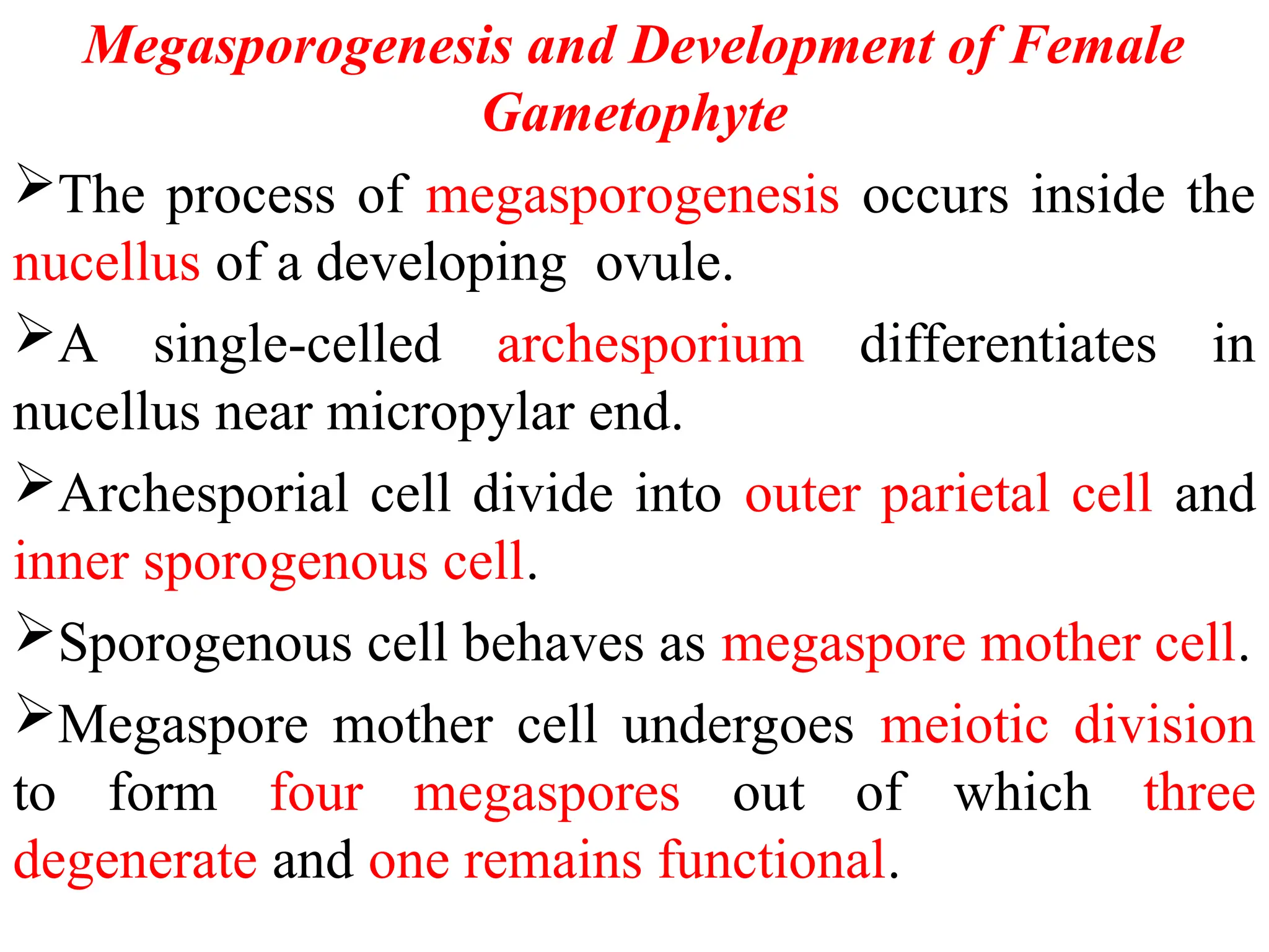
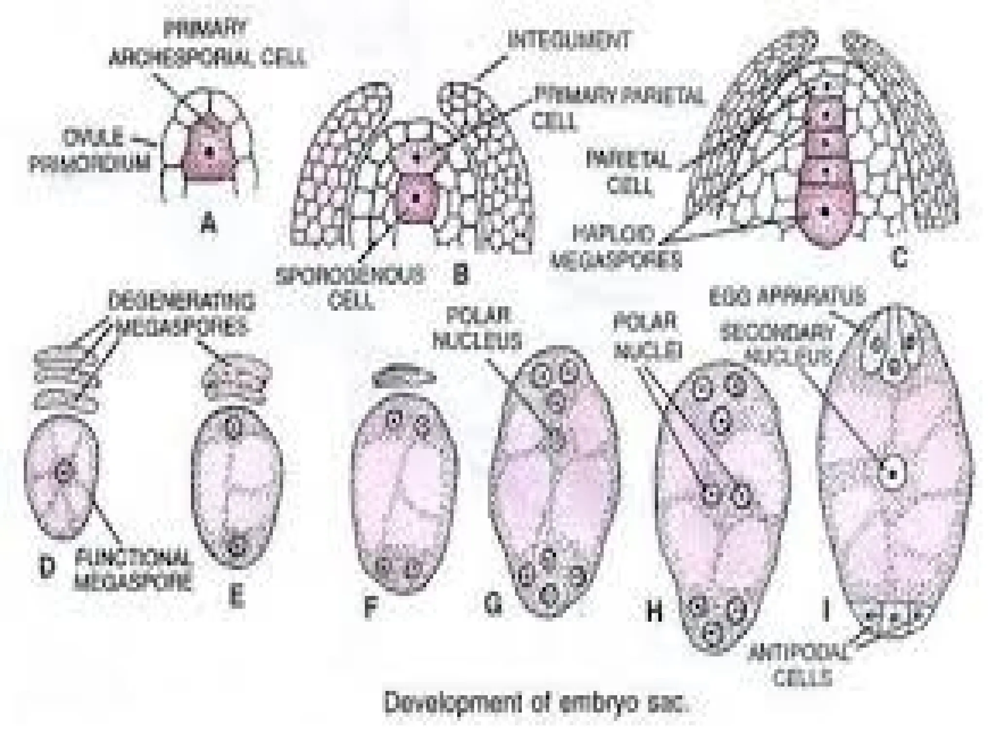
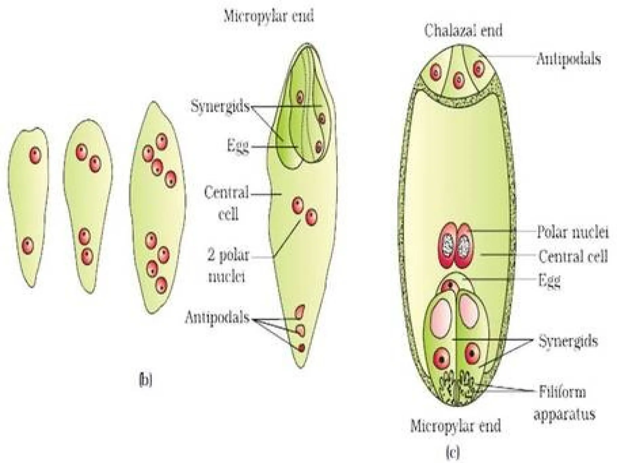
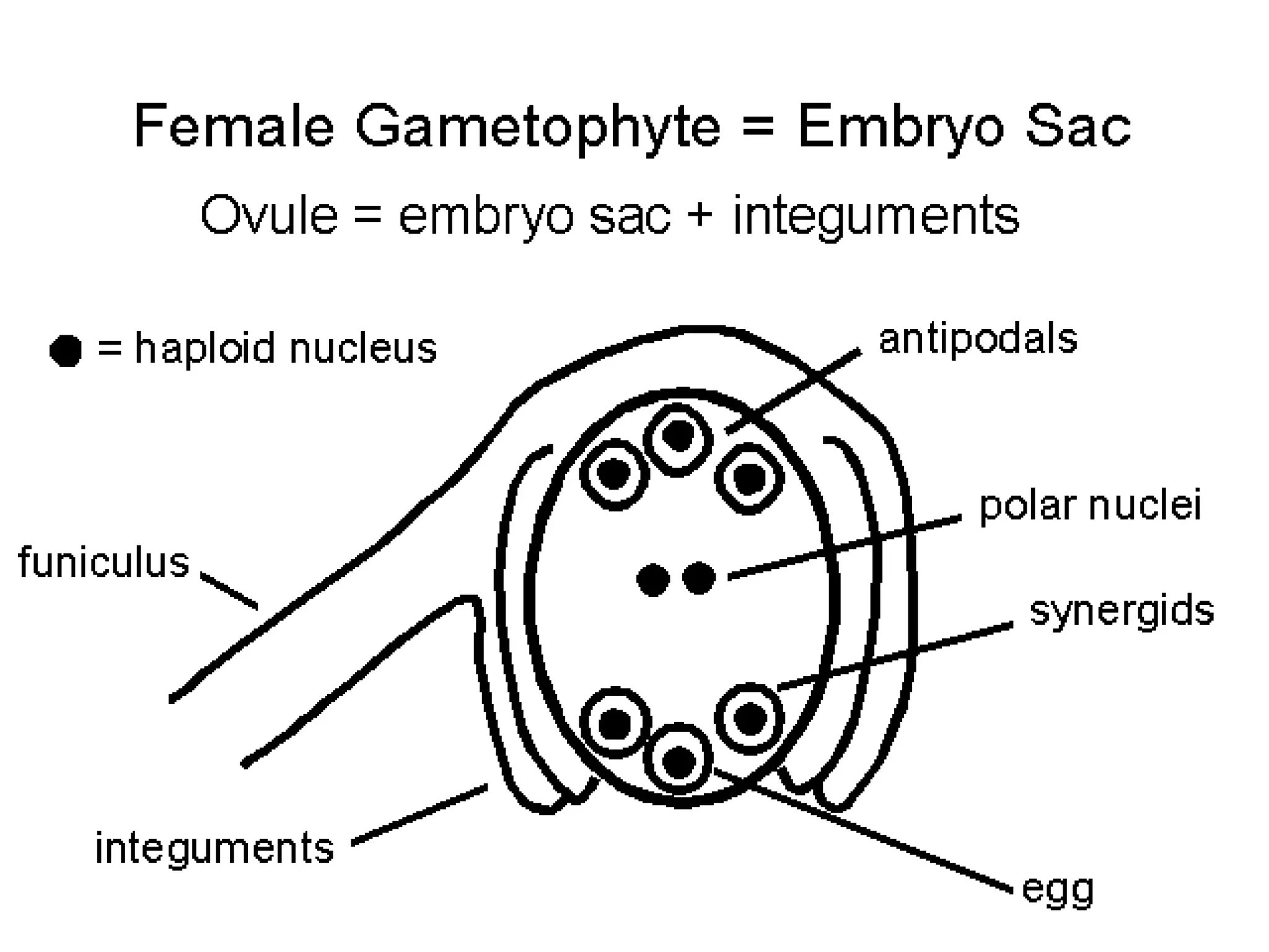
STRUCTURE
OF
OVULE
DR. HILAL LONE
The gynoecium represents the female
reproductive part of the flower.
The gynoecium may consist of a single pistil
or carpel (monocarpellary) or may have more
than one pistil or carpels (multicarpellary).
When there are more than one, the pistils may
be fused together (syncarpous) or may be free
(apocarpous).
Each pistil has three parts, the stigma, style
and ovary.
The stigma serves as a landing platform for pollen
grains.
The style is the elongated slender part beneath the
stigma.
The basal bulged part of the pistil is the ovary.
Inside the ovary is the ovarian cavity (locule) in
which ovules are present.
The ovules are borne on placenta as a small mound
of tissue.
The placenta is a cushion-shaped structure located
along the line of fusion of carpellary margins.
The ovules, which are enclosed in the ovary,
develop in to seeds after fertilization and the
wall of the ovary matures in to fruit.
A Megasporangium is a plant structure in
which megaspores are formed.
The megasporangium together with the
protective coats, the integuments, is called
ovule.
The megasporangium is attached to the
placenta, on the inner wall of the ovary, by a
stalk, called Funiculus.
Parts of the Ovule
Integuments
Micropyle
Nucellus
Obturator
Hypostase and Epistase
INTEGUMENTS
Unitegmic or Bitegmic
Unitegmic condition-Sympetalae or
Gamopetalae.
Bitegmic condition-Polypetalae and
Monocots.
Ategmic (without integument)-Members
of Olacaceae like Liriosma, Olax
imbricata, Ptychopetalum.
Integuments arise close to the base of the
homogenous tissue which forms the nucellus in a
mature ovule.
The inner integument is dermal in origin except in
Euphorbiaceae, where it is sub-dermal.
The outer integument is initiated either dermally or
sub-dermally.
The ovule begins to curve with the differentiation of
the integuments and it assumes the final shape by
the megaspore tetrad stage.
Although, integuments initiate later, they grow
faster than the nucellus and soon surround it almost
completely, except at the region of the micropyle.
In some members of Proteaceae (Macadamia),
the growth of the integument is so slow that
they do not enclose the nucellus until after
fertilization.
In Bitegmic ovules, the inner integument
differentiates earlier than the outer integument
but the latter usually overgrows the former.
In a fully formed ovule, the outer integument
is more massive and thicker as compared to
the inner integument.
The primordia of both the integuments
arise independently at the base of the
nucellus.
In Lannea and Rhus, the two integuments
arise as a result of splitting of the single
integumentary primordium.
The Unitemic condition of the ovule is
considered to be derived from fusion of
the two integuments, or by the
suppression/abortion of one integument.
In some taxa, especially belonging to the
family Cactaceae, a prominent air space is
present between the two integuments in the
chalazal region.
The occurrence of stomata on the outer
integument has been reported in Cleome,
Isomeris and Magnolia.
Chloroplasts are present in the cells of the
outer integument in several monocotyledons
such as Gladiolus, Lilium, Amaryllis and
Moringa.
More than two integuments (Supernumarary
integuments) like aril and caruncle have been
reported in some taxa.
An aril arises from the base of the ovule and it
completely covers the other two integuments
and thus forming a third integument, e.g.
Asphodelus, Trianthema.
In many Euphorbiaceae, an outgrowth called
Caruncle, arises from the tip of the outer
integument which turns backward and partially
envelops the ovules.
In most plants belonging to the Sympetalae with
Unitegmic, tenuinucellate ovules (much reduced and
represented by a single layer around the sporogenous
cell), the nucellate degenerates at an early stage of
ovule development, and the inner most layer of the
integument becomes specialized to perform the
nutritive function for the embryo sac. This specialized
tissue, present around the embryo sac is called
Endothelium or Integumenray Tapetum.
Their cells are radially elongated, store starch and
fats.
The cells of the endothelium are polyploid or
multinucleate in some taxa.
MICROPYLE
Small pore present at the apex of the ovule, mostly
formed by both integuments.
The part formed by the outer integument is known
as Exostome and by the inner integument is
Endostome.
In some families (Resedaceae) the Exostome and
Endostome are not in the same line, so that ziz-zag
path is formed.
In some families like Euphorbiaceae and
Podostemonaceae, only the outer integument forms
the micropyle.
The micropyle becomes filled with an exudate given
out by the nucellar cap and the inner integument and
forming a thin sheet across the exostome and sealing
it.
The exudate and thin sheet called Hymen ensures a
localized deposition of synergid-synthesized
chemotropic agents in the micropyle.
This may act as a stimulus for the pollen tube to
enter the micropyle.
In post-fertilization stages, a plug is formed that
occludes the micropyle. The plug probably has a
protective role against desiccation and pathogen
invasion.
OBTURATOR
Any ovular structure associated with directing the
growth of pollen tube toward the micropyle is
generally referred to as Obturator.
Shows great variation in their origin, morphology,
anatomy and development.
May originate from placenta (Euphorbiaceae and
Cuscutaceae) or funiculus (Acanthaceae,
Anacardiaceae, Lamiaceae, Magnoliaceae) or from
both.
Most common type is formed by local swelling of
funiculus.
NUCELLUS
Nucellus is a rounded or oval mass of
thin walled parenchymatous cells enclosed
by integuments. Nucellus represents the
wall of megasporangium.
Each ovule has only one nucellus.
Twin nucelli may occur as an
abnormality in Aegle marmelos and
Herminium angustifolium.
The archesporium differentiates
immediately below the nucellar epidermis.
In Sympetalae, the archesporial cell
directly functions as the megaspore mother
cell so that the sporogenous cell is also
hypodermal. Such ovules where the
sporogenous cell is hypodermal and the
nucellar tissue around it remains single-
layered, are called Tenuinucellate. This
condition of ovule generally occurs in more
advanced families and are usually
Unitegmic.
In Polypetalae and Monocotyledons, the hypodermal
archesporial cell divides transversely, cutting an outer
parietal cell and an inner sporogenous cell.
The parietal cell may either remain undivided or
undergo a few periclinal and anticlinal divisions so
that the sporogenous cell becomes embedded in the
massive nucellus.
All such ovules, where the sporogenous cell
becomes sub-hypodermal, either due to the formation
of parietal cells, or due to divisions in the nucellar
epidermis, or both, are called Crassinucellate.
These ovules are usually Bitegmic
According to Davis (1966), only those
ovules should be referred to as
Crassinucellate where the sporogenous cell,
becomes Sub-hypodermal due to the
occurrence of parietal cells.
She has proposed the term Pseudo-
Crassinucellate for all those ovules where
divisions in the nucellar epidermis are
responsible for the sub-epidermal nature of
the sporogenous cell.
The nucellus is usually confined within
the limits of inner integument, but it
extends in to the micropyle
(Caryophyllaceae) or beyond it forming a
Nucellar Beak (Euphorbiaceae).
The nucellus is mostly consumed by the
developing embryo sac or endosperm.
In some plants, it persists in the mature
seed as a nutritive tissue, called Perisperm.
Sometimes nucellar tissue breaks down
precociously and a large cavity, called pseudo-
embryo-sac, is formed around the embryo sac
(Podostemaceae).
The pseudo-embryo sac may be formed at the
megaspore mother cell stage or after
fertilization.
In the absence of endosperm in the
Podostemaceae, the pseudo-embryo sac, which
contains cytoplasm and free nuclei, nourishes
the developing embryo.
HYPOSTASE AND EPISTASE
Hypostase refers to a group of cells present right
below the embryo sac and above the vascular supply
to the funiculus.
They become thick-walled due to lignification, and
are poor in cytoplasmic contents.
The thick-walled cells of hypostase limit the
chalazal expansion of embryo sac, others regard it as a
tissue responsible for maintaining water balance in
dormant seeds and still others interpret it as a
glandular tissue which produce hormones required by
the embryo sac.
The Epistase is formed by the
nucellar epidermis above the embryo
sac (Castalia, Costus).
It forms a cap-like structure of
cutinized cells and is distinguishable
even during advanced stages of embryo
development.
Raphe
The portion of the funiculus that is united to
the ovule wall, commonly visible as a line or
ridge on the seed coat, is called Raphe.
Raphe is a ridge on a seed marking the line of
fusion between an anatropous (inverted) ovule
and the funicle.
Some seeds, a ridge caused by the fusion of
the funicle to the integument of the ovule is
called Raphe.
Megasporogenesis and Development of Female
Gametophyte
The process of megasporogenesis occurs inside the
nucellus of a developing ovule.
A single-celled archesporium differentiates in
nucellus near micropylar end.
Archesporial cell divide into outer parietal cell and
inner sporogenous cell.
Sporogenous cell behaves as megaspore mother cell.
Megaspore mother cell undergoes meiotic division
to form four megaspores out of which three
degenerate and one remains functional.
Functional megaspore develops in to female
gametophyte by sequential mitotic divisions.
Female gametophyte consists of 8-nucleate
Embryo Sac:
a) Three cells of embryo sac: 2 synergids
and 1 egg cell at micropylar end forms the egg
apparatus.
b) 3 antipodal cells are formed at the
chalazal end.
c) Central cell consists of 2 polar nuclei
THANK
YOU

Structure of Ovule.ppt ovule is female reproducgive cell

  • 1.
  • 2.
    The gynoecium representsthe female reproductive part of the flower. The gynoecium may consist of a single pistil or carpel (monocarpellary) or may have more than one pistil or carpels (multicarpellary). When there are more than one, the pistils may be fused together (syncarpous) or may be free (apocarpous). Each pistil has three parts, the stigma, style and ovary.
  • 5.
    The stigma servesas a landing platform for pollen grains. The style is the elongated slender part beneath the stigma. The basal bulged part of the pistil is the ovary. Inside the ovary is the ovarian cavity (locule) in which ovules are present. The ovules are borne on placenta as a small mound of tissue. The placenta is a cushion-shaped structure located along the line of fusion of carpellary margins.
  • 7.
    The ovules, whichare enclosed in the ovary, develop in to seeds after fertilization and the wall of the ovary matures in to fruit. A Megasporangium is a plant structure in which megaspores are formed. The megasporangium together with the protective coats, the integuments, is called ovule. The megasporangium is attached to the placenta, on the inner wall of the ovary, by a stalk, called Funiculus.
  • 10.
    Parts of theOvule Integuments Micropyle Nucellus Obturator Hypostase and Epistase
  • 11.
    INTEGUMENTS Unitegmic or Bitegmic Unitegmiccondition-Sympetalae or Gamopetalae. Bitegmic condition-Polypetalae and Monocots. Ategmic (without integument)-Members of Olacaceae like Liriosma, Olax imbricata, Ptychopetalum.
  • 12.
    Integuments arise closeto the base of the homogenous tissue which forms the nucellus in a mature ovule. The inner integument is dermal in origin except in Euphorbiaceae, where it is sub-dermal. The outer integument is initiated either dermally or sub-dermally. The ovule begins to curve with the differentiation of the integuments and it assumes the final shape by the megaspore tetrad stage. Although, integuments initiate later, they grow faster than the nucellus and soon surround it almost completely, except at the region of the micropyle.
  • 14.
    In some membersof Proteaceae (Macadamia), the growth of the integument is so slow that they do not enclose the nucellus until after fertilization. In Bitegmic ovules, the inner integument differentiates earlier than the outer integument but the latter usually overgrows the former. In a fully formed ovule, the outer integument is more massive and thicker as compared to the inner integument.
  • 15.
    The primordia ofboth the integuments arise independently at the base of the nucellus. In Lannea and Rhus, the two integuments arise as a result of splitting of the single integumentary primordium. The Unitemic condition of the ovule is considered to be derived from fusion of the two integuments, or by the suppression/abortion of one integument.
  • 16.
    In some taxa,especially belonging to the family Cactaceae, a prominent air space is present between the two integuments in the chalazal region. The occurrence of stomata on the outer integument has been reported in Cleome, Isomeris and Magnolia. Chloroplasts are present in the cells of the outer integument in several monocotyledons such as Gladiolus, Lilium, Amaryllis and Moringa.
  • 17.
    More than twointeguments (Supernumarary integuments) like aril and caruncle have been reported in some taxa. An aril arises from the base of the ovule and it completely covers the other two integuments and thus forming a third integument, e.g. Asphodelus, Trianthema. In many Euphorbiaceae, an outgrowth called Caruncle, arises from the tip of the outer integument which turns backward and partially envelops the ovules.
  • 19.
    In most plantsbelonging to the Sympetalae with Unitegmic, tenuinucellate ovules (much reduced and represented by a single layer around the sporogenous cell), the nucellate degenerates at an early stage of ovule development, and the inner most layer of the integument becomes specialized to perform the nutritive function for the embryo sac. This specialized tissue, present around the embryo sac is called Endothelium or Integumenray Tapetum. Their cells are radially elongated, store starch and fats. The cells of the endothelium are polyploid or multinucleate in some taxa.
  • 20.
    MICROPYLE Small pore presentat the apex of the ovule, mostly formed by both integuments. The part formed by the outer integument is known as Exostome and by the inner integument is Endostome. In some families (Resedaceae) the Exostome and Endostome are not in the same line, so that ziz-zag path is formed. In some families like Euphorbiaceae and Podostemonaceae, only the outer integument forms the micropyle.
  • 22.
    The micropyle becomesfilled with an exudate given out by the nucellar cap and the inner integument and forming a thin sheet across the exostome and sealing it. The exudate and thin sheet called Hymen ensures a localized deposition of synergid-synthesized chemotropic agents in the micropyle. This may act as a stimulus for the pollen tube to enter the micropyle. In post-fertilization stages, a plug is formed that occludes the micropyle. The plug probably has a protective role against desiccation and pathogen invasion.
  • 23.
    OBTURATOR Any ovular structureassociated with directing the growth of pollen tube toward the micropyle is generally referred to as Obturator. Shows great variation in their origin, morphology, anatomy and development. May originate from placenta (Euphorbiaceae and Cuscutaceae) or funiculus (Acanthaceae, Anacardiaceae, Lamiaceae, Magnoliaceae) or from both. Most common type is formed by local swelling of funiculus.
  • 25.
    NUCELLUS Nucellus is arounded or oval mass of thin walled parenchymatous cells enclosed by integuments. Nucellus represents the wall of megasporangium. Each ovule has only one nucellus. Twin nucelli may occur as an abnormality in Aegle marmelos and Herminium angustifolium.
  • 27.
    The archesporium differentiates immediatelybelow the nucellar epidermis. In Sympetalae, the archesporial cell directly functions as the megaspore mother cell so that the sporogenous cell is also hypodermal. Such ovules where the sporogenous cell is hypodermal and the nucellar tissue around it remains single- layered, are called Tenuinucellate. This condition of ovule generally occurs in more advanced families and are usually Unitegmic.
  • 28.
    In Polypetalae andMonocotyledons, the hypodermal archesporial cell divides transversely, cutting an outer parietal cell and an inner sporogenous cell. The parietal cell may either remain undivided or undergo a few periclinal and anticlinal divisions so that the sporogenous cell becomes embedded in the massive nucellus. All such ovules, where the sporogenous cell becomes sub-hypodermal, either due to the formation of parietal cells, or due to divisions in the nucellar epidermis, or both, are called Crassinucellate. These ovules are usually Bitegmic
  • 29.
    According to Davis(1966), only those ovules should be referred to as Crassinucellate where the sporogenous cell, becomes Sub-hypodermal due to the occurrence of parietal cells. She has proposed the term Pseudo- Crassinucellate for all those ovules where divisions in the nucellar epidermis are responsible for the sub-epidermal nature of the sporogenous cell.
  • 30.
    The nucellus isusually confined within the limits of inner integument, but it extends in to the micropyle (Caryophyllaceae) or beyond it forming a Nucellar Beak (Euphorbiaceae). The nucellus is mostly consumed by the developing embryo sac or endosperm. In some plants, it persists in the mature seed as a nutritive tissue, called Perisperm.
  • 32.
    Sometimes nucellar tissuebreaks down precociously and a large cavity, called pseudo- embryo-sac, is formed around the embryo sac (Podostemaceae). The pseudo-embryo sac may be formed at the megaspore mother cell stage or after fertilization. In the absence of endosperm in the Podostemaceae, the pseudo-embryo sac, which contains cytoplasm and free nuclei, nourishes the developing embryo.
  • 33.
    HYPOSTASE AND EPISTASE Hypostaserefers to a group of cells present right below the embryo sac and above the vascular supply to the funiculus. They become thick-walled due to lignification, and are poor in cytoplasmic contents. The thick-walled cells of hypostase limit the chalazal expansion of embryo sac, others regard it as a tissue responsible for maintaining water balance in dormant seeds and still others interpret it as a glandular tissue which produce hormones required by the embryo sac.
  • 34.
    The Epistase isformed by the nucellar epidermis above the embryo sac (Castalia, Costus). It forms a cap-like structure of cutinized cells and is distinguishable even during advanced stages of embryo development.
  • 36.
    Raphe The portion ofthe funiculus that is united to the ovule wall, commonly visible as a line or ridge on the seed coat, is called Raphe. Raphe is a ridge on a seed marking the line of fusion between an anatropous (inverted) ovule and the funicle. Some seeds, a ridge caused by the fusion of the funicle to the integument of the ovule is called Raphe.
  • 37.
    Megasporogenesis and Developmentof Female Gametophyte The process of megasporogenesis occurs inside the nucellus of a developing ovule. A single-celled archesporium differentiates in nucellus near micropylar end. Archesporial cell divide into outer parietal cell and inner sporogenous cell. Sporogenous cell behaves as megaspore mother cell. Megaspore mother cell undergoes meiotic division to form four megaspores out of which three degenerate and one remains functional.
  • 41.
    Functional megaspore developsin to female gametophyte by sequential mitotic divisions. Female gametophyte consists of 8-nucleate Embryo Sac: a) Three cells of embryo sac: 2 synergids and 1 egg cell at micropylar end forms the egg apparatus. b) 3 antipodal cells are formed at the chalazal end. c) Central cell consists of 2 polar nuclei
  • 43.