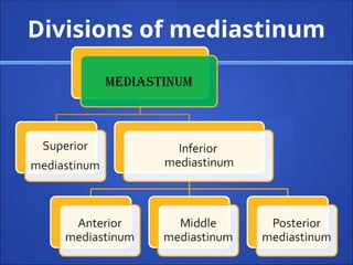
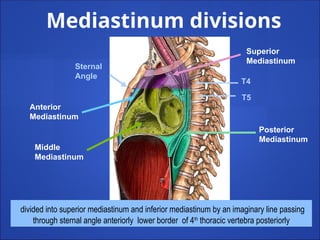
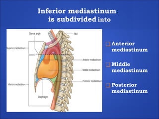
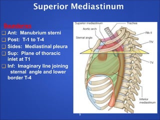
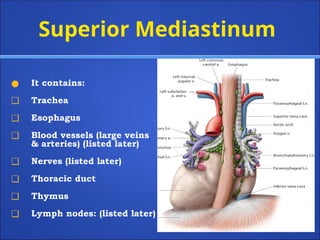
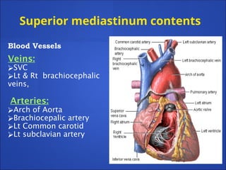
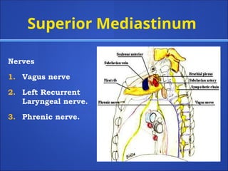
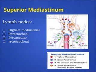
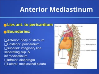
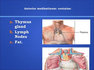
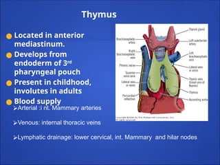
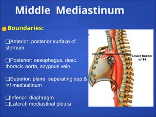
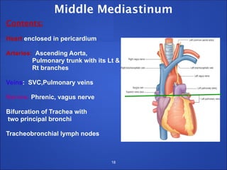
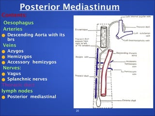
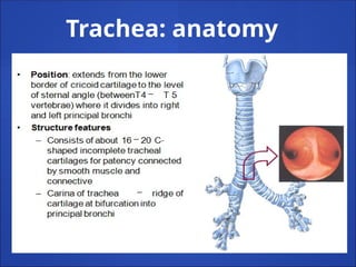
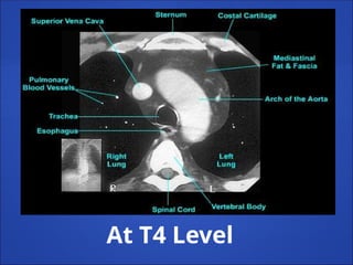
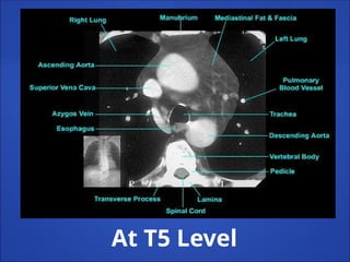
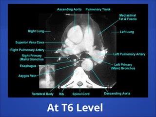
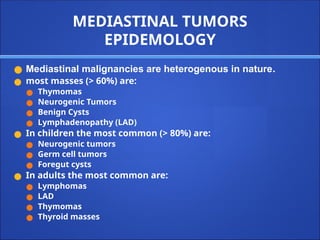
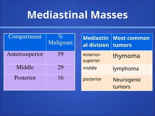
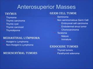
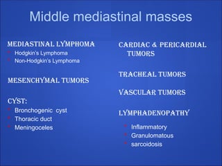
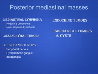
mediastinum
Dr. Deepak G
Assistant professor
Dept of Gen surgery
Introduction
●The mediastinum is the region in the chest
between the pleural cavities that contain the
heart and other thoracic viscera except the
lungs
●Boundaries
● Anterior - sternum
● Posterior - vertebral column and paravertebral
fascia
● Superior -thoracic inlet
● Inferior - diaphragm
● Lateral - parietal pleura
Sternal Angle
Thoracic inlet
Thoracic oulet
Boundaries of mediastinum
sternum
Thoracic vertebra
TS: Mediastinum
5
CS: Mediastinum
Divisions of mediastinum
Superior
Mediastinum
Posterior
Mediastinum
Anterior
Mediastinum
Middle
Mediastinum
Sternal
Angle
T4
T5
divided into superior mediastinum and inferior mediastinum by an imaginary line passing
through sternal angle anteriorly lower border of 4th
thoracic vertebra posteriorly
Mediastinum divisions
Inferior mediastinum:
is subdivided into
❑ Anterior
mediastinum
❑ Middle
mediastinum
❑ Posterior
mediastinum
Superior Mediastinum
Boundaries
❑ Ant: Manubrium sterni
❑ Post: T-1 to T-4
❑ Sides: Mediastinal pleura
❑ Sup: Plane of thoracic
inlet at T1
❑ Inf: Imaginary line joining
sternal angle and lower
border T-4
9
Superior Mediastinum
● It contains:
❑ Trachea
❑ Esophagus
❑ Blood vessels (large veins
& arteries) (listed later)
❑ Nerves (listed later)
❑ Thoracic duct
❑ Thymus
❑ Lymph nodes: (listed later)
Superior mediastinum contents
Blood Vessels
Veins:
⮚SVC
⮚Lt & Rt brachiocephalic
veins,
Arteries:
⮚Arch of Aorta
⮚Brachiocepalic artery
⮚Lt Common carotid
⮚Lt subclavian artery
Superior Mediastinum
Nerves
1. Vagus nerve
2. Left Recurrent
Laryngeal nerve.
3. Phrenic nerve.
Superior Mediastinum
Lymph nodes:
❑ Highest mediastinal
❑ Paratracheal
❑ Prevascular
❑ retrotracheal
Anterior Mediastinum
●Lies ant. to pericardium
●Boundaries:
❑Anterior: body of sternum
❑Posterior: pericardium
❑superior: imaginary line
separating sup. &
inf.mediastinum
❑Infreior: diaphragm
❑Lateral: mediastinal pleura
Anterior mediastinum: contains:
a. Thymus
gland
b. Lymph
Nodes
c. Fat.
Thymus
● Located in anterior
mediastinum.
● Develops from
endoderm of 3rd
pharyngeal pouch
● Present in childhood,
involutes in adults
● Blood supply
⮚Arterial :i nt. Mammary arteries
⮚Venous: internal thoracic veins
⮚Lymphatic drainage: lower cervical, int. Mammary and hilar nodes
Middle Mediastinum
●Boundaries:
❑Anterior: posterior surface of
sternum
❑Posterior: oesophagus, desc.
thoracic aorta, azygous vein
❑Superior: plane seperating sup.&
inf mediastinum
❑Inferior: diaphragm
❑Lateral: mediastinal pleura
Middle Mediastinum
Contents:
Heart enclosed in pericardium
Arteries: Ascending Aorta,
Pulmonary trunk with its Lt &
Rt branches
Veins: SVC,Pulmonary veins
Nerves: Phrenic, vagus nerve
Bifurcation of Trachea with
two principal bronchi
Tracheobronchial lymph nodes
18
Posterior Mediastinum
Boundaries:
Ant. Pericardium, Bifurcation of trachea
Post. T5 to T12
sup. Transverse thoracic plane
Inf. diaphragm
Sides: Mediastinal pleura
19
20
Posterior Mediastinum
Contents:
Oesophagus
Arteries
● Descending Aorta with its
brs
Veins
● Azygos
● Hemizygos
● Accessory hemizygos
Nerves:
● Vagus
● Splanchnic nerves
Thoracic duct
lymph nodes
● Posterior mediastinal
Trachea: anatomy
LENGTH:10-15 cm
❑ DIAMETER: 2cm in males &1.5
cm in females
❑ Lined by ciliated columnar
epithelium
❑Lower level at T6 on
inspiration & T4 on expiration
❑ Made of c shape rings
❑ 2 rings per cm
❑ The rings make tube convex
anterolateraly
❑ Posteriorly the gap is filled by
trachealis muscle.
NERVE SUPPLY:
●LYMPHATIC DRAINAGE
⮚ Pretracheal
⮚ paratracheal lymph node
● PARA SYMPHATHETIC:
vagus & recurrent
laryngeal nerves
(sensory & secreto-motor to
mucous membrane motor
to trachealis muscle)
● SYMPHATHETIC: -
middle cervical
ganglion (vasomotor)
Blood supply
●ARTERIAL SUPPLY
●Upper trachea
● Inferior thyroid artery
●Lower part
● Branches of the bronchial
artery
●VENOUS DRAINAGE
●Upper part :
left brachiocephalic vein
●Lower part:
● Inferior thyroid vein
Radiological antomy
CHEST X-RAY
27
Tracheobronchial anatomy
Tracheal Displacement Due to Goiter
Clues to locate mass to
mediastinum
Mediastinal masses : Masses in the lung
❖ not contain air bronchograms
❖ mediastinal mass will create
obtuse angles with the lung .
❖ Mediastinal lines will be
disrupted
– May contain air
bronchograms
– A lung mass abutts the
mediastinal surface and
creates acute angles with
the lung
LEFT: A lung mass abutts the mediastinal surface and creates acute angles
with the lung.
RIGHT: A mediastinal mass will sit under the surface of the mediastinum,
creating obtuse angles with the lung
Cervicothoracic sign
●The anterior mediastinum ends at the level of the
clavicles.
●The posterior mediastinum extends much higher.
●Therefore
● any mass that remains sharply outlined in the
apex of the thorax must be posterior and
entirely within the chest, and
● any mass that disappears at the clavicles must
be anterior and extends into neck
See sharp
margin
above
clavicle
Mass is in posterior mediastinum. because it remains sharply outlined in apex
of thorax, indicating that it is surrounded by lung.
This particular example is a ganglioneuroma
Thoracoabdominal sign
● A sharply marginated mediastinal mass seen through
the diaphragm must lie entirely within the chest.
● The posterior costophrenic sulcus extends far more
caudally than the anterior aspect of the lung
● Therefore
● Any mass that extends below the dome of the
diaphragm and remains sharply outlined must be in
the posterior compartments and surrounded by
lung, and
● Any mass that terminates at dome of diaphragm
must be anterior
Can you
see the
outline of
the
mass below
the
diaphragm?
Margin of mass is apparent and below diaphragm, therefore this must
be in the middle or posterior compartments where it is surrounded by
lung
This example is a ‘Lipoma’
Hilum overlay sign
● Principle of hilum overlay
● An anterior mediastinal mass will overlap the main
pulmonary arteries, therefore they will be seen within
the margins of the mass
Hilum can
be seen
through
mass
this must be an anterior
mediastinal mass
because it overlaps rather
than “pushes out” the
main pulmonary arteries
This particular example is a thymoma
VASCULAR ANATOMY
At T3 Level
At T4 Level
At T5 Level
At T6 Level
MEDIASTINAL TUMORS
EPIDEMOLOGY
● Mediastinal malignancies are heterogenous in nature.
● most masses (> 60%) are:
● Thymomas
● Neurogenic Tumors
● Benign Cysts
● Lymphadenopathy (LAD)
● In children the most common (> 80%) are:
● Neurogenic tumors
● Germ cell tumors
● Foregut cysts
● In adults the most common are:
● Lymphomas
● LAD
● Thymomas
● Thyroid masses
Mediastinal Masses
Compartment %
Malignant
Anterosuperior 59
Middle 29
Posterior 16
Mediastin
al division
Most common
tumors
Anterior-
superior
thymoma
middle lymphoma
posterior Neurogenic
tumors
Anterosuperior Masses
Thymus
• Thymoma
• Thymic carcinoma
• Thymic cyst
• Thymic carcinoid
• Thymolipoma
Mediastinal Lymphoma
• Hodgkin’s Lymphoma
• Non-Hodgkin’s Lymphoma
Mesenchymal tumors
Germ Cell Tumor
• Seminoma
• Non seminomatous Germ Cell
• Embryonal cell carcinoma
• Endodermal sinus tumor
• Choriocarcinoma
• Teratoma
• Mature
• Immature
Endocrine tumors
• Thyroid tumors
• Parathyroid adenoma
Middle mediastinal masses
Mediastinal Lymphoma
• Hodgkin’s Lymphoma
• Non-Hodgkin’s Lymphoma
Mesenchymal tumors
CYST:
• Bronchogenic cyst
• Thoracic duct
• Meningoceles
Cardiac & pericardial
tumors
Tracheal tumors
vascular tumors
Lymphadenopathy
• Inflammatory
• Granulomatous
• sarcoidosis
Posterior mediastinal masses
Mediastinal Lymphoma
• Hodgkin’s Lymphoma
• Non-Hodgkin’s Lymphoma
Mesenchymal tumors
Neurogenic tumors
• Peripheral nerves
• Symphathetic ganglia
• paraganglia
ENDOCRINE TUMORS
ESOPHAGEAL TUMORS
& CYSTS
Tumors of thymus
● Thymomas
● Thymic carcinomas
● Thymic lymphomas
● Carcinoids
● Thymolipomas
● Secondaries
Thymoma
Presentation
● Most common primary anterior mediastinal
tumor
● M=F, most >40
● Most patients are asymptomatic
● Half of patients suffer have associated
parathymic syndromes
• myasthenia gravis
• hypogammaglobulinemia
• pure red cell aplasia
● 1/3 have chest pain, cough or dyspnea on
presentation
● Myasthenia gravis occurs in 30-50% of pts with
thymoma. Hypogammaglobulinemia occurs in
10% of pts with thymoma
● Pure red cell aplasia occurs in 5%, but thymoma
occurs in 50% of pts with red cell aplasia
Thymoma
● lobulated mass in the anterior
mediastinum
thymoma
Invasive thymoma
● Encasement of
mediastinal
structures,
infiltration of fat
planes, and an
irregular interface
between the mass
and lung
parenchyma, are
highly suggestive of
invasion.
● Pleural thickening,
nodularity, or
effusion generally
indicates pleural
invasion by the
thymoma
Thymic Carcinoid
carcinoid tumors (neuroendocrine tumors) of the thymus are very
rare,
accounting for <5% of all neoplasms of the anterior mediastinum.
They originate from the normal thymic Kulchitsky cells, which belong
to the amine precursor uptake and decarboxylation (APUD) group
Presentation
● men aged 30 to 50 years
● (male/female ratio: 3:1)
● Rarely associated with carcinoid syndrome
● Associated endocrine abnormalities: Cushing’s syndrome due to
ectopic ACTH or MEN
● 73% have regional lymph node and/or distant osteoblastic bone
mets
● Thymic carcinoid tumor in a 22-year-old man with a 3-month history of
a persistent dry cough.
● Contrast-enhanced CT scan shows a heterogeneously enhancing
thymic mass .
● PET image shows intense FDG uptake by the mass
Thymic Carcinoma
Presentation
•M>F, 40s
•Thymic carcinomas are less common than thymomas, more aggressive
with a higher propensity for capsular invasion
•Early local invasion, widespread lymphatic and hematogenous
metastases
•Clinically, patients present initially with tussis, dyspnea, pleuritic chest
pain, phrenic nerve palsy, or superior vena cava syndrome
80% of patients with thymic carcinoma may have radiographic evidence
of invasion into adjacent structures in the mediastinum
40% may have evidence of mediastinal lymphadenopathy
•Distant metastases to regional lymphatics, bone, liver, kidney, and lung
are a common clinical feature
Thymic Carcinoma
● Thymic carcinomas
behave more
aggressively than
invasive thymomas
and are more likely
to metastasize to
distant sites
Thymic Lymphomas
●Lymphoma is the
most common
cause of an anterior
mediastinal mass in
children and the
second most
common cause of
an anterior
mediastinal mass in
adults.
cancers of the head and neck, abdomen, and pelvis can involve the thymus via lymphatic pathways
● Metastatic disease
to the thymus in a
10-year-old boy 2
years after
diagnosis of
alveolar
rhabdomyosarcom
a of the thigh.
Secondary Tumors of the Thymus
Mediastinal lymphoma
Primary Mediastinal Lymphoma
● 5-10% of patients with lymphoma present
with primary mediastinal lesions
● Primary mediastinal lymphoma represents
10-20% of primary mediastinal masses in
adults and are usually in the anterosuperior
compartment
● Usually present with fever, weight loss and
night sweats
● Pain, dyspnea, stridor, SVC syndrome due to
mass effects are uncommon
Primary Mediastinal Lymphoma
Two Types
● Primary Mediastinal Hodgkin’s Lymphoma
● Primary Mediastinal Non-Hodgkin’s
Lymphoma
• Poorly differentiated lymphoblastic
• Diffuse lymphocytic
• Primary Mediastinal B-cell Lymphoma
Primary Mediastinal Hodgkin’s Lymphoma
Presentation
● Incidental mediastinal mass on chest xray is
2nd most common presentation after
asymptomatic lymphadenopathy
● Mass is usually large, rarely causes retrosternal
chest pain, cough, dyspnea, effusions or SVC
syndrome
● Bimodal age distribution
● “B” symptoms: fever, weight loss (>10% body wt
in 6 months), night sweats
● Generalized pruritus present
A chest CT exam shows the mass to extend from the neck to the
diaphragm, compressing the tracheal and left mainstem bronchus leading to
left lower lobe atelectasis. The chest wall mass is partially eroding the
sternum.
Dx:Lymphoma, Hodgkin, Anterior Mediastinal, Sternal
Involvement
Two contiguous slices
from an enhanced chest
CT exam show a
homogenous, solid,
anterior mediastinal mass
and a large right pleural
effusion.
Dx-Lymphoma
Non-Hodgkin,
Anterior Mediastinal
Mediastinal Germ Cell Tumors
● Primary extragonadal germ cell tumors
comprise 2% to 5% of all germ cell tumors
● Approximately two thirds of these tumors
occur in the mediastinum
● The mediastinum is the most common site
of primary extragonadal germ cell tumors
in young adults
● Represent 10-15% of adult anterosuperior
mediastinal tumors
● they presumably arise from germ cells that
migrate along the urogenital ridge during
embryonic development .
● The embryologic urogenital ridge extends from
C6 to L4 and after malignant transformation of
displaced germ cells, explains the development of
primary germ cell tumors outside the gonads
Mediastinal Germ Cell Tumors
Three types
● Teratoma
● Seminoma
● Nonseminomatous Germ Cell Tumor
Mediastinal Teratomas
● Most common mediastinal germ cell tumor
● Three types:
• Mature: benign, well-differentiated
• Immature: contains >50% immature components,
may recur or metastasize
• Malignant: a mature teratoma that contains a focus
of carcinoma, sarcoma or malignant GCT
Mature Teratoma
● Occurs in children and young adults
● Usually asymptomatic, but if large enough, may
cause chest pain, dyspnea, cough or other
symptoms of mediastinal compression
● Contains derivatives of all three primitive germ
layers including
• Ectoderm: teeth, skin, hair
• Mesoderm: cartilage and bone
• Endoderm: bronchial, intestinal and pancreatic tissue
● Expectoration of hair (trichoptysis) is rare but
pathognomonic
Dx Teratoma,
Anterior Mediastinal
CT exam show a low
density mass in the
anterior mediastinum
with irregular walls with
calcium in it.
Mediastinal Seminoma
● Represents 40% of malignant mediastinal GCTs
● Afflicts Caucasian men in 20s-30s
● Only rarely represents a metastatic lesion from
a testicular primary tumor, but testicular USG
is usually performed to rule this out
● If any other germ cell tumor histology is
identified in the tumor, it is treated as a mixed
NSGCT
● AFP normal, β-HCG may be elevated in 10%
Mediastinal Seminoma
Presentation
● Slow growing tumor, usually symptomatic at
diagnosis
● Commonly presents with chest pain, dyspnea,
cough, weight loss
● Presents infrequently with SVC syndrome
● Bulky, lobulated, homogeneous mass, no
calcifications
● Usually not invasive, but many have metastasized
to regional lymph nodes, lung and/or bone by the
time of diagnosis
Mediastinal Nonseminomatous
Germ Cell Tumors
● Five Types
• Embryonal cell carcinoma
• Endodermal sinus tumor: elevated AFP
• Choriocarcinoma: elevated β-HCG
• Malignant Teratoma
• Mixed
Mediastinal Nonseminomatous
Germ Cell Tumors
● NSGCTs of the mediastinum have a worse
prognosis than mediastinal seminomas or
teratomas
● Occur in men in the 20-40 age group
● 20% of patients also have Klinefelter’s syndrome
Tracheal tumors
● Extremely rare tumors.
● Comprise of 0.1 to 0.4 %of all diagnosed malignancies
● Two types: squamous cell carcinoma M:F=3:1 Age:6th
decade
adenoid cystic carcinomas M:F=1:1
younger age
● Clinical feature: cough, dysnoea, dysphagia,stridor
hemoptysis, dysphonia
Clinical presentation of
mediastinal mass
Clinical Presentation
● Asymptomatic mass
● Incidental discovery – most common
● 50% of all mediastinal mass are asymptomatic
● 80% of such mass are benign
● More than half are malignant if with symptoms
Clinical Presentation
1Effects on Compression or invasion of
adjacent tissues
⮚ Chest pain, from traction on mediastinal mass, tissue
invasion, or bone erosion is common
⮚ Cough, because of extrinsic compression of the
trachea or bronchi, or erosion into the airway itself
⮚ Hemoptysis, hoarseness or stridor
⮚ Pleural effusion, invasion or irritation of pleural space
⮚ Dysphagia, invasion or direct invasioin of the
esophagus
⮚ Pericarditis or pericardial tamponade
⮚ Right ventricular outflow obstruction and cor
pulmonale
Clinical Presentation
2Effects on Compression of nerves
Hoarseness, invading or compressing the nerves
recurrent laryngeal nerve
● Horners syndrome, involvement of the sympathetic
ganglia
● Dyspnea, from phrenic nerve involvement causing
diaphragmatic paralysis
● Tachycardia, secondary to vagus nerve involvemenT
Clinical Presentation
● Superior vena cava
● Vulnerable to extrinsic compression and obstruction because
it is thin walled and its intravascular pressure is low.
● Superior vena cava syndrome
● Results from the increase venous pressure in the upper
thorax , head and neck
● characterized by dilation of the collateral veins in the upper
portion of the head and thorax and edema oand phlethora of
the face, neck and upper torso, suffusion and edema of the
conjunctiva and cerebral symptoms such as headache,
disturbance of consciousness and visual distortion
● Bronchogenic carcinoma and lymphoma are the most
common etiologies
Clinical Presentation
● Systemic symptoms and syndromes
● Fever, anorexia, weight loss and other non specific
symptoms of malignancy .
Mediastinal mass: pre treatment
evaluation
thankyou

mediastiniumnew-140731113229-phpapp02.pptx

  • 1.
    mediastinum Dr. Deepak G Assistantprofessor Dept of Gen surgery
  • 2.
    Introduction ●The mediastinum isthe region in the chest between the pleural cavities that contain the heart and other thoracic viscera except the lungs ●Boundaries ● Anterior - sternum ● Posterior - vertebral column and paravertebral fascia ● Superior -thoracic inlet ● Inferior - diaphragm ● Lateral - parietal pleura
  • 3.
    Sternal Angle Thoracic inlet Thoracicoulet Boundaries of mediastinum sternum Thoracic vertebra
  • 4.
  • 5.
  • 6.
    Superior Mediastinum Posterior Mediastinum Anterior Mediastinum Middle Mediastinum Sternal Angle T4 T5 divided into superiormediastinum and inferior mediastinum by an imaginary line passing through sternal angle anteriorly lower border of 4th thoracic vertebra posteriorly Mediastinum divisions
  • 7.
    Inferior mediastinum: is subdividedinto ❑ Anterior mediastinum ❑ Middle mediastinum ❑ Posterior mediastinum
  • 8.
    Superior Mediastinum Boundaries ❑ Ant:Manubrium sterni ❑ Post: T-1 to T-4 ❑ Sides: Mediastinal pleura ❑ Sup: Plane of thoracic inlet at T1 ❑ Inf: Imaginary line joining sternal angle and lower border T-4 9
  • 9.
    Superior Mediastinum ● Itcontains: ❑ Trachea ❑ Esophagus ❑ Blood vessels (large veins & arteries) (listed later) ❑ Nerves (listed later) ❑ Thoracic duct ❑ Thymus ❑ Lymph nodes: (listed later)
  • 10.
    Superior mediastinum contents BloodVessels Veins: ⮚SVC ⮚Lt & Rt brachiocephalic veins, Arteries: ⮚Arch of Aorta ⮚Brachiocepalic artery ⮚Lt Common carotid ⮚Lt subclavian artery
  • 11.
    Superior Mediastinum Nerves 1. Vagusnerve 2. Left Recurrent Laryngeal nerve. 3. Phrenic nerve.
  • 12.
    Superior Mediastinum Lymph nodes: ❑Highest mediastinal ❑ Paratracheal ❑ Prevascular ❑ retrotracheal
  • 13.
    Anterior Mediastinum ●Lies ant.to pericardium ●Boundaries: ❑Anterior: body of sternum ❑Posterior: pericardium ❑superior: imaginary line separating sup. & inf.mediastinum ❑Infreior: diaphragm ❑Lateral: mediastinal pleura
  • 14.
    Anterior mediastinum: contains: a.Thymus gland b. Lymph Nodes c. Fat.
  • 15.
    Thymus ● Located inanterior mediastinum. ● Develops from endoderm of 3rd pharyngeal pouch ● Present in childhood, involutes in adults ● Blood supply ⮚Arterial :i nt. Mammary arteries ⮚Venous: internal thoracic veins ⮚Lymphatic drainage: lower cervical, int. Mammary and hilar nodes
  • 16.
    Middle Mediastinum ●Boundaries: ❑Anterior: posteriorsurface of sternum ❑Posterior: oesophagus, desc. thoracic aorta, azygous vein ❑Superior: plane seperating sup.& inf mediastinum ❑Inferior: diaphragm ❑Lateral: mediastinal pleura
  • 17.
    Middle Mediastinum Contents: Heart enclosedin pericardium Arteries: Ascending Aorta, Pulmonary trunk with its Lt & Rt branches Veins: SVC,Pulmonary veins Nerves: Phrenic, vagus nerve Bifurcation of Trachea with two principal bronchi Tracheobronchial lymph nodes 18
  • 18.
    Posterior Mediastinum Boundaries: Ant. Pericardium,Bifurcation of trachea Post. T5 to T12 sup. Transverse thoracic plane Inf. diaphragm Sides: Mediastinal pleura 19
  • 19.
    20 Posterior Mediastinum Contents: Oesophagus Arteries ● DescendingAorta with its brs Veins ● Azygos ● Hemizygos ● Accessory hemizygos Nerves: ● Vagus ● Splanchnic nerves Thoracic duct lymph nodes ● Posterior mediastinal
  • 20.
  • 21.
    LENGTH:10-15 cm ❑ DIAMETER:2cm in males &1.5 cm in females ❑ Lined by ciliated columnar epithelium ❑Lower level at T6 on inspiration & T4 on expiration ❑ Made of c shape rings ❑ 2 rings per cm ❑ The rings make tube convex anterolateraly ❑ Posteriorly the gap is filled by trachealis muscle.
  • 22.
    NERVE SUPPLY: ●LYMPHATIC DRAINAGE ⮚Pretracheal ⮚ paratracheal lymph node ● PARA SYMPHATHETIC: vagus & recurrent laryngeal nerves (sensory & secreto-motor to mucous membrane motor to trachealis muscle) ● SYMPHATHETIC: - middle cervical ganglion (vasomotor)
  • 23.
    Blood supply ●ARTERIAL SUPPLY ●Uppertrachea ● Inferior thyroid artery ●Lower part ● Branches of the bronchial artery ●VENOUS DRAINAGE ●Upper part : left brachiocephalic vein ●Lower part: ● Inferior thyroid vein
  • 24.
  • 25.
  • 26.
  • 27.
    Clues to locatemass to mediastinum Mediastinal masses : Masses in the lung ❖ not contain air bronchograms ❖ mediastinal mass will create obtuse angles with the lung . ❖ Mediastinal lines will be disrupted – May contain air bronchograms – A lung mass abutts the mediastinal surface and creates acute angles with the lung
  • 28.
    LEFT: A lungmass abutts the mediastinal surface and creates acute angles with the lung. RIGHT: A mediastinal mass will sit under the surface of the mediastinum, creating obtuse angles with the lung
  • 29.
    Cervicothoracic sign ●The anteriormediastinum ends at the level of the clavicles. ●The posterior mediastinum extends much higher. ●Therefore ● any mass that remains sharply outlined in the apex of the thorax must be posterior and entirely within the chest, and ● any mass that disappears at the clavicles must be anterior and extends into neck
  • 30.
    See sharp margin above clavicle Mass isin posterior mediastinum. because it remains sharply outlined in apex of thorax, indicating that it is surrounded by lung. This particular example is a ganglioneuroma
  • 31.
    Thoracoabdominal sign ● Asharply marginated mediastinal mass seen through the diaphragm must lie entirely within the chest. ● The posterior costophrenic sulcus extends far more caudally than the anterior aspect of the lung ● Therefore ● Any mass that extends below the dome of the diaphragm and remains sharply outlined must be in the posterior compartments and surrounded by lung, and ● Any mass that terminates at dome of diaphragm must be anterior
  • 32.
    Can you see the outlineof the mass below the diaphragm? Margin of mass is apparent and below diaphragm, therefore this must be in the middle or posterior compartments where it is surrounded by lung This example is a ‘Lipoma’
  • 33.
    Hilum overlay sign ●Principle of hilum overlay ● An anterior mediastinal mass will overlap the main pulmonary arteries, therefore they will be seen within the margins of the mass
  • 34.
    Hilum can be seen through mass thismust be an anterior mediastinal mass because it overlaps rather than “pushes out” the main pulmonary arteries This particular example is a thymoma
  • 35.
  • 36.
  • 37.
  • 38.
  • 39.
  • 40.
    MEDIASTINAL TUMORS EPIDEMOLOGY ● Mediastinalmalignancies are heterogenous in nature. ● most masses (> 60%) are: ● Thymomas ● Neurogenic Tumors ● Benign Cysts ● Lymphadenopathy (LAD) ● In children the most common (> 80%) are: ● Neurogenic tumors ● Germ cell tumors ● Foregut cysts ● In adults the most common are: ● Lymphomas ● LAD ● Thymomas ● Thyroid masses
  • 41.
    Mediastinal Masses Compartment % Malignant Anterosuperior59 Middle 29 Posterior 16 Mediastin al division Most common tumors Anterior- superior thymoma middle lymphoma posterior Neurogenic tumors
  • 42.
    Anterosuperior Masses Thymus • Thymoma •Thymic carcinoma • Thymic cyst • Thymic carcinoid • Thymolipoma Mediastinal Lymphoma • Hodgkin’s Lymphoma • Non-Hodgkin’s Lymphoma Mesenchymal tumors Germ Cell Tumor • Seminoma • Non seminomatous Germ Cell • Embryonal cell carcinoma • Endodermal sinus tumor • Choriocarcinoma • Teratoma • Mature • Immature Endocrine tumors • Thyroid tumors • Parathyroid adenoma
  • 43.
    Middle mediastinal masses MediastinalLymphoma • Hodgkin’s Lymphoma • Non-Hodgkin’s Lymphoma Mesenchymal tumors CYST: • Bronchogenic cyst • Thoracic duct • Meningoceles Cardiac & pericardial tumors Tracheal tumors vascular tumors Lymphadenopathy • Inflammatory • Granulomatous • sarcoidosis
  • 44.
    Posterior mediastinal masses MediastinalLymphoma • Hodgkin’s Lymphoma • Non-Hodgkin’s Lymphoma Mesenchymal tumors Neurogenic tumors • Peripheral nerves • Symphathetic ganglia • paraganglia ENDOCRINE TUMORS ESOPHAGEAL TUMORS & CYSTS
  • 45.
    Tumors of thymus ●Thymomas ● Thymic carcinomas ● Thymic lymphomas ● Carcinoids ● Thymolipomas ● Secondaries
  • 46.
    Thymoma Presentation ● Most commonprimary anterior mediastinal tumor ● M=F, most >40 ● Most patients are asymptomatic ● Half of patients suffer have associated parathymic syndromes • myasthenia gravis • hypogammaglobulinemia • pure red cell aplasia
  • 47.
    ● 1/3 havechest pain, cough or dyspnea on presentation ● Myasthenia gravis occurs in 30-50% of pts with thymoma. Hypogammaglobulinemia occurs in 10% of pts with thymoma ● Pure red cell aplasia occurs in 5%, but thymoma occurs in 50% of pts with red cell aplasia
  • 48.
    Thymoma ● lobulated massin the anterior mediastinum
  • 49.
  • 50.
    Invasive thymoma ● Encasementof mediastinal structures, infiltration of fat planes, and an irregular interface between the mass and lung parenchyma, are highly suggestive of invasion. ● Pleural thickening, nodularity, or effusion generally indicates pleural invasion by the thymoma
  • 51.
    Thymic Carcinoid carcinoid tumors(neuroendocrine tumors) of the thymus are very rare, accounting for <5% of all neoplasms of the anterior mediastinum. They originate from the normal thymic Kulchitsky cells, which belong to the amine precursor uptake and decarboxylation (APUD) group Presentation ● men aged 30 to 50 years ● (male/female ratio: 3:1) ● Rarely associated with carcinoid syndrome ● Associated endocrine abnormalities: Cushing’s syndrome due to ectopic ACTH or MEN ● 73% have regional lymph node and/or distant osteoblastic bone mets
  • 52.
    ● Thymic carcinoidtumor in a 22-year-old man with a 3-month history of a persistent dry cough. ● Contrast-enhanced CT scan shows a heterogeneously enhancing thymic mass . ● PET image shows intense FDG uptake by the mass
  • 53.
    Thymic Carcinoma Presentation •M>F, 40s •Thymiccarcinomas are less common than thymomas, more aggressive with a higher propensity for capsular invasion •Early local invasion, widespread lymphatic and hematogenous metastases •Clinically, patients present initially with tussis, dyspnea, pleuritic chest pain, phrenic nerve palsy, or superior vena cava syndrome 80% of patients with thymic carcinoma may have radiographic evidence of invasion into adjacent structures in the mediastinum 40% may have evidence of mediastinal lymphadenopathy •Distant metastases to regional lymphatics, bone, liver, kidney, and lung are a common clinical feature
  • 54.
    Thymic Carcinoma ● Thymiccarcinomas behave more aggressively than invasive thymomas and are more likely to metastasize to distant sites
  • 55.
    Thymic Lymphomas ●Lymphoma isthe most common cause of an anterior mediastinal mass in children and the second most common cause of an anterior mediastinal mass in adults.
  • 56.
    cancers of thehead and neck, abdomen, and pelvis can involve the thymus via lymphatic pathways ● Metastatic disease to the thymus in a 10-year-old boy 2 years after diagnosis of alveolar rhabdomyosarcom a of the thigh. Secondary Tumors of the Thymus
  • 57.
  • 58.
    Primary Mediastinal Lymphoma ●5-10% of patients with lymphoma present with primary mediastinal lesions ● Primary mediastinal lymphoma represents 10-20% of primary mediastinal masses in adults and are usually in the anterosuperior compartment ● Usually present with fever, weight loss and night sweats ● Pain, dyspnea, stridor, SVC syndrome due to mass effects are uncommon
  • 59.
    Primary Mediastinal Lymphoma TwoTypes ● Primary Mediastinal Hodgkin’s Lymphoma ● Primary Mediastinal Non-Hodgkin’s Lymphoma • Poorly differentiated lymphoblastic • Diffuse lymphocytic • Primary Mediastinal B-cell Lymphoma
  • 60.
    Primary Mediastinal Hodgkin’sLymphoma Presentation ● Incidental mediastinal mass on chest xray is 2nd most common presentation after asymptomatic lymphadenopathy ● Mass is usually large, rarely causes retrosternal chest pain, cough, dyspnea, effusions or SVC syndrome ● Bimodal age distribution ● “B” symptoms: fever, weight loss (>10% body wt in 6 months), night sweats ● Generalized pruritus present
  • 61.
    A chest CTexam shows the mass to extend from the neck to the diaphragm, compressing the tracheal and left mainstem bronchus leading to left lower lobe atelectasis. The chest wall mass is partially eroding the sternum. Dx:Lymphoma, Hodgkin, Anterior Mediastinal, Sternal Involvement
  • 62.
    Two contiguous slices froman enhanced chest CT exam show a homogenous, solid, anterior mediastinal mass and a large right pleural effusion. Dx-Lymphoma Non-Hodgkin, Anterior Mediastinal
  • 63.
    Mediastinal Germ CellTumors ● Primary extragonadal germ cell tumors comprise 2% to 5% of all germ cell tumors ● Approximately two thirds of these tumors occur in the mediastinum ● The mediastinum is the most common site of primary extragonadal germ cell tumors in young adults ● Represent 10-15% of adult anterosuperior mediastinal tumors
  • 64.
    ● they presumablyarise from germ cells that migrate along the urogenital ridge during embryonic development . ● The embryologic urogenital ridge extends from C6 to L4 and after malignant transformation of displaced germ cells, explains the development of primary germ cell tumors outside the gonads Mediastinal Germ Cell Tumors
  • 65.
    Mediastinal Germ CellTumors Three types ● Teratoma ● Seminoma ● Nonseminomatous Germ Cell Tumor
  • 66.
    Mediastinal Teratomas ● Mostcommon mediastinal germ cell tumor ● Three types: • Mature: benign, well-differentiated • Immature: contains >50% immature components, may recur or metastasize • Malignant: a mature teratoma that contains a focus of carcinoma, sarcoma or malignant GCT
  • 67.
    Mature Teratoma ● Occursin children and young adults ● Usually asymptomatic, but if large enough, may cause chest pain, dyspnea, cough or other symptoms of mediastinal compression ● Contains derivatives of all three primitive germ layers including • Ectoderm: teeth, skin, hair • Mesoderm: cartilage and bone • Endoderm: bronchial, intestinal and pancreatic tissue ● Expectoration of hair (trichoptysis) is rare but pathognomonic
  • 68.
    Dx Teratoma, Anterior Mediastinal CTexam show a low density mass in the anterior mediastinum with irregular walls with calcium in it.
  • 69.
    Mediastinal Seminoma ● Represents40% of malignant mediastinal GCTs ● Afflicts Caucasian men in 20s-30s ● Only rarely represents a metastatic lesion from a testicular primary tumor, but testicular USG is usually performed to rule this out ● If any other germ cell tumor histology is identified in the tumor, it is treated as a mixed NSGCT ● AFP normal, β-HCG may be elevated in 10%
  • 70.
    Mediastinal Seminoma Presentation ● Slowgrowing tumor, usually symptomatic at diagnosis ● Commonly presents with chest pain, dyspnea, cough, weight loss ● Presents infrequently with SVC syndrome ● Bulky, lobulated, homogeneous mass, no calcifications ● Usually not invasive, but many have metastasized to regional lymph nodes, lung and/or bone by the time of diagnosis
  • 71.
    Mediastinal Nonseminomatous Germ CellTumors ● Five Types • Embryonal cell carcinoma • Endodermal sinus tumor: elevated AFP • Choriocarcinoma: elevated β-HCG • Malignant Teratoma • Mixed
  • 72.
    Mediastinal Nonseminomatous Germ CellTumors ● NSGCTs of the mediastinum have a worse prognosis than mediastinal seminomas or teratomas ● Occur in men in the 20-40 age group ● 20% of patients also have Klinefelter’s syndrome
  • 73.
    Tracheal tumors ● Extremelyrare tumors. ● Comprise of 0.1 to 0.4 %of all diagnosed malignancies ● Two types: squamous cell carcinoma M:F=3:1 Age:6th decade adenoid cystic carcinomas M:F=1:1 younger age ● Clinical feature: cough, dysnoea, dysphagia,stridor hemoptysis, dysphonia
  • 74.
  • 75.
    Clinical Presentation ● Asymptomaticmass ● Incidental discovery – most common ● 50% of all mediastinal mass are asymptomatic ● 80% of such mass are benign ● More than half are malignant if with symptoms
  • 76.
    Clinical Presentation 1Effects onCompression or invasion of adjacent tissues ⮚ Chest pain, from traction on mediastinal mass, tissue invasion, or bone erosion is common ⮚ Cough, because of extrinsic compression of the trachea or bronchi, or erosion into the airway itself ⮚ Hemoptysis, hoarseness or stridor ⮚ Pleural effusion, invasion or irritation of pleural space ⮚ Dysphagia, invasion or direct invasioin of the esophagus ⮚ Pericarditis or pericardial tamponade ⮚ Right ventricular outflow obstruction and cor pulmonale
  • 77.
    Clinical Presentation 2Effects onCompression of nerves Hoarseness, invading or compressing the nerves recurrent laryngeal nerve ● Horners syndrome, involvement of the sympathetic ganglia ● Dyspnea, from phrenic nerve involvement causing diaphragmatic paralysis ● Tachycardia, secondary to vagus nerve involvemenT
  • 78.
    Clinical Presentation ● Superiorvena cava ● Vulnerable to extrinsic compression and obstruction because it is thin walled and its intravascular pressure is low. ● Superior vena cava syndrome ● Results from the increase venous pressure in the upper thorax , head and neck ● characterized by dilation of the collateral veins in the upper portion of the head and thorax and edema oand phlethora of the face, neck and upper torso, suffusion and edema of the conjunctiva and cerebral symptoms such as headache, disturbance of consciousness and visual distortion ● Bronchogenic carcinoma and lymphoma are the most common etiologies
  • 79.
    Clinical Presentation ● Systemicsymptoms and syndromes ● Fever, anorexia, weight loss and other non specific symptoms of malignancy .
  • 80.
    Mediastinal mass: pretreatment evaluation
  • 84.

Editor's Notes

  • #47 1/3 have chest pain, cough or dyspnea on presentation Myasthenia gravis occurs in 30-50% of pts with thymoma. All pts should have antiacetylcholine receptor antibodies measured prior to surgery and subsequently Hypogammaglobulinemia occurs in 10% of pts with thymoma Pure red cell aplasia occurs in 5%, but thymoma occurs in 50% of pts with red cell aplasia