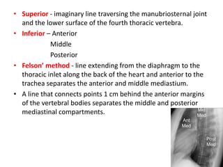
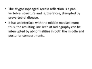
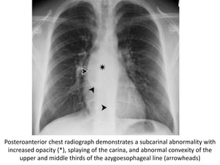
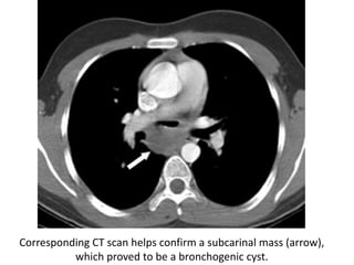
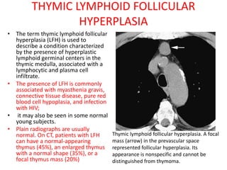
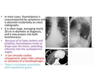
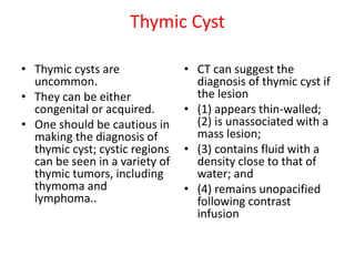
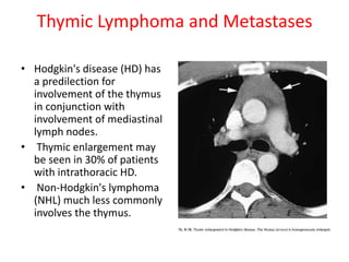
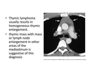
The document describes the anatomy and imaging features of the mediastinum and mediastinal masses. It defines the mediastinum and its subdivisions. Key points include:
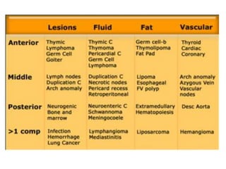
- The mediastinum is divided into anterior, middle, and posterior compartments
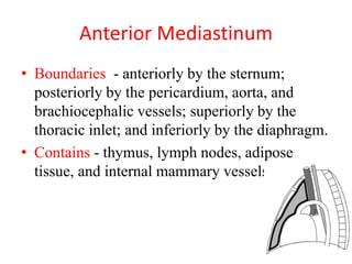
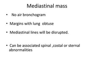
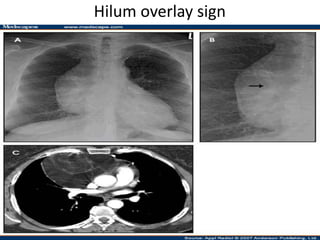
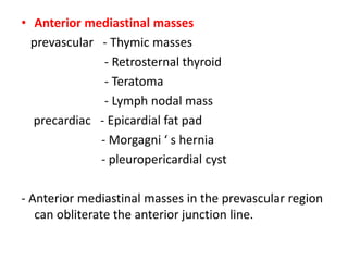
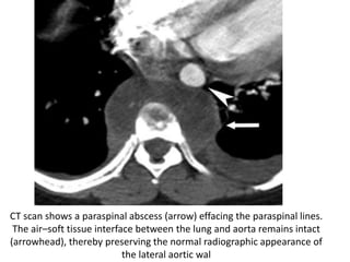
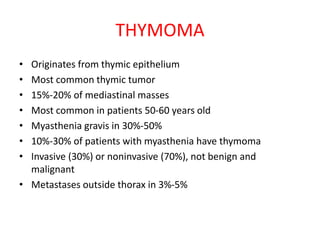
- Common anterior mediastinal masses include thymomas and lymphadenopathy which can displace mediastinal lines
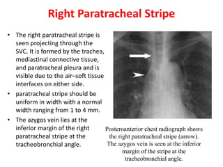
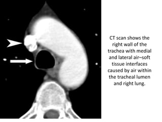
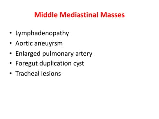
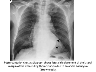
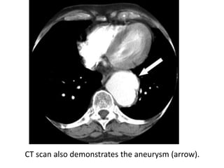
- Middle mediastinal masses include enlarged lymph nodes and vascular structures
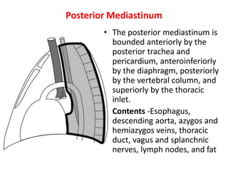
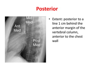
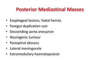
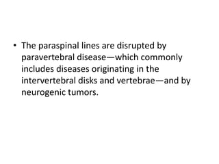
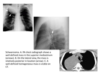
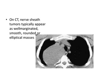
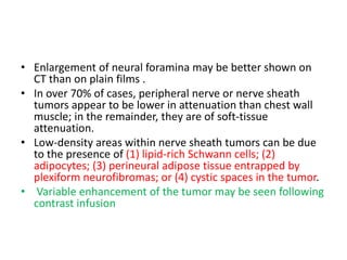
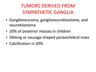
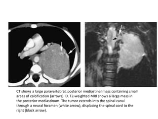
- Posterior mediastinal masses involve the esophagus and include neurogenic tumors
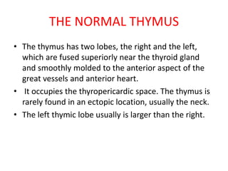
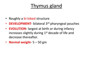
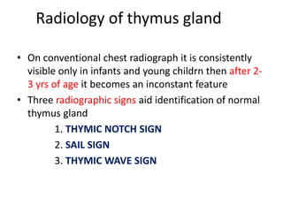
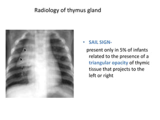
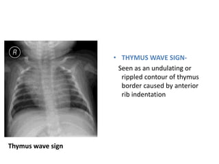
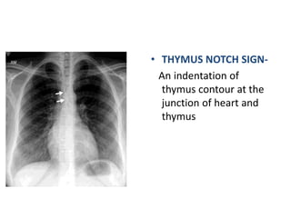
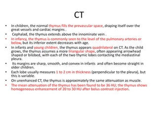
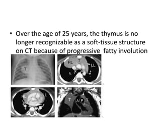
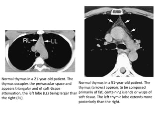
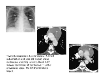
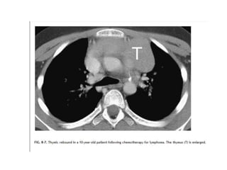
- Normal thymus anatomy and imaging signs are described as well as thymic hyperplasia and rebound
Radioanatomy of mediastinum and approach to mediastinal massesAkankshaMalviya3
The document discusses the anatomy, divisions, and contents of the mediastinum. It describes approaches to evaluating mediastinal masses through investigations such as chest x-rays, CT, MRI, and biopsy. Differential diagnoses are provided for masses in various mediastinal compartments, including the anterior mediastinum where the five most common masses are thymoma, thyroid masses, lymphoma, teratomas, and aortic aneurysms. Evaluation of mediastinal masses involves determining the location and narrowing the differential diagnosis through imaging studies and biopsy.
Lecture 4-5. Radiological semiotics of lung and mediastinum pathology..pptssuser61c85b
The document discusses general radiological signs used to analyze lung and mediastinum pathology on x-rays. It outlines 9 basic x-ray syndromes of lung pathology including total opacity, limited opacity, round opacity, ring-like shadows, nodules, diffuse nodular dissemination, changes to pulmonary markings, changes to lung hilum, and extensive lucency. Differential diagnoses and distinguishing characteristics are provided for each syndrome. CT and MRI are also briefly discussed as advanced imaging techniques that provide additional diagnostic information compared to basic x-rays.
This document discusses mediastinal masses, beginning with an overview of the mediastinum and its divisions. It then focuses on anterior mediastinal masses, describing various pathologies that can occur including thymoma, thymolipoma, thymic hyperplasia, and thymic cysts. For each pathology, it discusses incidence, associations, pathology, and radiographic features visible on plain radiographs and CT scans. Key signs on imaging include mass location and characteristics, presence of cystic or fatty components, calcification, and effects on surrounding structures.
The document discusses the anatomy and divisions of the mediastinum. It is divided into superior and inferior mediastinum by an imaginary line passing through the sternal angle anteriorly and lower border of the 4th thoracic vertebra posteriorly. The inferior mediastinum is further divided into anterior, middle and posterior mediastinum. Each compartment contains different structures like blood vessels, nerves and lymph nodes. Common tumors in different mediastinal compartments are also mentioned. Radiological anatomy and signs to locate mediastinal masses are described along with epidemiology of mediastinal tumors.
Presentation1.pptx radiological imaging of mediastinal masses .Abdellah Nazeer
This document discusses common mediastinal masses categorized by their location in the mediastinum - anterior, middle, or posterior. Anterior masses typically displace the anterior junctional line and obliterate the cardiophrenic angle. Common anterior masses include retro-sternal goiter, enlarged lymph nodes, thymoma, and germ cell tumors. Middle mediastinal masses widen the para-tracheal stripes and displace the azygo-esophageal recess on the right side. Lymph node enlargement, aortic arch aneurysm, enlarged pulmonary artery, and bronchogenic cysts are listed as common middle masses. Posterior mediastinal masses include neurogenic tumors, descending thoracic aortic aneurysms,
This document summarizes the anatomy and radiographic appearance of structures in the mediastinum. It describes the boundaries and contents of the anterior, middle, and posterior mediastinum. Key structures discussed include the anterior and posterior junction lines, right paratracheal stripe, azygoesophageal recess, and paraspinal lines. Common masses and abnormalities that can involve each mediastinal compartment are also reviewed.
This presentation is almost a complete Pictoral view of Radiograph chest.
This presentation will help radiologist in daily reporting.
This presentation will help physicians, surgeons, anesthetist and almost all medical professionals in diagnosing commonly presenting cardiac diseases.
This will also help all in preparaing TOACS examination.
This document discusses imaging of pathologies of the aortic vessels. It begins with the normal anatomy of the thoracic and abdominal aorta, including branches. It then discusses various pathologies such as aneurysms, dissections, and intramural hematomas. Imaging features of these conditions on various modalities such as CT, MRI, and angiography are provided. Complications related to the pathologies and classifications are also summarized.
Chest trauma can involve injuries to multiple structures in the chest cavity. Common injuries include pneumothorax, hemothorax, rib fractures, lung contusions and lacerations. On chest x-ray, a pneumothorax appears as a thin white line along the edge of the lung with no lung markings extending past it. A tension pneumothorax causes mediastinal shift away from the affected side. CT is useful for evaluating lung injuries like contusions, which appear as non-segmental areas of opacity, and lacerations, which may form pneumatoceles. Proper diagnosis requires understanding the mechanisms and radiographic appearances of various chest trauma injuries.
This document discusses the anatomy and radiological imaging of mediastinal masses. It begins with an introduction to the mediastinum and its boundaries. It then describes the different divisions of the mediastinum and contents of each region. Specific anatomical structures discussed include the anterior junction line, right paratracheal stripe, azygoesophageal recess, and paraspinal lines. Common masses that can occur in each mediastinal compartment are mentioned. Radiographic findings that suggest the location of a mediastinal mass are also described.
The document discusses mediastinal masses. It begins by defining the mediastinum and its boundaries. It then discusses approaches to evaluating mediastinal masses including distinguishing their location based on chest x-ray findings. Common masses are then reviewed for each compartment - anterior, middle, and posterior. Anterior masses include thymoma, teratoma, thyroid goiter, and lymphoma. Middle masses include adenopathy, primary neoplasms, aneurysms, and abnormalities. Posterior masses are often neural tumors, but can also include vertebral abscesses or vascular structures. Example cases are provided to demonstrate key radiographic findings.
Imaging in mediastinal masses by Dr. Milan SilwalMilan Silwal
This document provides an overview of imaging methods used to evaluate mediastinal pathologies. It discusses the relevant anatomy of the mediastinum and its divisions. Common imaging modalities like CT, MRI, ultrasound, and PET are described for assessing mediastinal masses and diffuse diseases. The document outlines an approach to characterizing mediastinal abnormalities by first localizing the mass within the mediastinum, then characterizing it with CT or MRI to determine benign vs malignant nature. Anterior, middle and posterior compartment masses have distinguishing features on imaging.
Purpose of this presentation is to educate non radiologist about basic CT anatomy of abdominal viscera and basic interpretation of very common diseases
Radiological imaging of mediastinal massesPankaj Kaira
1. CT is the most important tool for evaluating mediastinal masses and characterizing their nature and extent.
2. Thymomas are the most common primary mediastinal neoplasm, typically occurring in patients over 40 and appearing on CT as well-defined solid masses in the anterior mediastinum that can demonstrate calcification.
3. CT is useful for staging thymomas and identifying features like invasion of surrounding tissues or distant metastases that indicate more advanced disease.
1. The document describes the five deep neck spaces: anterior visceral space, carotid space, retropharyngeal space, posterior cervical space, and perivertebral space.
2. Each space is defined by its anatomical boundaries and contents, which can include viscera, blood vessels, nerves, lymph nodes, and pathologies such as tumors, infections, cysts.
3. CT and MRI images are presented to illustrate normal anatomy as well as various diseases found within the deep neck spaces, such as abscesses, aneurysms, lymphadenopathy, tumors.
This document discusses several congenital lung lesions and neonatal chest issues seen in infants. It provides descriptions of cystic adenomatoid malformation, pulmonary sequestration, bronchogenic cyst, congenital lobar emphysema, congenital diaphragmatic hernia, and bronchial atresia. For each condition, it summarizes the location, radiographic features on imaging such as x-ray and CT, and pathological findings. It also briefly discusses other neonatal issues like surfactant deficient disease and meconium aspiration syndrome.
This document provides guidelines for performing and interpreting a normal chest x-ray. It describes recommended projections including PA, AP, lateral, and oblique views. Proper positioning, centering, penetration, and rotation are discussed. Key anatomical structures are defined, such as the mediastinum, heart, lungs, fissures, hila, and diaphragm. Common variations and pathological findings are outlined. The document aims to serve as a reference for radiologists in evaluating and diagnosing chest x-ray images.
This document provides an overview of CT chest imaging, including the different types of CT chest scans, chest anatomy visualized on CT, and common abnormalities seen on CT chest exams. It discusses standard CT chest, HRCT, low dose CT, CT angiography, and combined PET/CT scans. It details the mediastinal compartments and lung segments seen on CT. It also provides examples of abnormalities such as pulmonary nodules/masses, pulmonary embolism, interstitial lung disease patterns, emphysema, atelectasis, pneumothorax, pleural effusions, and cardiomegaly. Virtual bronchoscopy and CT-guided biopsy procedures are also summarized.
The document discusses various airway diseases seen on imaging. It covers benign and malignant tracheal tumors including papilloma, hamartoma, lipoma and squamous cell carcinoma. It also discusses chronic bronchial diseases such as COPD, emphysema, asthma and bronchiectasis. Non-neoplastic tracheal diseases described include saber sheath trachea, tracheopathia osteoplastica and tracheobronchomalacia. The document provides detailed information on the pathology and radiographic features of many common and rare airway diseases.
X RAY CHEST IN EVALUATION OF MEDIASTINAL MASSESMrudang Raval
The document discusses several anatomical lines in the mediastinum that divide it theoretically rather than physically. It describes the locations and normal appearances of the anterior junction line, posterior junction line, azygoesophageal line, para-aortic line, and paraspinal lines. Key points include that the anterior junction line is always concave towards the lung, while the posterior junction line extends above the sternoclavicular notch. The azygoesophageal line forms an inverted S curve behind the esophagus. The document also notes how locations of masses and abnormalities can be identified relative to these anatomical lines and structures.
Radiographic Presentation of Congenital Heart DiseaseTarique Ajij
1. The document discusses the radiographic presentation of various congenital heart diseases including atrial septal defects, ventricular septal defects, patent ductus arteriosus, atrioventricular septal defects, pulmonic stenosis, aortic stenosis, coarctation of the aorta, tetralogy of Fallot, Ebstein's anomaly, transposition of the great arteries, truncus arteriosus, and total anomalous pulmonary venous connection.
2. Key findings on chest x-rays are described such as enlargement of specific heart chambers, changes in pulmonary vascularity, positioning of the great vessels, and rib notching.
3. Diagnosis is made through precordial examination, echocard
Radiological imaging of pleural diseases Pankaj Kaira
The document discusses the anatomy, imaging, and common diseases of the pleura. It begins by describing the normal anatomy of the pleural layers and thickness. Common pleural diseases are then reviewed, including pleural effusions, pneumothorax, hemothorax, and empyema. Imaging findings on chest x-ray, ultrasound, CT, and MRI are provided for diagnosing and characterizing various pleural conditions. Key signs that help differentiate pleural, pulmonary, and extra-pleural masses are also outlined.
This document provides information about cholangiocarcinoma, a malignant tumor arising from the biliary tree. It discusses the incidence, clinical presentation, locations, growth patterns, staging, and radiographic features. Cholangiocarcinoma is usually seen in the elderly and presents with painless jaundice. It can be located in the hilar region or peripherally. On imaging, it may appear as a mass, infiltrate along bile ducts, or have an intraductal growth pattern. Staging uses the Bismuth-Corlette classification. Key radiographic findings include dilated intrahepatic ducts, hilar lesions causing central obstruction without a clear mass, and encasement of portal veins
This document provides guidance on interpreting a normal chest x-ray. It outlines the key factors to consider, including orientation, inspiration, penetration, and rotation. It describes the normal radiographic anatomy, including the lungs, heart, diaphragm, mediastinum, and other structures. A proper technique is important to avoid artifacts that could be mistaken for pathology. The document emphasizes performing the examination with good inspiration in the PA orientation for optimal visualization of structures.
This document discusses imaging of the chest in trauma patients. It describes the use of chest radiographs and CT scans to evaluate for fractures, pneumothorax, hemothorax, pulmonary contusions, diaphragm injuries, and vascular injuries. Specific signs on imaging that indicate various injuries are outlined. CT is highlighted as more sensitive than chest x-ray for detecting many injuries like small pneumothoraces or diaphragm injuries. The document provides an overview of diagnostic imaging of common chest trauma injuries.
Presentation1.pptx, radiological imaging of large bowel diseasesAbdellah Nazeer
This document discusses congenital anomalies and diseases of the large bowel. It begins by describing congenital abnormalities that can cause obstruction in neonates and the importance of radiological imaging to diagnose the location and cause. It then discusses different types of intestinal obstructions and how radiography and contrast enema exams are used to further evaluate obstructions and make a specific diagnosis. Examples of different congenital anomalies and diseases seen on imaging are also presented, including colonic atresia, meconium ileus, Hirschsprung disease, and colon cancer.
This document discusses the diagnostic workup for mediastinal tumors. It begins by describing the different types of tumors that can occur in the anterior, middle, and posterior mediastinum. Signs and symptoms are non-specific but may include cough, chest pain, and weight loss. Chest x-ray is often the initial test, while CT scan provides more detail on location and tissue characteristics. MRI and nuclear imaging can further characterize masses. Tissue sampling methods include needle biopsy, mediastinoscopy, and thoracoscopy. Surgical procedures like mediastinotomy or thoracotomy may be needed in some cases. Laboratory tests help identify certain tumor types. A multidisciplinary approach is typically needed for diagnosis and treatment planning of
This document discusses the gloved finger sign and cervicothoracic sign in radiology. It begins by defining the gloved finger sign as branching finger-like opacities seen on imaging that represent dilated bronchi filled with mucus radiating from the hila. This sign is commonly seen in allergic bronchopulmonary aspergillosis. It then defines the cervicothoracic sign, where a mediastinal mass that projects above the clavicles is retrotracheal and posterior, while one that fades at the clavicles is anterior. Determining the location of mediastinal masses using this sign is discussed through examples.
Radiological mapping of mediastinum.pptxrambhoopal1
1. The mediastinum is divided into superior, anterior, middle and posterior compartments by planes. The superior mediastinum is above the plane from the sternal angle to T4, while the inferior mediastinum is below the plane and further divided.
2. Interface lines and stripes appear on chest radiographs where air in the lungs abuts mediastinal structures. Key lines include the anterior and posterior junction lines, azygoesophageal line, and right/left paratracheal stripes.
3. Anterior mediastinal masses can obliterate the anterior junction line while posterior masses can obliterate posterior lines. Location and relationship to structures provides clues to the nature of mediast
The document describes the anatomy and divisions of the mediastinum. It notes that the mediastinum is the central partition in the chest that contains structures like the heart, great vessels, trachea and esophagus. It divides the mediastinum into superior, anterior, middle and posterior compartments. The anterior mediastinum contains the thymus gland while the middle mediastinum contains the heart and pericardium. Various radiographic signs of mediastinal structures are also described.
The document discusses radiographic imaging techniques for evaluating mediastinal masses. It begins by explaining that computed tomography (CT) is the most important tool for characterizing mediastinal abnormalities based on tissue attenuation. Magnetic resonance imaging (MRI) also allows excellent evaluation of mediastinal tumors due to its soft tissue contrast. The document then describes several radiographic signs and landmarks that can help locate a mediastinal mass to a specific compartment. These include the hilum overlay sign, effects on nearby structures, and relationships to lines like the paratracheal stripe or azygoesophageal recess. Thymoma is discussed as a common anterior mediastinal tumor that appears on CT as a homogeneous soft tissue mass, sometimes with calc
This document provides an overview of how to interpret a normal chest x-ray by describing the anatomical structures that should be visualized and what is considered normal for each structure. It discusses evaluating the bony thorax, soft tissues, mediastinum, lungs and diaphragm and how they change with age from infancy to childhood to older age. Key anatomical structures and their expected appearance on a normal chest x-ray are defined.
This document provides an overview of the radiological anatomy of the thorax as seen on various imaging modalities like plain X-ray, CT, MRI, ultrasound and nuclear medicine. It describes the lungs, mediastinum, bony cage and soft tissues of the thorax. It also discusses the anatomy of specific structures like the heart, lungs, bronchial tree, pulmonary arteries and veins. Standard views used for chest X-rays and important lines and stripes seen are explained. CT windows and segmental anatomy of the lungs are outlined as well.
The middle mediastinum contains structures like the heart, great vessels, trachea and lymph nodes. Common masses include lymphadenopathy, bronchogenic cysts, duplication cysts and aneurysms. Aortic aneurysms appear as round masses that may erode bone or involve nerves. Bronchogenic cysts are usually round and contain fluid. Duplication cysts may contain gastric mucosa. The document describes anatomy of the middle mediastinum and structures within it, common masses found and how they appear on imaging. It also discusses some pitfalls in interpretation like variants involving great vessels.
The document discusses various anatomical structures and pathologies related to the pleura and lungs. It describes the major interlobar fissures that separate the lobes of the lungs, including the major oblique fissure and minor horizontal fissure. It also discusses accessory fissures and the azygos fissure. Common pleural abnormalities are described such as parietal and visceral pleural thickening. Normal anatomical structures like fat pads and muscles that can mimic pleural abnormalities on imaging are also reviewed. Finally, the document discusses the appearance of pleural effusions on chest x-rays and CT scans, including how effusions present differently in upright versus supine patients.
This document discusses various radiology methods used to examine the respiratory system including x-rays, CT scans, fluoroscopy, tomography, angiography, ultrasound, MRI, PET scans, and endoscopy. It provides details on positioning and normal anatomy seen on chest x-rays. It also describes signs seen on chest imaging such as the silhouette sign, air bronchogram sign, snowball sign, and signs of mass effect or volume loss that can help determine the location and characteristics of abnormalities.
RADIOIMAGING IN MEDIASTINAL MASSES YN.pptxdypradio
The document provides information on radiographic imaging of mediastinal masses. It discusses the anatomy of the mediastinum and defines its compartments. Common masses are described based on their location in the anterior, middle or posterior mediastinum. Anterior masses include thymoma, thymic cyst, germ cell tumors such as teratomas and seminomas. Middle mediastinal masses include lymph nodes, cysts and vascular structures. Posterior masses are often neurogenic tumors like schwannomas. Specific radiographic signs and features of different masses are outlined to aid in diagnosis.
Thoracic anatomy on various imaging modalitiesDev Lakhera
1. The document describes the normal thoracic anatomy as seen on various imaging modalities like chest radiography and computed tomography. It details the anatomy of structures like the trachea, bronchi, lungs, blood vessels, mediastinum and heart.
2. Key anatomical structures are defined, such as the lobes and fissures of the lungs, segments and zones. Common variations are also mentioned.
3. Imaging features of mediastinal lymph nodes, bones and soft tissues are provided. Different "windows" used for optimal visualization of soft tissue and lung structures on CT are explained.
4. Axial, sagittal and coronal reformats of CT images demonstrate the 3D anatomy of medi
Cross sectional anatomy of chest by Dr. Milan Silwal, Resident, NAMS, Kathman...Milan Silwal
The document provides information on the cross sectional anatomy of the chest, including the boundaries and divisions of the thorax and mediastinum. It describes the contents and boundaries of the superior, anterior, middle, and posterior mediastinum. It also discusses the lungs, bronchopulmonary segments, and six representative chest CT scan levels that are used to interpret mediastinal anatomy. Finally, it presents six clinical cases pertaining to conditions that may appear on chest imaging.
Contouring of CTV Lymph nodes in NSCLC (Part III)Mohammed Fathy
This document provides definitions for the borders of different lymph node stations in the lung according to various mapping guidelines, including the International Association for the Study of Lung Cancer (IASLC), Japan Esophageal Society (JES), and the Japan Lung Cancer Society (JLCS) and Japanese Society for Radiation Oncology (JASTRO) Consensus-based CT atlas. It describes the anatomical landmarks used to delineate each lymph node station in detail.
This document provides an overview of the radiological anatomy of the tracheobronchial tree and techniques for demonstrating it. It begins with discussing the gross anatomy of the trachea and bronchi, their relations, blood supply and divisions. It then covers their appearance on various imaging modalities like plain radiography, CT, MRI, radionuclide scans and ultrasound. The techniques used for obtaining images with these different modalities are also described.
Rad Seminar CHEST IMAGING By Dr Siraj.pptxImanuIliyas
The document provides an overview of chest radiographic anatomy and an approach to interpreting chest x-rays (CXRs). It discusses the normal anatomy seen on CXRs including the lungs, heart, blood vessels, trachea, bronchi, ribs and diaphragm. CT anatomy is also reviewed. The presentation outlines a systematic approach to interpreting CXRs which involves identifying structures, assessing technical adequacy, and interpreting findings based on knowledge of normal radiographic anatomy.
The mediastinum is the central compartment of the thoracic cavity that contains the heart, lungs, esophagus, and other structures. It is divided into superior, anterior, middle, and posterior compartments. The superior mediastinum contains the thymus gland, major blood vessels like the aorta and superior vena cava, nerves like the vagus and phrenic nerves, and the esophagus. The middle mediastinum contains the heart surrounded by the pericardium. The anterior and posterior mediastinum contain the esophagus and major blood vessels.
The subclavian artery becomes the axillary artery after passing through the thoracic outlet. The thoracic outlet connects the neck to the thorax and contains many structures including the subclavian vessels and brachial plexus. Thoracic outlet syndrome occurs when these structures are compressed as they pass through the thoracic outlet, most commonly due to scalene muscle abnormalities. It can cause neurogenic, venous, or arterial symptoms in the upper limb that are exacerbated by arm movements above the head. Treatment depends on the type of thoracic outlet syndrome but may include surgery or stenting.
The document describes 10 mediastinal lines seen on chest radiographs:
1. The anterior junction line forms where the lungs meet in the anterior mediastinum.
2. The posterior junction line lies above the anterior line and is formed by the lungs meeting behind the esophagus.
3. The paraortic line follows the descending aorta on its left side when the left lower lobe is aerated.
4. The right paratracheal line outlines the right side of the trachea and may indicate adjacent disease when thickened.
The skin is the largest organ and its health plays a vital role among the other sense organs. The skin concerns like acne breakout, psoriasis, or anything similar along the lines, finding a qualified and experienced dermatologist becomes paramount.
Adhd Medication Shortage Uk - trinexpharmacy.comreignlana06
The UK is currently facing a Adhd Medication Shortage Uk, which has left many patients and their families grappling with uncertainty and frustration. ADHD, or Attention Deficit Hyperactivity Disorder, is a chronic condition that requires consistent medication to manage effectively. This shortage has highlighted the critical role these medications play in the daily lives of those affected by ADHD. Contact : +1 (747) 209 – 3649 E-mail : sales@trinexpharmacy.com
These lecture slides, by Dr Sidra Arshad, offer a simplified look into the mechanisms involved in the regulation of respiration:
Learning objectives:
1. Describe the organisation of respiratory center
2. Describe the nervous control of inspiration and respiratory rhythm
3. Describe the functions of the dorsal and respiratory groups of neurons
4. Describe the influences of the Pneumotaxic and Apneustic centers
5. Explain the role of Hering-Breur inflation reflex in regulation of inspiration
6. Explain the role of central chemoreceptors in regulation of respiration
7. Explain the role of peripheral chemoreceptors in regulation of respiration
8. Explain the regulation of respiration during exercise
9. Integrate the respiratory regulatory mechanisms
10. Describe the Cheyne-Stokes breathing
Study Resources:
1. Chapter 42, Guyton and Hall Textbook of Medical Physiology, 14th edition
2. Chapter 36, Ganong’s Review of Medical Physiology, 26th edition
3. Chapter 13, Human Physiology by Lauralee Sherwood, 9th edition
Kosmoderma Academy, a leading institution in the field of dermatology and aesthetics, offers comprehensive courses in cosmetology and trichology. Our specialized courses on PRP (Hair), DR+Growth Factor, GFC, and Qr678 are designed to equip practitioners with advanced skills and knowledge to excel in hair restoration and growth treatments.
- Video recording of this lecture in English language: https://youtu.be/Pt1nA32sdHQ
- Video recording of this lecture in Arabic language: https://youtu.be/uFdc9F0rlP0
- Link to download the book free: https://nephrotube.blogspot.com/p/nephrotube-nephrology-books.html
- Link to NephroTube website: www.NephroTube.com
- Link to NephroTube social media accounts: https://nephrotube.blogspot.com/p/join-nephrotube-on-social-media.html
Osteoporosis - Definition , Evaluation and Management .pdfJim Jacob Roy
Osteoporosis is an increasing cause of morbidity among the elderly.
In this document , a brief outline of osteoporosis is given , including the risk factors of osteoporosis fractures , the indications for testing bone mineral density and the management of osteoporosis
Histololgy of Female Reproductive System.pptxAyeshaZaid1
Dive into an in-depth exploration of the histological structure of female reproductive system with this comprehensive lecture. Presented by Dr. Ayesha Irfan, Assistant Professor of Anatomy, this presentation covers the Gross anatomy and functional histology of the female reproductive organs. Ideal for students, educators, and anyone interested in medical science, this lecture provides clear explanations, detailed diagrams, and valuable insights into female reproductive system. Enhance your knowledge and understanding of this essential aspect of human biology.
Travel Clinic Cardiff: Health Advice for International TravelersNX Healthcare
Travel Clinic Cardiff offers comprehensive travel health services, including vaccinations, travel advice, and preventive care for international travelers. Our expert team ensures you are well-prepared and protected for your journey, providing personalized consultations tailored to your destination. Conveniently located in Cardiff, we help you travel with confidence and peace of mind. Visit us: www.nxhealthcare.co.uk
5-hydroxytryptamine or 5-HT or Serotonin is a neurotransmitter that serves a range of roles in the human body. It is sometimes referred to as the happy chemical since it promotes overall well-being and happiness.
It is mostly found in the brain, intestines, and blood platelets.
5-HT is utilised to transport messages between nerve cells, is known to be involved in smooth muscle contraction, and adds to overall well-being and pleasure, among other benefits. 5-HT regulates the body's sleep-wake cycles and internal clock by acting as a precursor to melatonin.
It is hypothesised to regulate hunger, emotions, motor, cognitive, and autonomic processes.
2. • The mediastinum is defined as the tissue
compartment located between the two lungs,
posterior to the sternum, anterior to the
vertebral column, and extending from the
thoracic inlet to the diaphragm.
3. • Superior - imaginary line traversing the manubriosternal joint
and the lower surface of the fourth thoracic vertebra.
• Inferior – Anterior
Middle
Posterior
• Felson’ method - line extending from the diaphragm to the
thoracic inlet along the back of the heart and anterior to the
trachea separates the anterior and middle mediastium.
• A line that connects points 1 cm behind the anterior margins
of the vertebral bodies separates the middle and posterior
mediastinal compartments.
4. Anterior Mediastinum
• Boundaries - anteriorly by the sternum;
posteriorly by the pericardium, aorta, and
brachiocephalic vessels; superiorly by the
thoracic inlet; and inferiorly by the diaphragm.
• Contains - thymus, lymph nodes, adipose
tissue, and internal mammary vessels
5.
6. Anterior Junction Line
• The line is formed by the anterior apposition of the
lungs and consists of the four layers of pleura
separating the lungs behind the upper two-thirds of
the sternum.
• The line runs obliquely from upper right to lower left
and does not extend above the manubriosternal
junction.
• Contains variable amount of fat.
Posteroanterior chest radiograph
demonstrates the anterior junction line
CT scan shows the four layers of pleura that
constitute the anterior junction line
8. Mediastinal mass
• No air bronchogram
• Margins with lung obtuse
• Mediastinal lines will be disrupted.
• Can be associated spinal ,costal or sternal
abnormalities
10. ISilhouette sign
• If an intrathoracic opacity is situated in
anatomic contact with a border of heart or
aorta ,will obscure that border.
A radioopacity causes obliteration of rt border of
heart , is anterior in location ,Anterior
mediastinum.
If it overlaps but does not obliterates , it lies in
posterior or middle mediastinum.
12. Hilum overlay sign
• Differentiates cardic enlargement from
mediastinal mass
• In mediastinal mass if Hilar vessels are seen
through the mass indicates that the mass
does not arise from hilum
• For accuracy the film should be true frontal
,slight obliquity may project normal
pulmonary artery medialy
14. Cervicothoracic sign
Well defined mass seen above the clavicle is
always situated in posterior compartment ,the
anterior compartment mass being in contact
with soft tissue rather than aerated lung is ill
defined
18. • Anterior mediastinal masses
prevascular - Thymic masses
- Retrosternal thyroid
- Teratoma
- Lymph nodal mass
precardiac - Epicardial fat pad
- Morgagni ‘ s hernia
- pleuropericardial cyst
- Anterior mediastinal masses in the prevascular region
can obliterate the anterior junction line.
19. Middle Mediastinum
• Boundaries - anteriorly by
the pericardium, posteriorly
by the pericardium and
posterior tracheal wall,
superiorly by the thoracic
inlet, and inferiorly by the
diaphragm.
• Contains - heart and
pericardium; the ascending
and transverse aorta; SVC
and IVC; the brachiocephalic
vessels; the pulmonary
vessels; the trachea and
main bronchi; lymph nodes;
and the phrenic, vagus, and
left recurrent laryngeal
nerves.
20. Right Paratracheal Stripe
• The right paratracheal stripe is
seen projecting through the
SVC. It is formed by the trachea,
mediastinal connective tissue,
and paratracheal pleura and is
visible due to the air–soft tissue
interfaces on either side.
• paratracheal stripe should be
uniform in width with a normal
width ranging from 1 to 4 mm.
• The azygos vein lies at the
inferior margin of the right
paratracheal stripe at the
tracheobronchial angle.
Posteroanterior chest radiograph shows
the right paratracheal stripe (arrow).
The azygos vein is seen at the inferior
margin of the stripe at the
tracheobronchial angle.
21. CT scan shows the
right wall of the
trachea with medial
and lateral air–soft
tissue interfaces
caused by air within
the tracheal lumen
and right lung.
22. The right paratracheal stripe can be widened due to
abnormality of any of its components, from the
tracheal mucosa to the pleural space.
24. Posterior Mediastinum
• The posterior mediastinum is
bounded anteriorly by the
posterior trachea and
pericardium, anteroinferiorly
by the diaphragm, posteriorly
by the vertebral column, and
superiorly by the thoracic
inlet.
• Contents -Esophagus,
descending aorta, azygos and
hemiazygos veins, thoracic
duct, vagus and splanchnic
nerves, lymph nodes, and fat
25. Posterior
• Extent: posterior to a
line 1 cm behind the
anterior margin of the
vertebral column,
anterior to the chest
wall
27. Azygoesophageal Recess
• The azygoesophageal recess is the interface between the right
lung and the mediastinal reflection, with the esophagus lying
anteriorly and the azygos vein posteriorly within the
mediastinum.
• On X-ray,it appears as a line –
- in its upper 1/3rd , it deviates to the right at the level of the
carina to accommodate the azygos vein arching forward.
- middle 1/3rd , the line has a variable appearance: It is usually
straight.
- lower 1/3rd , usually straight. ( air in esophagus)
29. CT scan shows the
azygoesophageal recess
(white arrow) formed
by the esophagus
anteriorly (black arrow)
and the azygos vein
posteriorly
(arrowhead).
30. • The azygoesophageal recess reflection is a pre-
vertebral structure and is, therefore, disrupted by
prevertebral disease.
• It has an interface with the middle mediastinum;
thus, the resulting line seen at radiography can be
interrupted by abnormalities in both the middle and
posterior compartments.
31. Posteroanterior chest radiograph demonstrates a subcarinal abnormality with
increased opacity (*), splaying of the carina, and abnormal convexity of the
upper and middle thirds of the azygoesophageal line (arrowheads)
32. Corresponding CT scan helps confirm a subcarinal mass (arrow),
which proved to be a bronchogenic cyst.
33. Posterior Junction Line
• Seen above the level of the azygos vein and aorta
and that is formed by the apposition of the lungs
posterior to the esophagus.
• usually extend from third to fifth thoracic vertebrae.
• posterior junction line can be seen above the
suprasternal notch and lies almost vertical, whereas
the anterior junction line deviates to the left.
34. Collimated posteroanterior chest radiograph shows the posterior
junction line (arrow) projecting through the tracheal air column.
35. CT scan shows the posterior junction line (arrow), which is formed by
the interface between the lungs posterior to the mediastinum and
consists of four pleural layers.
36. Posteroanterior chest radiograph shows a mass (arrow) obliterating
the posterior junction line. Note that the mass extends above the
level of the clavicle and has a well-demarcated outline due to the
interface with adjacent lung (arrowhead).
37. CT scan helps confirm the posterior location of the mass (arrow),
which proved to be a bronchogenic cyst.
38. Paraspinal Lines
• The paraspinal lines are created by the interface
between lung and the pleural reflections over the
vertebral bodies.
• The left paraspinal line is much more commonly seen
than the right. The descending aorta holds the pleural
reflection off the vertebral body, allowing the lung–
soft tissue interface to be more tangential to the x-ray
beam.
• The left paraspinal line runs parallel to the lateral
margin of the vertebral bodies and can lie anywhere
medial to the lateral wall of the descending aorta
39. On a collimated
posteroanterior
chest radiograph,
the left
paraspinal line
(arrow) is seen
separate and
distinct from the
vertebral body
(black
arrowhead) and
the descending
thoracic aorta
40. CT scan shows the left paraspinal line. The descending aorta holds the pleural
reflection (arrow) away from the vertebral body, which allows the lung–soft
tissue interface to be more tangential to the x-ray beam and therefore
visualized as a line
41. • The paraspinal lines are disrupted by
paravertebral disease—which commonly
includes diseases originating in the
intervertebral disks and vertebrae—and by
neurogenic tumors.
42. Posteroanterior chest radiograph shows a mass (arrow) effacing the left
paraspinal line. The lateral wall of the descending aorta is seen as a separate
entity (arrowhead).
43. CT scan shows a paraspinal abscess (arrow) effacing the paraspinal lines.
The air–soft tissue interface between the lung and aorta remains intact
(arrowhead), thereby preserving the normal radiographic appearance of
the lateral aortic wal
44. Posteroanterior chest radiograph shows lateral displacement of the lateral
margin of the descending thoracic aorta due to an aortic aneurysm
(arrowheads).
46. THE NORMAL THYMUS
• The thymus has two lobes, the right and the left,
which are fused superiorly near the thyroid gland
and smoothly molded to the anterior aspect of the
great vessels and anterior heart.
• It occupies the thyropericardic space. The thymus is
rarely found in an ectopic location, usually the neck.
• The left thymic lobe usually is larger than the right.
47. Thymus gland
• Roughly a bi-lobed structure
• DEVELOPMENT- bilateral 3rd pharyngeal pouches
• EVOLUTION- largest at birth or during infancy
increases slightly during 1st decade of life and
decrease thereafter.
• Normal weight- 5 – 50 gm
48. Radiology of thymus gland
• On conventional chest radiograph it is consistently
visible only in infants and young childrn then after 2-
3 yrs of age it becomes an inconstant feature
• Three radiographic signs aid identification of normal
thymus gland
1. THYMIC NOTCH SIGN
2. SAIL SIGN
3. THYMIC WAVE SIGN
49. Radiology of thymus gland
Sail sign
• SAIL SIGN-
present only in 5% of infants
related to the presence of a
triangular opacity of thymic
tissue that projects to the
left or right
50. Thymus wave sign
• THYMUS WAVE SIGN-
Seen as an undulating or
rippled contour of thymus
border caused by anterior
rib indentation
51. • THYMUS NOTCH SIGN-
An indentation of
thymus contour at the
junction of heart and
thymus
52. CT
• In children, the normal thymus fills the prevascular space, draping itself over the
great vessels and cardiac margins .
• Cephalad, the thymus extends above the innominate vein .
• In infancy, the thymus is commonly seen to the level of the pulmonary arteries or
below, but its inferior extent decreases with age.
• In infants and young children, the thymus appears quadrilateral on CT. As the child
grows, the thymus assumes a more triangular shape, often appearing arrowhead
shaped or bilobed, with each of the two thymic lobes contacting the mediastinal
pleura.
• Its margins are sharp, smooth, and convex in infants and often become straight in
older children.
• Each lobe usually measures 1 to 2 cm in thickness (perpendicular to the pleura), but
this is variable.
• On unenhanced CT, the thymus is approximately the same attenuation as muscle.
• The mean attenuation of the thymus has been found to be 36 HU; the thymus shows
homogeneous enhancement of 20 to 30 HU after bolus contrast injection.
53. • Over the age of 25 years, the thymus is no
longer recognizable as a soft-tissue structure
on CT because of progressive fatty involution
54. Normal thymus in a 21-year-old patient. The
thymus occupies the prevascular space and
appears triangular and of soft-tissue
attenuation, the left lobe (LL) being larger than
the right (RL).
Normal thymus in a 51-year-old patient. The
thymus (arrows) appears to be composed
primarily of fat, containing islands or wisps of
soft tissue. The left thymic lobe extends more
posteriorly than the right.
55. THYMIC LYMPHOID FOLLICULAR
HYPERPLASIA
• The term thymic lymphoid follicular
hyperplasia (LFH) is used to
describe a condition characterized
by the presence of hyperplastic
lymphoid germinal centers in the
thymic medulla, associated with a
lymphocytic and plasma cell
infiltrate.
• The presence of LFH is commonly
associated with myasthenia gravis,
connective tissue disease, pure red
blood cell hypoplasia, and infection
with HIV;
• it may also be seen in some normal
young subjects.
• Plain radiographs are usually
normal. On CT, patients with LFH
can have a normal-appearing
thymus (45%), an enlarged thymus
with a normal shape (35%), or a
focal thymus mass (20%)
Thymic lymphoid follicular hyperplasia. A focal
mass (arrow) in the prevascular space
represented follicular hyperplasia. Its
appearance is nonspecific and cannot be
distinguished from thymoma.
56. THYMIC HYPERPLASIA
• Thymic hyperplasia, as distinguished from LFH, is defined by an
increase in size of the thymus associated with an otherwise normal
gross and histologic appearance.
• It may be associated with hyperthyroidism (Graves' disease,
sarcoidosis, red blood cell aplasia, and other entities.
• It most commonly is seen in association with recovery from
chemotherapy, stress, or burns, a phenomenon known as thymic
rebound (described below).
• Chest radiographs in adults are usually normal.
• In children, thymic enlargement may be seen.
• CT demonstrates increased thickness of the thymic lobes but an
otherwise normal-appearing thymus
57. Thymic hyperplasia in Graves' disease. A. Chest
radiograph in a 40-year-old woman shows
mediastinal widening (arrows). B and C. CT
shows enlargement of the thymus (T) in the
prevascular space. The left thymic lobe is
largest
58. Thymic Rebound
• The thymus involutes during periods of stress (e.g.,
burns, chemotherapy) and may decrease significantly
in size
• the thymus will generally regrow to its original size
within several months of the stressful episode
• The presence of lymph node enlargement in
association with thymic enlargement should suggest
recurrent tumor, while isolated thymic enlargement
should suggest thymic rebound
59.
60. THYMOMA
• Originates from thymic epithelium
• Most common thymic tumor
• 15%-20% of mediastinal masses
• Most common in patients 50-60 years old
• Myasthenia gravis in 30%-50%
• 10%-30% of patients with myasthenia have thymoma
• Invasive (30%) or noninvasive (70%), not benign and
malignant
• Metastases outside thorax in 3%-5%
61. Plain Radiographs
• Most thymomas occur near the junction of the heart and great
vessels, although they may be seen superiorly to the level of the
clavicles and inferiorly to the level of the costophrenic angles.
• Thymomas typically appear sharply marginated, smooth or lobulated
in contour, and usually project to one side of the mediastinum.
• They usually range in size from 5 to 10 cm in diameter when visible on
radiographs, and may obscure the right or left heart border,
depending on their location and size.
• Dense calcification may be seen in the periphery of the mass or
throughout its substance.
62.
63. • On lateral radiographs,
thymoma often produces
a distinct opacity in the
inferior aspect of the
retrosternal clear space,
the relatively lucent
triangular region
posterior to the sternum
and anterior to the aortic
arch, main pulmonary
artery, and heart
64. CT
• In nearly all cases,
thymomas occur in the
prevascular space and
displace the great
vessels (aorta and its
branches, superior vena
cava and its branches,
and main pulmonary
artery) posteriorly
CT shows a cystic
(fluid attenuation)
mass in the right
cardiophrenic
angle. The mass
shows dense
calcification,
including
calcification of its
capsule. A
noninvasive cystic
thymoma was
found at surgery.
65. • Thymoma or other
thymic neoplasm may
be distinguished from
thymic hyperplasia if
enlargement of the
thymus is grossly
asymmetric, if the
thymus has a lobular
contour, or if a focal
rounded lesion is visible
Invasive thymoma with calcification in a
patient with myasthenia gravis. A. CT shows a
lobulated mass (T) in the prevascular space,
typical of thymoma. Medially, the mass invades
mediastinal fat (large arrow). A small pleural
nodule (small arrow) is also seen, resulting
from pleural invasion. Compression of the
superior vena cava is also present. B. At a
lower level, calcification is visible.However, follicular thymic hyperplasia can
result in a focal thymic mass up to 5 cm in
diameter
66. • Approximately 80% of
thymomas occur at the
base of the heart, as
shown on CT .
• They usually appear as
homogeneous soft-tissue
attenuation masses that
are sharply demarcated
and oval, round, or
lobulated and do not
conform to the normal
shape of the thymus
67. It is often difficult to distinguish invasive and noninvasive
thymoma on CT
• Findings highly suggestive
of invasion include
• pericardial thickening
contiguous with the
tumor, pleural thickening,
nodules, or effusion,
• encasement of
mediastinal structures,
• fat infiltration
• an irregular interface
between the tumor and
lung.
Invasive thymoma with calcification. CT shows
a lobulated thymoma with multiple
calcifications. The presence of pleural effusion
strongly suggests invasion
68. Invasive thymoma with pleural metastases. A.
PA chest radiograph shows a mass at the left
cardiophrenic angle (arrows). B. CT shows a
homogeneous rounded mass (T) representing
thymoma. C. At a lower level, CT shows focal
pleural thickening or nodules (arrows), typical
of pleural metastases. Pleural nodules are
usually unassociated with pleural effusion. This
finding indicates invasion.
69. Noninvasive thymoma. A. CT shows a homogeneous thymoma (T)
separated from the aorta (A) and pulmonary artery (P) by a layer of fat.
B. Oblique sagittal reconstruction shows separation of the thymoma (T)
from the ascending aorta (A).
70. THYMIC CARCINOMA
• Originates from thymic epithelium
• 20% of thymic epithelial tumors
• Average age 50
• Unlike thymoma, appears malignant histologically
• Invasion common, distant metastases in 50%-65%
• Myasthenia rare
• Appearance indistinguishable from thymoma
unless metastases are visible
71. • Thymic carcinoma usually results
in a mass 5 to 15 cm in diameter.
• A large mass with or without
areas of low attenuation is typical
.
• Calcification may be present .
• Thymic carcinoma cannot be
distinguished from thymoma on
CT unless enlarged lymph nodes
are visible in the mediastinum or
distant metastases are evident.
• Thymic carcinoma is less likely
than thymoma to result in pleural
implants.
72. THYMIC CARCINOID TUMOR
• Originates from neural
crest cells.
• Average age 45
• Usually malignant
• Cushing's syndrome in
25%-40%
• MEN in 20%
• Mimics thymoma
radiographically
• Dense enhancement
possibly seen on CT
73. • This tumor is more
aggressive than
thymoma, being
malignant in most cases,
and superior vena cava
obstruction is much more
common with thymic
carcinoid than with
thymoma.
• The 5-year survival rate is
about 65%. Metastases
may be visible.
74. Thymolipoma
• Consists of fat and
thymic tissue
• 5% of thymic tumors
• Benign
• Often young patients
• Usually asymptomatic
• Large droopy
mediastinal mass
containing fat and
strands of tissue
75. • In most cases, thymolipoma is
unaccompanied by symptoms and
is detected incidentally on chest
radiographs.
• It is often large, averaging nearly
20 cm in diameter at diagnosis,
and it may project into both
hemithoraces.
• Because of its fatty content and
pliability, thymolipoma tends to
drape over the heart, extending
inferiorly into the cardiophrenic
angles.
• It can simulate cardiac
enlargement, lower lobe collapse,
or elevation of a hemidiaphragm .
• There is no known association
with myasthenia gravis.
76. Thymic Cyst
• Thymic cysts are
uncommon.
• They can be either
congenital or acquired.
• One should be cautious in
making the diagnosis of
thymic cyst; cystic regions
can be seen in a variety of
thymic tumors, including
thymoma and
lymphoma..
• CT can suggest the
diagnosis of thymic cyst if
the lesion
• (1) appears thin-walled;
(2) is unassociated with a
mass lesion;
• (3) contains fluid with a
density close to that of
water; and
• (4) remains unopacified
following contrast
infusion
77. Thymic Lymphoma and Metastases
• Hodgkin's disease (HD) has
a predilection for
involvement of the thymus
in conjunction with
involvement of mediastinal
lymph nodes.
• Thymic enlargement may
be seen in 30% of patients
with intrathoracic HD.
• Non-Hodgkin's lymphoma
(NHL) much less commonly
involves the thymus.
78. • Thymic lymphoma
usually results in
homogeneous thymic
enlargement.
• thymic mass with mass
or lymph node
enlargement in other
areas of the
mediastinum is
suggestive of this
diagnosis
79. GERM CELL TUMORS
• Germ cell tumors account for about 10% of primary
mediastinal masses and arise from primitive germ cells
that have arrested their embryologic migration in the
mediastinum, frequently within the thymus.
• They are more common in the anterior mediastinum
• Germ cell tumors include benign and malignant
teratoma, seminoma, embryonal carcinoma,
endodermal sinus (yolk sac) tumor, choriocarcinoma,
and mixed cell types.
• Overall, more than 80% of germ cell tumors are benign
80. TERATOMA
• Contains elements of all germinal layers
• Mature teratomas (contain mature tissues)
Benign
60%-75% of mediastinal germ cell tumors
Occur in children and young adults
Dermoid cyst
• Immature teratomas (contain immature tissue)
Often have benign course in infants or young children
Often aggressive and malignant in adults
• Malignant teratoma (contains frankly malignant tissues)
Poor prognosis
Usually men
Anterior mediastinal mass; posterior in 5%
Smooth, rounded, or lobulated mass
81. • Mature teratoma
• Well defined, smooth,
cystic
• Teeth and bone rare
• Fluid in 90%; fat in 75%;
calcification in 50%
• Immature or malignant
teratoma
• Nodular or poorly
defined
• Fat in 40%
• Compression or
invasion of mediastinal
structures
• Enhancing capsule
82. Mature mediastinal teratoma. A. PA chest radiograph shows a smooth,
sharply marginated left mediastinal mass (arrows). It obscures the left heart
border, indicating its anterior location. B. Lateral view shows increased
density of the retrosternal clear space (arrow), but a discrete mass is not
visible.
83. • Benign lesions are
• typically well defined,
smooth, and cystic, and
90% contain fat.
• Malignant teratomas
are nodular or poorly
defined, they are more
likely to appear solid,
and a smaller
proportion (40%)
contains fat
. Mature mediastinal teratoma. A large mass is
compressing right cardiac structures. It
contains calcifications, areas of fat (arrow), and
cystic or soft-tissue regions.
84. SEMINOMA
• 30% of germ cell tumors
• Mean age 29
• Large, lobulated, anterior mediastinal mass
• Homogeneous in attenuation
• Good prognosis; 5-year survival rate 50%-75%
85. Mediastinal seminoma. A. PA radiograph shows a large, lobulated, and
poorly marginated mediastinal mass (arrows) obscuring the right heart
border. A small right pleural effusion is also visible. B. Lateral view shows a
large anterior mediastinal mass (arrows).
86. NONSEMINOMATOUS GERM CELL
TUMOR
• Cell types
Embryonal carcinoma
Endodermal sinus (yolk sac) tumor
Choriocarcinoma
Mixed types
• Large, lobulated, anterior mediastinal mass associated with
pleural effusion because of local invasion of lung and
pleura. ,
• Inhomogeneous in attenuation, including ill-defined areas
of low attenuation secondary to necrosis and hemorrhage
or cystic areas
• Poor prognosis
87. THYROID GLAND AND THYROID
ENLARGEMENT
• The thyroid gland is located in close
approximation to the thoracic inlet, and
thyroid enlargement is commonly associated
with extension into the mediastinum.
• Multinodular goiter is the most common
condition in which a mediastinal thyroid
abnormality is detected; carcinoma and
thyroiditis are relatively rare causes.
88. MEDIASTINAL THYROID MASS
• 10% of mediastinal masses
• Almost always connected to cervical thyroid
• Anterior in 75%-90%; posterior in 10%-25%
• Masses high in attenuation on CT; densely
enhance with contrast
• Cystic or inhomogeneous
• Punctate calcifications
• Distinguishing goiter and carcinoma difficult
89. Mediastinal goiter. A mass in the superior
mediastinum (white arrows) is sharply defined
inferiorly and poorly defined above the
clavicles. The trachea is displaced to the left
(black arrow).
Cystic cervical and mediastinal goiter. A. Chest
radiograph shows displacement of the trachea
to the right (arrow) and poorly defined
superior mediastinal widening at the level of
the thoracic inlet. B. CT shows an enhancing
multicystic mass (arrows) at the level of the
thoracic inlet. The trachea (T) is displaced to
the right and slightly narrowed.
90. • Recognizing that a mediastinal mass originates
from the thyroid gland depends on
• (1) demonstration of a communication with the
cervical portion of the thyroid gland on
contiguous slices
• (2) high attenuation of at least a portion of the
mass
• (3) marked enhancement after contrast injection
• (4) prolonged enhancement (more than 2
minutes).
91. Hodgkin's Disease
• HD has a predilection for thoracic involvement, and up to 85% of
patients with HD present with mediastinal adenopathy.
• HD most often involves the superior mediastinal (prevascular,
paratracheal, aortopulmonary) lymph nodes
• Multiple node groups are commonly involved in patients with HD
• In addition to superior mediastinal node groups, sites of lymph
node enlargement, in order of decreasing frequency, include hilar
nodes, subcarinal nodes, cardiophrenic angle (paracardiac) lymph
nodes, internal mammary nodes, and posterior mediastinal nodes.
• Enlargement of lymph nodes in a single node group can be seen in
some patients with HD, most commonly in the anterior
(prevascular) mediastinum.
• This often indicates the presence of nodular sclerosing HD, which
accounts for 50% to 80% of adult HD cases.
92. Superior mediastinal lymph nodes in Hodgkin's
disease. A. PA radiograph shows a mass
(arrows) in the region of the aortopulmonary
window. B. An anterior mediastinal mass
(arrows) is visible on lateral chest radiograph.
93. Involvement of multiple lymph node groups in
Hodgkin's disease. A and B. Involvement of
pretracheal (small arrows) and prevascular
(large arrows) nodes is common in Hodgkin's
disease. C. At a lower level, paracardiac lymph
node enlargement is also seen (arrows).
94. Non-Hodgkin's Lymphoma
• The term non-Hodgkin's lymphoma refers to a
diverse group of diseases, varying in radiologic
findings, clinical presentation, course, and
prognosis. In comparison to HD, these tumors
are less common as causes of thoracic disease.
95. Bronchogenic Cysts
• 60% of foregut duplication cysts
• Wall contains respiratory epithelium, smooth muscle,
mucous glands, or cartilage
• 50% subcarinal; 20% paratracheal; 10% retrocardiac
• Bronchogenic cysts can be present in any part of the
mediastinum but are most commonly located in the
middle or posterior mediastinum, near the carina (50%),
• Round, smooth, sharply defined
• Wall thin or invisible on CT; wall may calcify
• Fluid contents variable in attenuation on CT (0-40 HU)
96. • On plain radiographs,
bronchogenic cysts
appear as
1) smooth, sharply
marginated, round or
elliptical masses.
2) They may be large and
displace the trachea or
bronchi.
3) Subcarinal cysts may
result in convexity in the
superior aspect of the
azygoesophageal recess.
97. • On CT, bronchogenic cysts
usually appear rounded or
elliptical, smooth in
contour;
• The wall of a bronchogenic
cyst appears thin or is
imperceptible.
• Half of bronchogenic cysts
are of water attenuation
• lack of enhancement on
scans obtained following
intravenous contrast
infusion Bronchogenic cyst. A. A large subcarinal mass (arrows) is visible on
chest radiograph. B. Unenhanced CT shows a large subcarinal
bronchogenic cyst (C, white arrows), appearing slightly less dense
than soft tissue. Focal calcification of the cyst wall is visible (black
arrow) C. Coronal T1-weighted MRI shows a large subcarinal cyst.
Signal from the cyst (C) is due to its protein content. D. Transaxial T2-
weighted, fat-saturated image shows high signal intensity. This is
typical. C, cyst.
98. Neurogenic Tumors
• Neurogenic tumors account for about 10% to 20% of primary mediastinal
masses in adults and 30% to 35% of mediastinal tumors in children.
• Seventy-five percent of posterior mediastinal masses are neurogenic
tumors.
• Tumors may arise from peripheral nerves and nerve sheath (neurofibroma,
schwannoma, malignant peripheral nerve-sheath tumors) or sympathetic
ganglia (ganglioneuroma, ganglioneuroblastoma, neuroblastoma).
• The most common cell types are schwannoma (35%), ganglioneuroma
(25%), neuroblastoma (15% to 20%), ganglioneuroblastomas (7% to 15%),
and neurofibroma (5% to 10%), but the incidence varies with the patient's
age.
• Eighty percent are benign.
• Nearly 85% of tumors in children are of ganglionic origin, while in adults
more than 75% are nerve sheath tumors.
• Specifically, schwannoma and neurofibroma are more common in adults,
while ganglioneuroblastoma and neuroblastoma are more common in
children
99. PERIPHERAL NERVE SHEATH TUMORS
• Neurilemmoma (schwannoma), neurofibroma, and
neurogenic sarcoma
• Most common in adults
• Round, elliptical, or lobulated paravertebral mass
• Often one or two interspaces in length
• Rib or vertebral abnormalities in 50%
• Lower attenuation than muscle in 70%
• One third of patients with neurofibromas have
neurofibromatosis
• Plexiform neurofibroma: extensive fusiform or
infiltrating mass
100. • On plain radiographs, nerve
sheath tumors typically appear
as sharply marginated round,
elliptical, or lobulated
paravertebral masses,
although they may be well
seen only on the frontal
radiograph
• They tend to be limited to one
or two interspaces in length
but in some patients can be
large.
• Associated rib or vertebral
deformity or enlargement of a
neural foramen is visible in
about 50% of cases Schwannoma. PA chest radiograph shows a
well-defined right mediastinal mass associated
with deformity of right-sided ribs.
101. Schwannoma. A. PA chest radiograph shows a
well-defined mass in the superior mediastinum
(arrows). B. On the lateral view, the mass is
relatively posterior in location (arrow). C. A
well-defined homogeneous mass is visible on
CT.
102. • On CT, nerve sheath
tumors typically appear
as wellmarginated,
smooth, rounded or
elliptical masses
103. • Enlargement of neural foramina may be better shown on
CT than on plain films .
• In over 70% of cases, peripheral nerve or nerve sheath
tumors appear to be lower in attenuation than chest wall
muscle; in the remainder, they are of soft-tissue
attenuation.
• Low-density areas within nerve sheath tumors can be due
to the presence of (1) lipid-rich Schwann cells; (2)
adipocytes; (3) perineural adipose tissue entrapped by
plexiform neurofibromas; or (4) cystic spaces in the tumor.
• Variable enhancement of the tumor may be seen following
contrast infusion
104. TUMORS DERIVED FROM
SYMPATHETIC GANGLIA
• Ganglioneuroma, ganglioneuroblastoma, and
neuroblastoma
• 20% of posterior masses in children
• Oblong or sausage-shaped paravertebral mass
• Calcification in 20%
105. Ganglioneuroma
• Ganglioneuroma, a benign tumor made up of
Schwann cells, collagen, and ganglion cells, is
a common neoplasm in teenagers and young
adults.
• On plain radiographs, this tumor often appear
as an oblong or sausage-shaped mass in a
paravertebral location; they tend to be longer
than nerve sheath tumors occurring in a
similar location
106. • Ganglioneuroma cannot be distinguished from
schwannoma or neurofibroma on the basis of its
CT appearance.
• Calcification is seen in about 20% of cases, and
vertebral abnormalities may be seen.
• On MRI, ganglioneuroma may appear
homogenous in intensity on -T1- and
• T2-weighted images or may show a unique
whorled appearance of layers of varying intensity
107. neuroblastoma
• Approximately 15% of neuroblastomas arise in
the mediastinum, and almost all are located
posteriorly.
• Mediastinal neuroblastoma is seen almost
exclusively in young children under the age of
5 years.
• It is malignant
108. CT
• neuroblastomas appear as soft-tissue attenuation masses,
but up to 40% contain speckled or curvilinear calcifications .
• They are most common in the paravertebral regions and
may extend superiorly and inferiorly for several
centimeters.
• Neuroblastoma often shows inhomogeneous enhancement
following contrast injection.
• CT and MR may be used to help determine the presence
and extent of mediastinal or vertebral column invasion.
• Invasion of the extradural spinal canal is common, even in
the absence of neurologic signs and symptoms
109. Mediastinal neuroblastoma in a 16-month-old
child. PA (A) and lateral (B) chest radiographs
show a large superior, posterior mediastinal
mass
110. CT shows a large paravertebral, posterior mediastinal mass containing small
areas of calcification (arrows). D. T2-weighted MRI shows a large mass in
the posterior mediastinum. The tumor extends into the spinal canal
through a neural foramen (white arrow), displacing the spinal cord to the
right (black arrow).