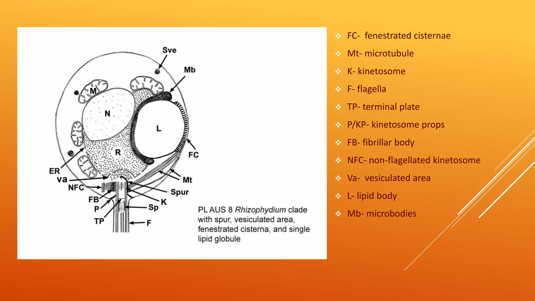
This maybe of help for UG+PG Botany students studying mycology. It's about the general account of class Chytridiomycetes. Good for quick revision and information.
*Critics are very welcomed*
Introduction,In some fungi ,true sexual cycle comprising of nuclear fusion and meiosis is absent.
These fungi derive the benefits of sexuality through a cycle know as parasexuaL cycle.
First Reported by- Gudio Pontecorvo and J.A.Roper(1952)
Parasexual cycle was reported in
Aspergillus nidulans,the imperfect stage of Emericella nidulans.
Since then parasexual cycle has been discovered not only in several members of Deutromycetes but also in fungi belonging to Ascomycetes and Basidiomycetes.
DEFINETION - Parasexuality is defined as a cycle in which Plasmogamy, Karyogamy and Meiosis [Haploidization] take place in sequence but not at a specified time or at specified points in the life cycle of an organism.
Generally parasexual cycle occurs in those fungi in which true sexual cycle does not take place.
Parasexualcycle also know as Somatic recombination. PASEXUALITY ALSO REPORTED IN SOME ORGANISMS- Aspergillus nigar, Penicillium crysogenum, STEPS OF PARASEXUAL CYCLE - 1) ESTABLISHMENT OF HETEROKARYOSIS, 2) Formation of Heterozygous DIPLOIDS, 3) occasional mitotic crossing-over during multiplication of diploid nuclei, 4)occasional haplodization through aneuploidy , COMPARISION BETWEEN SEXUAL AND PARASEXUAL CYCLE, IMPORTANCE OF PARASEXUALITY, C0NCLUSION
Heterothallic species have sexes that reside in different individuals. . The term is applied particularly to distinguish heterothallic fungi, which require two compatible partners to produce sexual spores, from homothallic ones, which are capable of sexual reproduction from a single organism.
Detail about Basidiomycetes.In this detail about its Ecosystem Relationship,Symbiotic Relationships,General characters,Basidiospores,Life cycle and its Fruiting body.
Contact Email: mzeeshan_93@yahoo.com
Chlamydomonas is unicellular, motile green algae. In this presentation the systematic position, occurrence, structure and different types of reproduction is being explained. palmella stage in vegetative reproduction is one of the outstanding character found among the other algae.
Algae are chlorophyll bearing autotrophic bodies with thalloid plant body. Thallus may be unicellular to multicellular, microscopic or macroscopic in structure.
1) Strategies and structuresIn Protozoans the method of movement .pdfaptelecom16999
1) Strategies and structures:
In Protozoans the method of movement is determined by the type of organism and the
surrounding environment. Protozoans mainly move by cell extension, flagella or pseudopodia
and cilia, the movement as per the presence of structure can be classified as ciliary, flagellar and
amoeboid movement.
Ciliates : Ciliates form the largest group of protozoa. These organisms vary in size and often live
in watery environments, including oceans, marshes, bays and streams. Ciliates move using tiny
cilia, which are hair-like strands that act as sensors and tiny limbs.
Flagella are longer and less numerous that cilia, they use their long tail like flagella to move.
Amoebas : In these two cytoskeleton get polymerized . This creates a vacancy and cytoplasmice
material flow to cover the vacancy created. When amoeba moves cytoplasm moves to the arm
like extension called pseudopodium. This pseudopodium extends and enlarge and hence this
push the animal body towards that respective direction.
2) A) Flagellates can live as single cells, in colonies, or as parasites.
Commonly live in niche\'s of water.
They conduct photosynthesis and have a cell wall.
They contain flagella for propulsion or to create a current to bring in food.
They can inhabit the reproductive tract, alimentary canal, tissue sites and also the blood stream,
lymph vessels and cerebrospinal canal.
B) Pseudopods : Also called as false feet , are projections that can appear and disappear from the
organism\'s body. These are used for movement and to engulf prey and digest them using
enzymes.
C) Apicomplexa : Unicellular and spore forming, most of them possess a unique form of
organelle that comprises a type of plastid called an apicoplast, and an apical complex structure.
They have apicoplast(non photosynthetic plastid) , mitochondria and nuclear genomes.
Lack of cilia, sexual reproduction, use micropores for feeding, and the production of oocysts
containing sporozoites as the infective form.
They have unique gliding capability which enables them to cross through tissues and enter and
leave their host cells. This gliding ability is made possible by the use of adhesions and small
static myosin motors.
3) Key characteristics of fungi :
Fungi are unicellular or multicellular.
Most of the fungi grow as tubular filaments called hyphae
They are haploid.
Fungus are heterotrophs (they can obtain nutrients by absorption) . They absorb food and secrete
enzymes to digest complex molecules
Propogate by spores
Asexual or sexual reproduction
They can be multinucleated
Fungi are achlorophyllous (lack of cholorphyll pigment)
Both Fungi and protists belong to same kingdom but fungi is different from protist, protists are
able to live in an anaerobic environment without oxygen but fungi need aerobic respiration to
survive.
Protists are unicellular but fungi are multicellular. Protists are autotrophic (make their own
energy) and heterotrophic (rely on outside source to get energy), but fungi a.
Cyanophyceae or Myxophyceae is a group of prokaryotic organisms, commonly called blue-green algae. Since their cellular organization is typically prokaryotic, the current trend is to consider them not as true algae, but as monerans. Accordingly, they are now grouped under Sub-kingdom Cyanobacteria of Kingdom Monera. The name Cyanophyceae or blue-green algae denotes the presence of the blue-green pigment phycocyanin; the name Myxophyceae refers to the presence of the carotenoid pigment myxoxanthin.
(i) Most members are filamentous forms, but some are unicellular and some others are colonial.(ii) Filamentous forms consist of a linear row of cells, called trichome, enclosed by a common sheath. Trichome may have some large and thick-walled cells, called heterocysts.(iii) Cells are typically prokaryotic, without membrane-bound cell organelles and an organized nucleus.
(iv) Chief pigments are chlorophyll-a, phycocyanin, allophycocyanin, phycoerythrin, myxoxanthin, oscillaxanthin, ß-carotene and leutein.(v) Reserve food include cyanophysean starch (glycogen) and cyanophysin (a protein).(vi) Protoplast is differentiated into outer chromoplasm and inner centroplasm. (vii) Flagella are altogether absent at any stage.(viii) Reproduction is asexual
Microbiology - Algae
Algae is an informal term for a large and diverse group of photosynthetic eukaryotic organisms. It is a polyphyletic grouping that includes species from multiple distinct clades.
Algae are sometimes considered plants and sometimes considered "protists" (a grab-bag category of generally distantly related organisms that are grouped on the basis of not being animals, plants, fungi, bacteria, or archaeans).
Introduction:
RNA interference (RNAi) or Post-Transcriptional Gene Silencing (PTGS) is an important biological process for modulating eukaryotic gene expression.
It is highly conserved process of posttranscriptional gene silencing by which double stranded RNA (dsRNA) causes sequence-specific degradation of mRNA sequences.
dsRNA-induced gene silencing (RNAi) is reported in a wide range of eukaryotes ranging from worms, insects, mammals and plants.
This process mediates resistance to both endogenous parasitic and exogenous pathogenic nucleic acids, and regulates the expression of protein-coding genes.
What are small ncRNAs?
micro RNA (miRNA)
short interfering RNA (siRNA)
Properties of small non-coding RNA:
Involved in silencing mRNA transcripts.
Called “small” because they are usually only about 21-24 nucleotides long.
Synthesized by first cutting up longer precursor sequences (like the 61nt one that Lee discovered).
Silence an mRNA by base pairing with some sequence on the mRNA.
Discovery of siRNA?
The first small RNA:
In 1993 Rosalind Lee (Victor Ambros lab) was studying a non- coding gene in C. elegans, lin-4, that was involved in silencing of another gene, lin-14, at the appropriate time in the
development of the worm C. elegans.
Two small transcripts of lin-4 (22nt and 61nt) were found to be complementary to a sequence in the 3' UTR of lin-14.
Because lin-4 encoded no protein, she deduced that it must be these transcripts that are causing the silencing by RNA-RNA interactions.
Types of RNAi ( non coding RNA)
MiRNA
Length (23-25 nt)
Trans acting
Binds with target MRNA in mismatch
Translation inhibition
Si RNA
Length 21 nt.
Cis acting
Bind with target Mrna in perfect complementary sequence
Piwi-RNA
Length ; 25 to 36 nt.
Expressed in Germ Cells
Regulates trnasposomes activity
MECHANISM OF RNAI:
First the double-stranded RNA teams up with a protein complex named Dicer, which cuts the long RNA into short pieces.
Then another protein complex called RISC (RNA-induced silencing complex) discards one of the two RNA strands.
The RISC-docked, single-stranded RNA then pairs with the homologous mRNA and destroys it.
THE RISC COMPLEX:
RISC is large(>500kD) RNA multi- protein Binding complex which triggers MRNA degradation in response to MRNA
Unwinding of double stranded Si RNA by ATP independent Helicase
Active component of RISC is Ago proteins( ENDONUCLEASE) which cleave target MRNA.
DICER: endonuclease (RNase Family III)
Argonaute: Central Component of the RNA-Induced Silencing Complex (RISC)
One strand of the dsRNA produced by Dicer is retained in the RISC complex in association with Argonaute
ARGONAUTE PROTEIN :
1.PAZ(PIWI/Argonaute/ Zwille)- Recognition of target MRNA
2.PIWI (p-element induced wimpy Testis)- breaks Phosphodiester bond of mRNA.)RNAse H activity.
MiRNA:
The Double-stranded RNAs are naturally produced in eukaryotic cells during development, and they have a key role in regulating gene expression .
Earliest Galaxies in the JADES Origins Field: Luminosity Function and Cosmic ...Sérgio Sacani
We characterize the earliest galaxy population in the JADES Origins Field (JOF), the deepest
imaging field observed with JWST. We make use of the ancillary Hubble optical images (5 filters
spanning 0.4−0.9µm) and novel JWST images with 14 filters spanning 0.8−5µm, including 7 mediumband filters, and reaching total exposure times of up to 46 hours per filter. We combine all our data
at > 2.3µm to construct an ultradeep image, reaching as deep as ≈ 31.4 AB mag in the stack and
30.3-31.0 AB mag (5σ, r = 0.1” circular aperture) in individual filters. We measure photometric
redshifts and use robust selection criteria to identify a sample of eight galaxy candidates at redshifts
z = 11.5 − 15. These objects show compact half-light radii of R1/2 ∼ 50 − 200pc, stellar masses of
M⋆ ∼ 107−108M⊙, and star-formation rates of SFR ∼ 0.1−1 M⊙ yr−1
. Our search finds no candidates
at 15 < z < 20, placing upper limits at these redshifts. We develop a forward modeling approach to
infer the properties of the evolving luminosity function without binning in redshift or luminosity that
marginalizes over the photometric redshift uncertainty of our candidate galaxies and incorporates the
impact of non-detections. We find a z = 12 luminosity function in good agreement with prior results,
and that the luminosity function normalization and UV luminosity density decline by a factor of ∼ 2.5
from z = 12 to z = 14. We discuss the possible implications of our results in the context of theoretical
models for evolution of the dark matter halo mass function.
Slide 1: Title Slide
Extrachromosomal Inheritance
Slide 2: Introduction to Extrachromosomal Inheritance
Definition: Extrachromosomal inheritance refers to the transmission of genetic material that is not found within the nucleus.
Key Components: Involves genes located in mitochondria, chloroplasts, and plasmids.
Slide 3: Mitochondrial Inheritance
Mitochondria: Organelles responsible for energy production.
Mitochondrial DNA (mtDNA): Circular DNA molecule found in mitochondria.
Inheritance Pattern: Maternally inherited, meaning it is passed from mothers to all their offspring.
Diseases: Examples include Leber’s hereditary optic neuropathy (LHON) and mitochondrial myopathy.
Slide 4: Chloroplast Inheritance
Chloroplasts: Organelles responsible for photosynthesis in plants.
Chloroplast DNA (cpDNA): Circular DNA molecule found in chloroplasts.
Inheritance Pattern: Often maternally inherited in most plants, but can vary in some species.
Examples: Variegation in plants, where leaf color patterns are determined by chloroplast DNA.
Slide 5: Plasmid Inheritance
Plasmids: Small, circular DNA molecules found in bacteria and some eukaryotes.
Features: Can carry antibiotic resistance genes and can be transferred between cells through processes like conjugation.
Significance: Important in biotechnology for gene cloning and genetic engineering.
Slide 6: Mechanisms of Extrachromosomal Inheritance
Non-Mendelian Patterns: Do not follow Mendel’s laws of inheritance.
Cytoplasmic Segregation: During cell division, organelles like mitochondria and chloroplasts are randomly distributed to daughter cells.
Heteroplasmy: Presence of more than one type of organellar genome within a cell, leading to variation in expression.
Slide 7: Examples of Extrachromosomal Inheritance
Four O’clock Plant (Mirabilis jalapa): Shows variegated leaves due to different cpDNA in leaf cells.
Petite Mutants in Yeast: Result from mutations in mitochondrial DNA affecting respiration.
Slide 8: Importance of Extrachromosomal Inheritance
Evolution: Provides insight into the evolution of eukaryotic cells.
Medicine: Understanding mitochondrial inheritance helps in diagnosing and treating mitochondrial diseases.
Agriculture: Chloroplast inheritance can be used in plant breeding and genetic modification.
Slide 9: Recent Research and Advances
Gene Editing: Techniques like CRISPR-Cas9 are being used to edit mitochondrial and chloroplast DNA.
Therapies: Development of mitochondrial replacement therapy (MRT) for preventing mitochondrial diseases.
Slide 10: Conclusion
Summary: Extrachromosomal inheritance involves the transmission of genetic material outside the nucleus and plays a crucial role in genetics, medicine, and biotechnology.
Future Directions: Continued research and technological advancements hold promise for new treatments and applications.
Slide 11: Questions and Discussion
Invite Audience: Open the floor for any questions or further discussion on the topic.
Professional air quality monitoring systems provide immediate, on-site data for analysis, compliance, and decision-making.
Monitor common gases, weather parameters, particulates.
(May 29th, 2024) Advancements in Intravital Microscopy- Insights for Preclini...Scintica Instrumentation
Intravital microscopy (IVM) is a powerful tool utilized to study cellular behavior over time and space in vivo. Much of our understanding of cell biology has been accomplished using various in vitro and ex vivo methods; however, these studies do not necessarily reflect the natural dynamics of biological processes. Unlike traditional cell culture or fixed tissue imaging, IVM allows for the ultra-fast high-resolution imaging of cellular processes over time and space and were studied in its natural environment. Real-time visualization of biological processes in the context of an intact organism helps maintain physiological relevance and provide insights into the progression of disease, response to treatments or developmental processes.
In this webinar we give an overview of advanced applications of the IVM system in preclinical research. IVIM technology is a provider of all-in-one intravital microscopy systems and solutions optimized for in vivo imaging of live animal models at sub-micron resolution. The system’s unique features and user-friendly software enables researchers to probe fast dynamic biological processes such as immune cell tracking, cell-cell interaction as well as vascularization and tumor metastasis with exceptional detail. This webinar will also give an overview of IVM being utilized in drug development, offering a view into the intricate interaction between drugs/nanoparticles and tissues in vivo and allows for the evaluation of therapeutic intervention in a variety of tissues and organs. This interdisciplinary collaboration continues to drive the advancements of novel therapeutic strategies.
This pdf is about the Schizophrenia.
For more details visit on YouTube; @SELF-EXPLANATORY;
https://www.youtube.com/channel/UCAiarMZDNhe1A3Rnpr_WkzA/videos
Thanks...!
Richard's aventures in two entangled wonderlandsRichard Gill
Since the loophole-free Bell experiments of 2020 and the Nobel prizes in physics of 2022, critics of Bell's work have retreated to the fortress of super-determinism. Now, super-determinism is a derogatory word - it just means "determinism". Palmer, Hance and Hossenfelder argue that quantum mechanics and determinism are not incompatible, using a sophisticated mathematical construction based on a subtle thinning of allowed states and measurements in quantum mechanics, such that what is left appears to make Bell's argument fail, without altering the empirical predictions of quantum mechanics. I think however that it is a smoke screen, and the slogan "lost in math" comes to my mind. I will discuss some other recent disproofs of Bell's theorem using the language of causality based on causal graphs. Causal thinking is also central to law and justice. I will mention surprising connections to my work on serial killer nurse cases, in particular the Dutch case of Lucia de Berk and the current UK case of Lucy Letby.
2. Idea about the origin of chytrids and how are
they related to the higher fungi
Here ‘mya’ means million years ago
So accordingly it clearly depicts that chytrid have
originated from a separate lineage/ from
different ancestors than those of it’s higher fungi.
Though the ancestors of ascomycetes and
basidiomycetes are same but chytrids have
separate ancestors
3. INTRODUCTION
Fungi of this class are called as ‘Chytrids’
Grow aerobically in soil, mud, water. Though some species inhabits estuaries and sea
Reproduce using zoospores with single whiplash type flagellum; exception- Neocallimastigales
have multiflagella
Many members are saprotrophs i.e. utilizes chitin, keratin and cellulose of plants; whilst species of
Caulochytrium grows as mycoparasite on the mycelium and conidia of terrestrial fungi
terrestrial fungi
Some are biotropic parasite of filamentous algae and diatoms and severely deplete their growth;
species of Synchytrium and Olpidium are too biotropic parasite but of vascular plants
4. THALLUS STRUCTURE
Generally the biotropic
parasite have Holocarpic
thalli, but usually they
have Eucarpic thalli.
The former is that type of thallus where the
whole thallus is contained within a single
cell and there is no differentiation between
the vegetative and reproductive structures
The latter Eucarpic thalli, are
differentiated into a reproductive
and vegetative parts- Sporangia
and Rhizoids.
If one sporangia-
monocentric; here
rhizoids radiate from a
single portion
Epibiotic- rhizoids inside
the host cell and
sporangia outside
Endobiotic- both rhizoids
and sporangia inside the
host cell
If many sporangia-
polycentric; here rhizoids
are branched, forming
Rhizomycelium
Some species shows both
mono- and poly- centric
characters.
7. CELL WALL
X-ray diffraction and other microscopic techniques have shown presence of chitin- a polymer of
NAGA, as well as cellulose.
As chitin is present in other higher classes of fungi like basidiomycetes and ascomycetes.
Whereas oomycetes have presence of cellulose in their cell wall. This clearly depicts that the
chytrids shows homology with the higher and advanced classes, thus is of evolutionary
importance.
Members of hypochytrdiomycota like Hypochitrium and Rhizidiomyces shows presence of both
chitin and cellulose in their cell wall.
8. Asexual reproduction
Zoospore in sporangia
Autospore in sporangia
Fragmentation- polycentric and resting spores
Vegetative stage is generally haploid
11. THE ZOOSPORE
Round or pear shaped sac containing one or more exit papillae
Divided into two types: -
Inoperculate- no operculum; Olpidium, Cladochytrium etc. where the discharge tubes
penetrates the exterior of the host cell and then dissolves
Operculate- operculum present i.e. a special line of weakness which breaks open at the tip of
the discharge tube. Eg. Chytridium, Nowakoskiella etc.
Zoospores are though similar in size but sporangia number and sizes varies within species, thus is
of taxonomic importance.
13. CONTINUE…
This varying number of sporangia and their sizes depends upon the nutrition and conditions they
grow around. This conclusion is supported by a study on Rhizophlyctis rozea. It has been seen
that in culture media with less carbs, sporangia containing only one or two zoospores have been
observed. Whereas cellulose rich media contained large sporangia with hundred of such spores.
The zoospores are separated from each other by a spongy fluid which may absorb water during
maturation and help in the sporangia to burst open
Structure of the zoospores though vary among different genera but are similar within same
genera (D. J. S. Barr,1990) thus it is of taxonomic importance.
14. The flagellar structure t.s.
The concentration of mitochondria is high towards the base of the kinetosome
Another complex which helps in the movement and directing the flagella of zoospore is known as
MLC (microbody lipid complex) consisting of a large lipid granule, microbodies, mitochondria,
smooth cisternae and Rumposome (fenestrated cisternae)
Dormant kinetosome is also found in many members of this family
15. The flagella is of whiplash type as similar as those of eukaryotes (study the structure in details of
eukaryotic flagella)
a smooth membrane enclosing the cylindrical shaft, along with the long axoneme with 9 doublets
and two centrally placed singlet microtubular rings
The flagellum consists of three main portions- the flagellum proper, the kinetosome and the
transitional zone.
The evolutionary significance of the dormant kinetosome is that, maybe the ancestors of chytrids
were biflagellate and during the course of evolution they might have lost it
Sometimes along with the doublets of A- and B- rings there maybe a presence of third ring or the
C- ring of microtubule.
18. The zoospore germination:
Generally the zoospore either absorbs or cut off it’s flagella from their body before encysting
themselves.
Holocarpic parasites encyst on the host surface and injects its cytoplasmic content inside the host cell
In monocentric parasites the zoospore forms cyst and develops rhizoids from one point. The cyst itself
matures and enlarges to form zoosporangium. Sometimes prosporangium also develops.
In polycentric parasites, the zoospores forms limited rhizomycelium, which again later on give rise to
more swollen structure which ultimately gives rise to more rhizomycelium. Germination maybe from
one point monopolar (Chytridiales) and bipolar (Blastocladiales).
19. Isomorphic alternation of generation
The gametothallic(haploid) and sporothallic(diploid) bodies morphology is same.
The sexual reproduction are of two types- Gametogamy and Gamentangio-gametangiogamy
Gametogamy are of: -
Isogamy
Anisogamy
Oogamy
Somatogamy
21. A proper diagram illustrating the sexual and asexual reproduction of Allomyces
22. Somatogamy process: -
Rhizoids from uninucleate contributing thallus together fuses and forms anastomosis.
Cytoplasm and nuclei from both these contributing thallus comes to the point of anastomosis,
which ultimately becomes a globular resting body
These resting body is binucleate and full of cytosol causing the contributing thalli to become
empty
Two nuclei later fuses to form a diploid body which ultimately gives rise to a sporangium which
are not genetically dissimilar than their parents.
24. Conjugation takes place only when a small donor produces a conjugation tube to a larger recipient
gametangium.
Following the nuclear fusion between the two gametangium, the larger recipient forms a thick
covering outside forming a diploid resting sporangium.
After maturation the sporangium forms presporangium and gives rise to thin walled
meiosporangium, where obviously the meosis takes place, giving rise to haploid spores.
A variant of this type of sexual diferrentiation (gametangio-gametangigamy) is seen in
Rhizophydium, where copulation between the gametangium of a rhizoid forming thallus and a
forming thallus and a motile gamete that encyst directly on the gametangium.
27. Members of Chytridiomycetes and their hosts: -
Synchitrium endobioticum- black wart of potato
Olpidium brassicae- zoospores are vectors for virus causing big vein disease of lettuce
lettuce
Colleomyces spp.- pathogen of freshwater invertebrates such as copepods and mosquito larvae
mosquito larvae
Neocallimastigales- present in gut of herbivorous mammals, and feed the herbage