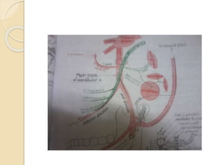
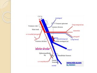
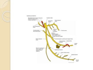
This document describes the anatomy and branches of the mandibular nerve (CN V3). It originates from the trigeminal ganglion and pons and exits the skull through the foramen ovale. Its main branches innervate the muscles of mastication and provide sensory innervation to the lower face and oral cavity. The anterior and posterior divisions each give off motor and sensory branches with specific distributions.
4. It is the 3rd division of the trigeminal nerve.
Origin:
It is a mixed nerve formed of 2 roots.
a)Sensory root :arises from the trigeminal
ganglion& runs forward& laterally toward
foramen ovale.
b) Motor root: a smaller root which arises
from the trigeminal motor nucleus in the
pons & runs deep to the trigeminal
ganglion to join the sensory root in the
foramen ovale
5. Course& relations
1)The 2 roots unite together in the foramen
ovale to form the main trunk mandibular
n.(mixed).
2)The main trunk descends through the f. ovale
to reach the infra temporal fossa where it is
related :laterally to lat. Pterygoid m.
madially to otic ganglion& tensor palati
m.
posterolaterally to middle meningeal art.
3)The main n. trunk gives 2 branches then
divides into 2 divisions (small ant.& large
post.) about 12cm below the f. ovale.
6. Branches of the mand. nerve
A)Branches of the main nerve trunk.
B)Branches from the anterior division.
C)Branches from the posterior division.
7.
8.
9. Branches of the main nerve trunk
2 branches (one motor &one sensory):
(11)Nervous spinosus (sensory): it enters the
cranial
cavity through the foramen spinosum to supply
the
dura of the middle cranial fossa .
2) Nerve to medial pterygoid (motor): supplies
this muscle through its deep surface.
It also gives a branch which traverse the otic
ganglion without relay to supply the tensor
palati & tensor tympani muscles.
10.
11. Branches of the anterior
division
3motor&1 sensory:
1)Masseteric n. :passes laterally above the
upper border of the lat. Pterygoid m. then
through mand. Notch to enter the deep
surface of the masseter m.
2) N. to lat. Pterygoid m: enter the deep
surface of lat.pterygoid m.
3) 2 deep temporal nerves pass laterally
above the upper border of the lat.
Pterygoid to enter the deep surface of
the temporalis m.
12.
13. 4) Buccul n.(sensory):
Passes bet. The 2 heads of the lat.
Pterygoid m.
Then it descends superficial to the lower
head of lat. Pterygoid & medial to the
coronoid process of the mandible, to
appear bet. Masseter & buccinator
muscles.
It supplies:
The skin covering the buccinator muscle.
The mucous membrane of the cheeck &
gum oppsite to the molar teeth.
14.
15. c)Brancnes from the posterior
division
3 sensory & 1 motor:
(1Auriculo-temporal n. (sensory)
Arises by 2 roots which surround the
middle meningeal a.
Passes backwards medial to the neck of
the mandible,then behind the tmj
,ascends deep to the parotid gland to
appear at its upper pole behind the
superficial temporal vessels.
16. It supplies:
The tempromandibular joint.
Sensory fibers to the parenchyma of the
parotid gland
Skin of the upper 23 of outer surface of
the auricle +scalp above it(temporal
region).
Skin lining the external auditory meatus,
tragus of ear+ outer surface of the ear
drum.
It caries symp.& parasymp. Secretory
fibers from the otic ganglion to the
17. Lingual nerve (sensory)
Origin: post. Div. of mand. N.
Course & relations:
It lies deep to the lat. Pterygoid m. ,here it
is joined by the chorda tympani n. (br. Of
facial n. carrying taste ¶sympathatic
fibers)
It emerges from the undercover of the
lower border of the lat. Pterygoid m.
,infront of the inferior alveolar n.
Then it descends bet. The ramus of the
mand.(superficially)& the med. Pterygoid
m. (deeply)
18. It passes along a groove on the inner
surface of the socket of the last molar
tooth just undercover the mucosa of the
gum (dangerous area during tooth
extractin).
Then it runs superficial to the styloglossus
m.
Then it crosses superficial to the
hyoglossus m. Here the nerve lies deep
to the submandibular salivary gland&
submandibular ganglion hangs from it.
Finally it passes deep to the mylohyoid
m.& ascends superficial to genioglossus
m.
19. Here the lingual n. has a tripple relation
with the submandibular duct :
1st it passes lat. To the duct,
Then it curves below the duct,
Finally it ascends med. To the duct
Booth the nerve & the duct pass forward &
upward bet. Genioglossus m. & subling.
Salivary gl. To reach the tongue.
Termination: it ends by dividing into
terminal branches which enter the side of
the tongue under its mucous membrane.
20. Distribution:
It carries general& taste sensations from
the ant. 23 of the tongue.
It carries general sensations from the
floor of the mouth& gums.
It carries secretomotor parasympathatic
fibers from chorda tympani to the
submand. & subling. Glands (relaying
1st in the submandibular ganglion)
21.
22.
23. Inferior alveolar nerve
It is the largest branch of the post. Division
of mandibular nerve.
Course & relations:
It begins deep to the lateral pterygoid m.
then emerges from undercover of its
lower border where the lingual n. lies
infront of it
It descends bet. The ramus of the mand.
(laterally) & med. Pterygoid m. medially.
It enters the mandibular foramen infront of
the inf. Alveolar art. & runs in the
mandibular canal suppling the molar&
premolar teeth
24. It ends opposite to the mental foramen
by dividing into 2 branches:
Mental n : comes out of mental foramen
& it is sensory to the chin .
Incisive n. : continues inside the
mandible to supply the lower canine &
incisor teeth.
25.
26. Nerve to mylohyoid(motor)
It contains all the motor fibers of the
post. Division of mandibular n.
It arises as a branch of inf. Alv. N.
before it enters the mandibular
foramen.
It runs in the mylohyoid groove on the
inner surface of ramus of mandible.
It ends by supplying the mylohyoid m. +
anterior belly of digastric m.