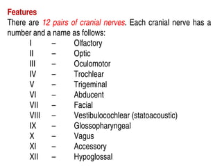
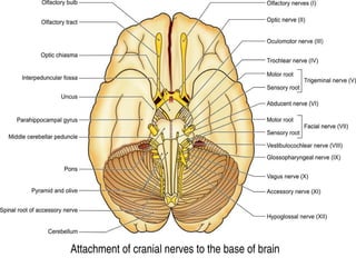
Features
There are 12pairs of cranial nerves. Each cranial nerve has a
number and a name as follows:
I – Olfactory
II – Optic
III – Oculomotor
IV – Trochlear
V – Trigeminal
VI – Abducent
VII – Facial
VIII – Vestibulocochlear (statoacoustic)
IX – Glossopharyngeal
X – Vagus
XI – Accessory
XII – Hypoglossal
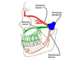
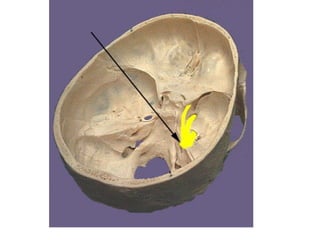
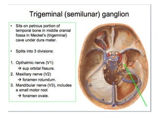
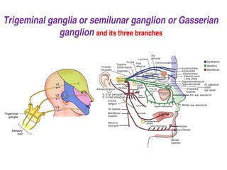
Trigeminal ganglia orsemilunar ganglion or Gasserian
ganglion and its three branches
8.
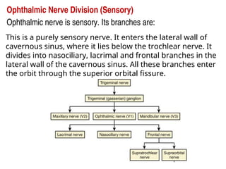
Ophthalmic Nerve Division(Sensory)
Ophthalmic nerve is sensory. Its branches are:
This is a purely sensory nerve. It enters the lateral wall of
cavernous sinus, where it lies below the trochlear nerve. It
divides into nasociliary, lacrimal and frontal branches in the
lateral wall of the cavernous sinus. All these branches enter
the orbit through the superior orbital fissure.
9.
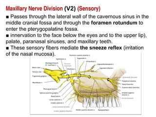
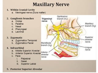
Maxillary Nerve Division(V2) (Sensory)
■ Passes through the lateral wall of the cavernous sinus in the
middle cranial fossa and through the foramen rotundum to
enter the pterygopalatine fossa.
■ innervation to the face below the eyes and to the upper lip),
palate, paranasal sinuses, and maxillary teeth.
■ These sensory fibers mediate the sneeze reflex (irritation
of the nasal mucosa).
11.
■ Major branchesof this complex nerve include the following:
•1. Meningeal branch - dura mater of the middle cranial fossa.
•2. Pterygopalatine (communicating) nerve connects sensory fibers that pass
through the pterygopalatine ganglion and join branches off the ganglion.
•3. Posterior–superior alveolar nerve leaves the pterygopalatine fossa to
innervate the cheeks, gums, molar teeth, as well as the maxillary sinus.
•4. Zygomatic nerve courses through the zygomatic bone in the maxillary sinus
and divides into the zygomaticofacial and zygomaticotemporal nerves.
•5. Infraorbital nerve is the anterior continuation of the maxillary nerve and
gives rise to the middle and anterior–superior alveolar nerves that supply the
maxillary sinus, teeth, and gums. It then emerges through the infraorbital
foramen and divides in the face into the inferior palpebral, lateral nasal, and
superior labial branches.
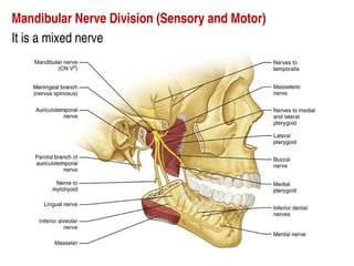
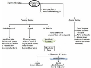
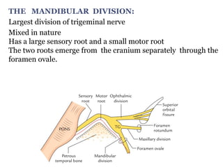
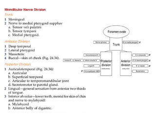
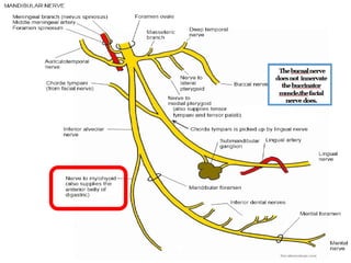
THE MANDIBULAR DIVISION:
Largestdivision of trigeminal nerve
Mixed in nature
Has a large sensory root and a small motor root
The two roots emerge from the cranium separately through the
foramen ovale.
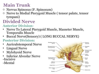
17.
Main Trunk
o NervusSpinosus (F. Spinosum)
o Nerve to Medial Pterygoid Muscle ( tensor palate, tensor
tympani)
Divided Nerve
Anterior Division-
• Nerve To Lateral Pterygoid Muscle, Masseter Muscle,
Temporalis Muscle
• Buccal Nerve(Sensory) ( LONG BUCCAL NERVE)
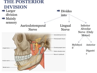
Posterior Division:
• Auriculotemporal Nerve
• Lingual Nerve
• Mylohyoid Nerve
• Inferior Alveolar Nerve
-Incisive
-Mental
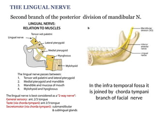
THE LINGUAL NERVE
Secondbranch of the posterior division of mandibular N.
In the infra temporal fossa it
is joined by chorda tympani
branch of facial nerve
21.
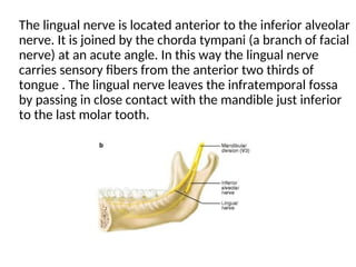
The lingual nerveis located anterior to the inferior alveolar
nerve. It is joined by the chorda tympani (a branch of facial
nerve) at an acute angle. In this way the lingual nerve
carries sensory fibers from the anterior two thirds of
tongue . The lingual nerve leaves the infratemporal fossa
by passing in close contact with the mandible just inferior
to the last molar tooth.
22.
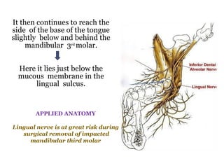
It then continuesto reach the
side of the base of the tongue
slightly below and behind the
mandibular 3rd molar.
Here it lies just below the
mucous membrane in the
lingual sulcus.
APPLIED ANATOMY
Lingual nerve is at great risk during
surgical removal of impacted
mandibular third molar
23.
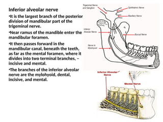
Inferior alveolar nerve
•Itis the largest branch of the posterior
division of mandibular part of the
trigeminal nerve.
•Near ramus of the mandible enter the
mandibular foramen.
•It then passes forward in the
mandibular canal, beneath the teeth,
as far as the mental foramen, where it
divides into two terminal branches, –
incisive and mental.
•The branches of the inferior alveolar
nerve are the mylohyoid, dental,
incisive, and mental.
24.
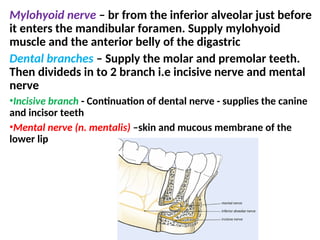
Mylohyoid nerve –br from the inferior alveolar just before
it enters the mandibular foramen. Supply mylohyoid
muscle and the anterior belly of the digastric
Dental branches – Supply the molar and premolar teeth.
Then divideds in to 2 branch i.e incisive nerve and mental
nerve
•Incisive branch - Continuation of dental nerve - supplies the canine
and incisor teeth
•Mental nerve (n. mentalis) –skin and mucous membrane of the
lower lip
25.
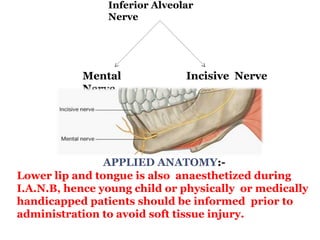
Inferior Alveolar
Nerve
APPLIED ANATOMY:-
Lowerlip and tongue is also anaesthetized during
I.A.N.B, hence young child or physically or medically
handicapped patients should be informed prior to
administration to avoid soft tissue injury.
Mental
Nerve
Incisive Nerve
26.
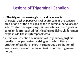
Lesions of TrigeminalGanglion
• i. The trigeminal neuralgia or tic doloureux is
characterized by paroxysms of acute pain in the sensory
area of one of the divisions of the trigeminal nerve on one
side. To stop the agonizing pain sometimes the trigeminal
ganglion is approached for injecting medicine via foramen
ovale inside the infratemporal fossa.
•ii. The viral infection of neurons of trigeminal ganglion
results in herpes zoster or shingles in which there is
eruption of painful blisters in cutaneous distribution of
any one or more of the main divisions of the trigeminal
nerve.
27.
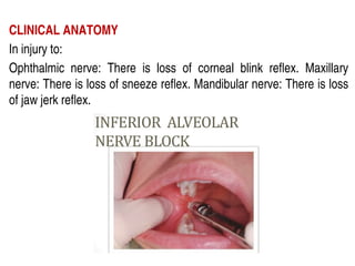
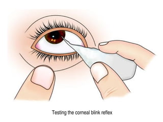
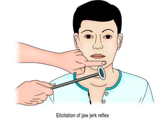
CLINICAL ANATOMY
In injuryto:
Ophthalmic nerve: There is loss of corneal blink reflex. Maxillary
nerve: There is loss of sneeze reflex. Mandibular nerve: There is loss
of jaw jerk reflex.


![Trigeminal nerve - 1st year class [Autosaved][1].pptx](https://cdn.slidesharecdn.com/ss_thumbnails/trigeminalnerve-1styearclassautosaved1-251217164702-3b80e5cf-thumbnail.jpg?width=640&height=640&fit=bounds)


![ONFH[AVN HIP] -TRIPLE REGIME -A NOVAL SURGICAL CONCEPT .pptx](https://cdn.slidesharecdn.com/ss_thumbnails/onfhavnhip2026koaconcalicutdrgokuldevdrmashraf-260210064517-213ec005-thumbnail.jpg?width=640&height=640&fit=bounds)


