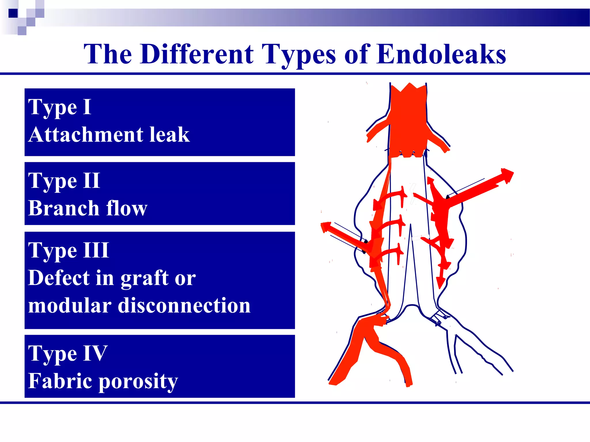
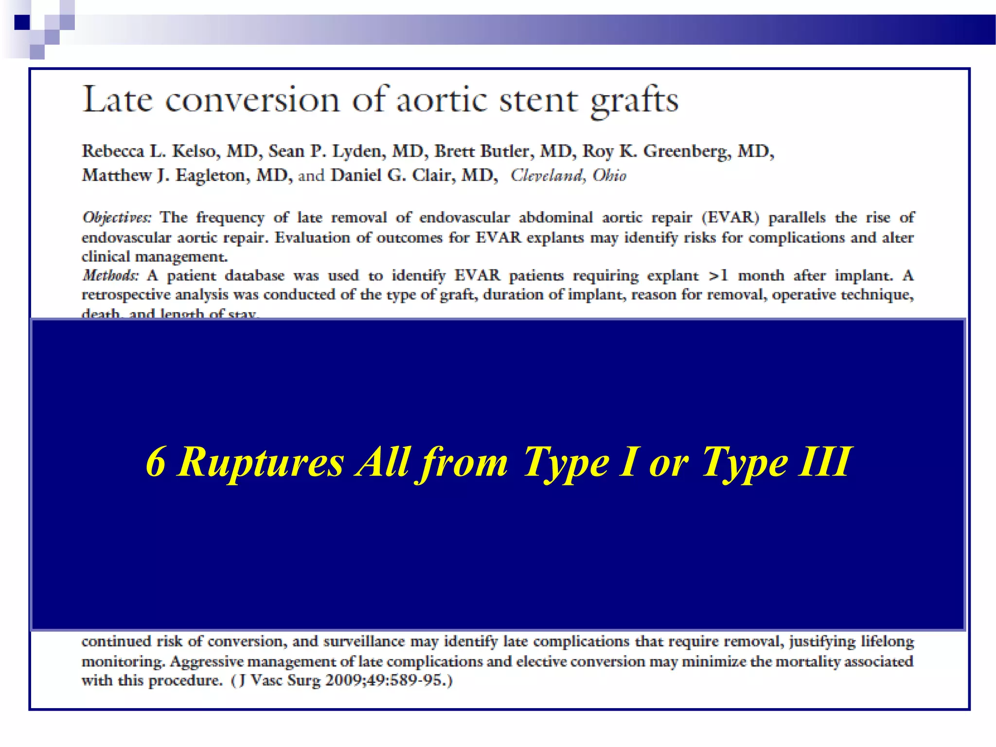
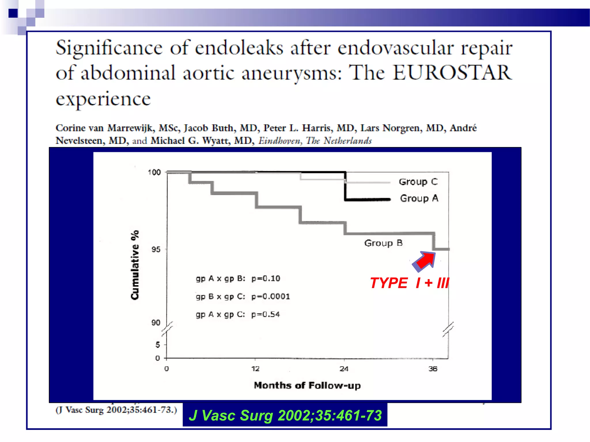
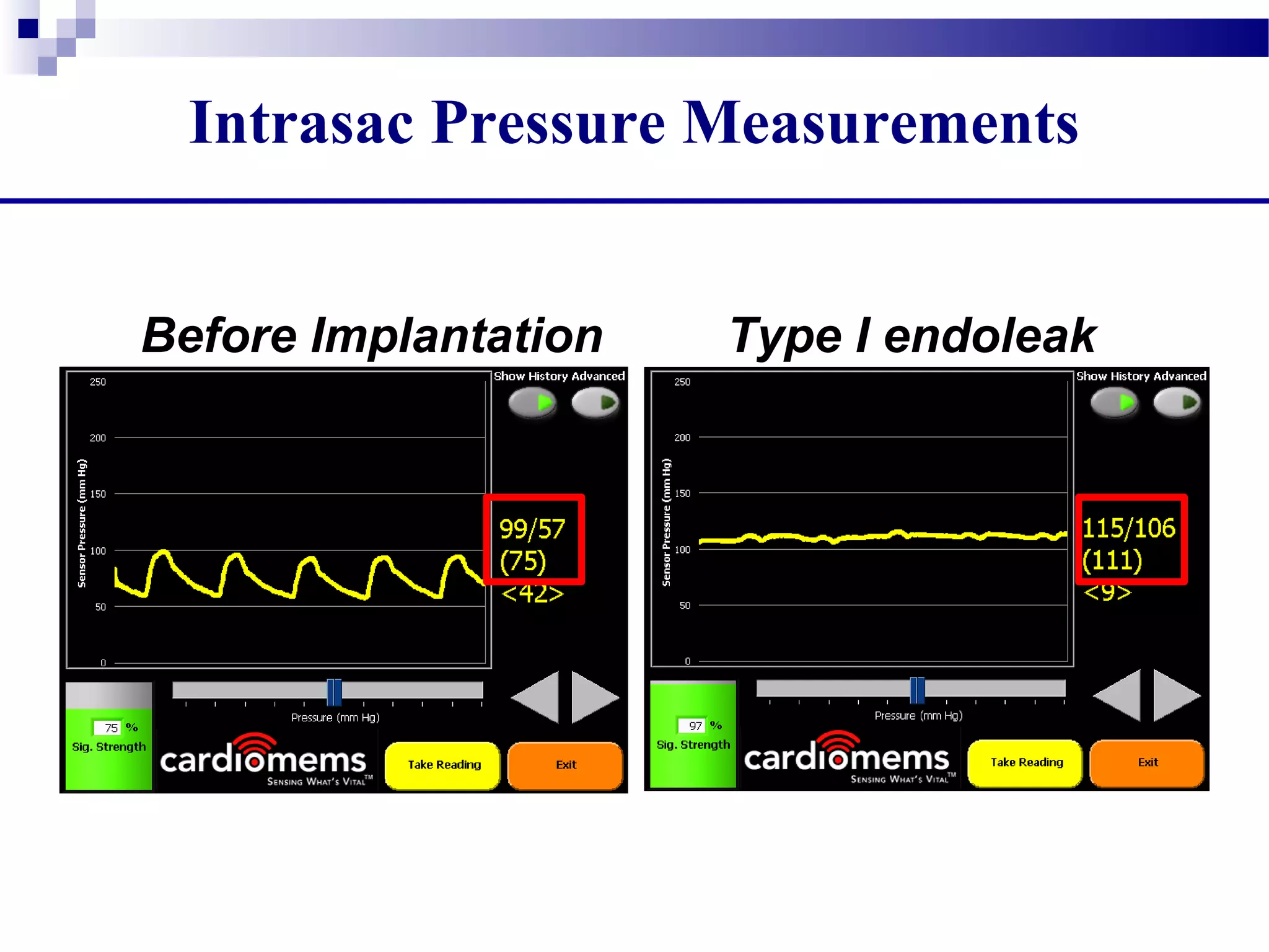
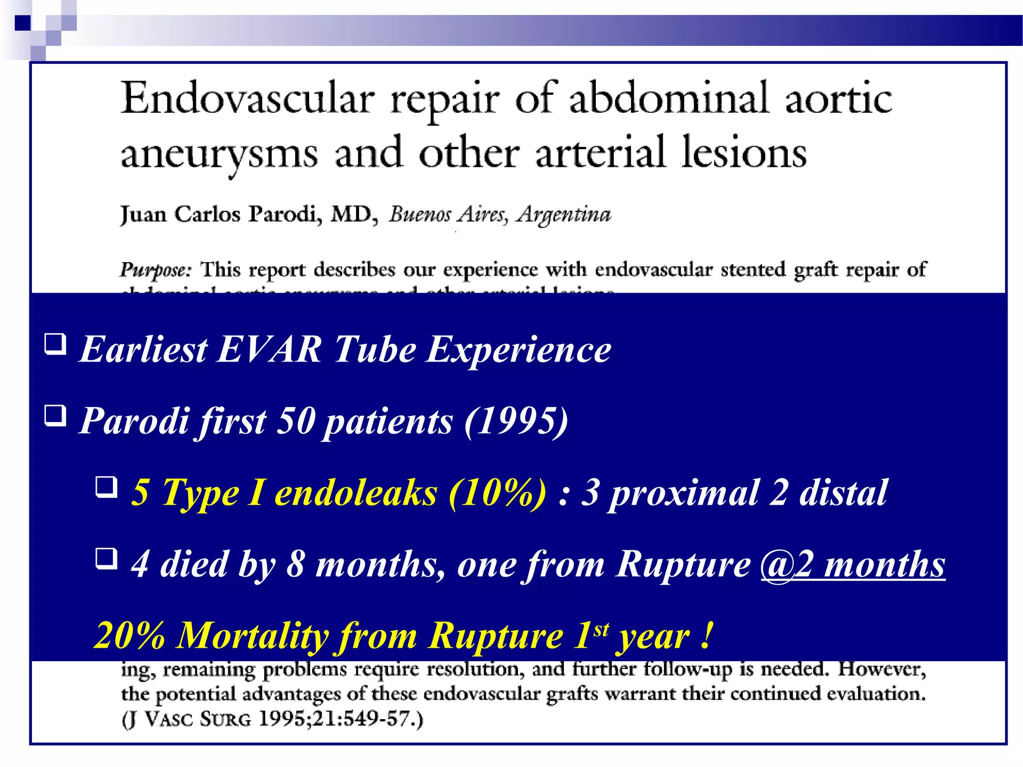
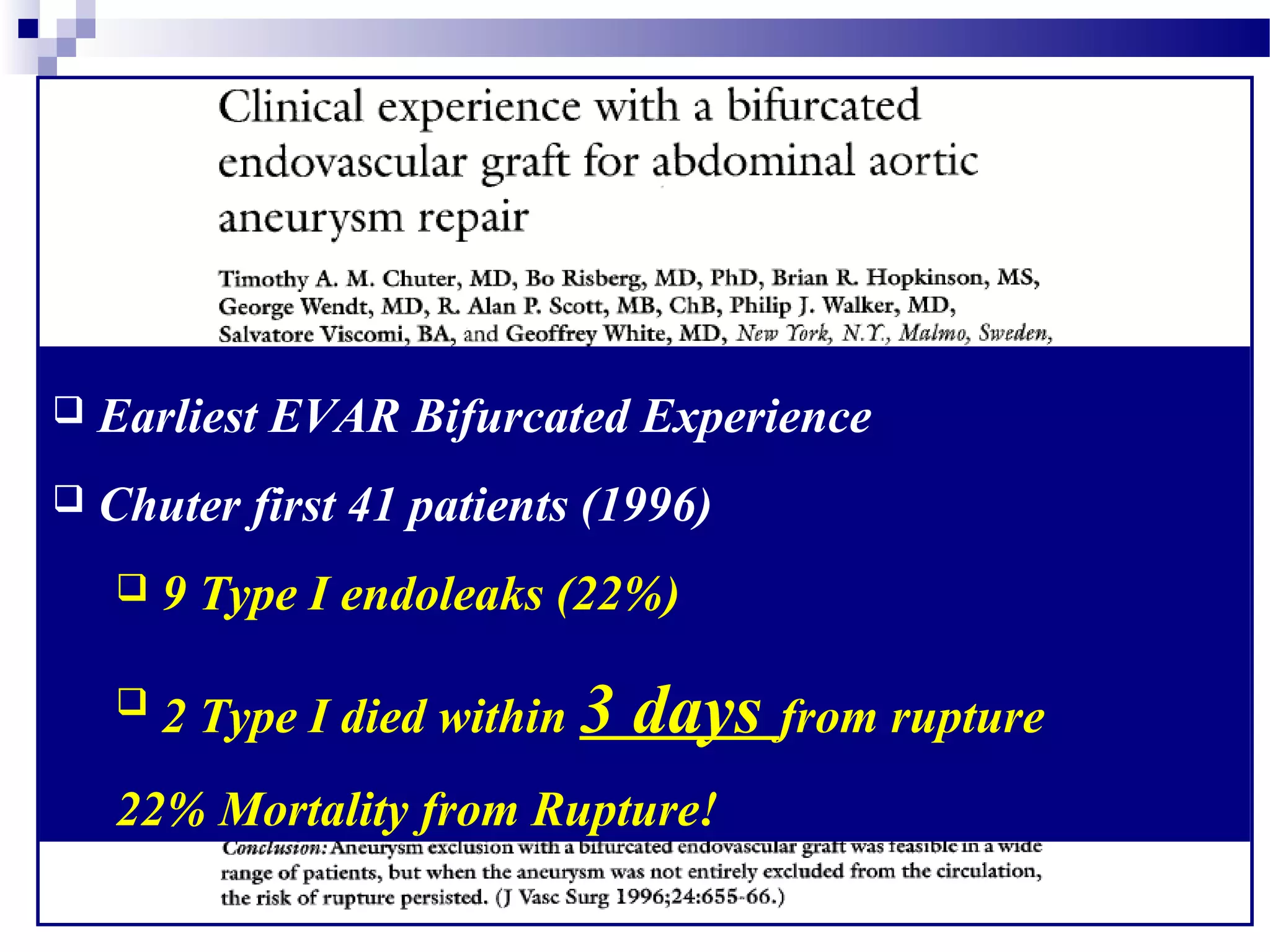
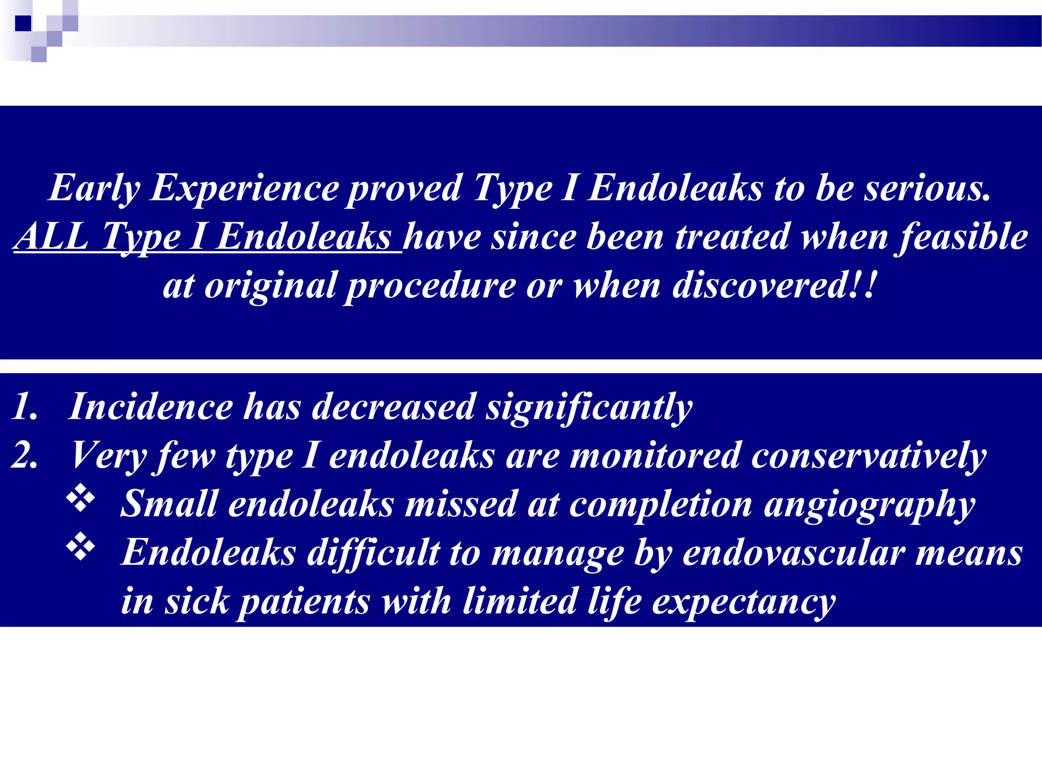
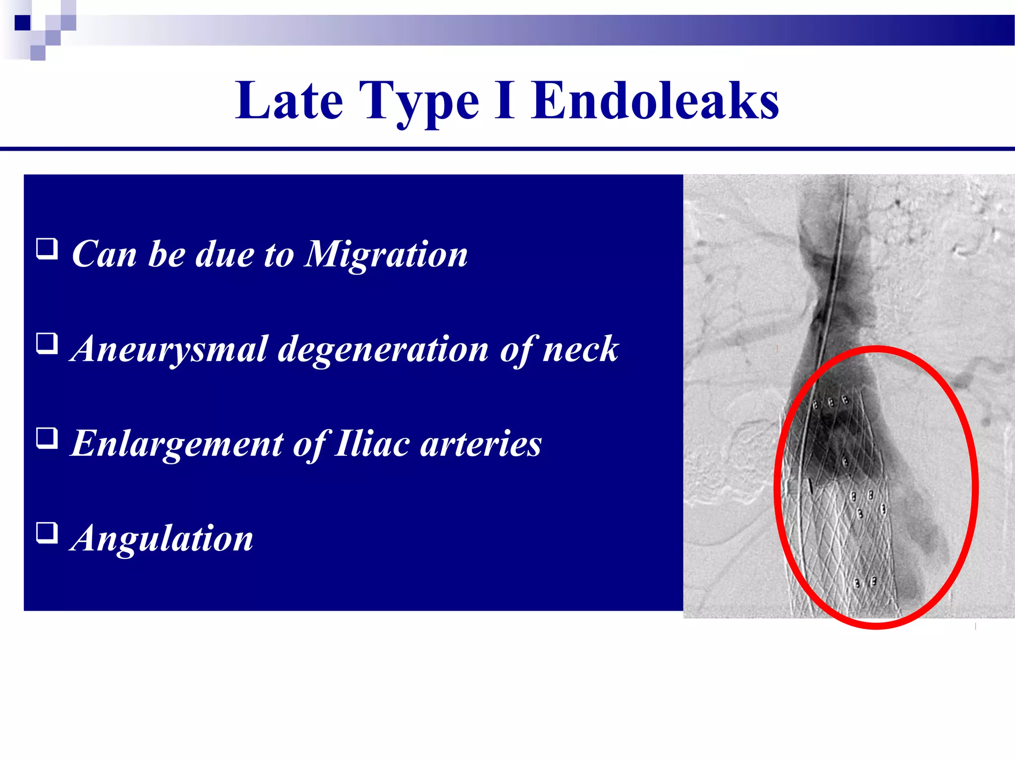
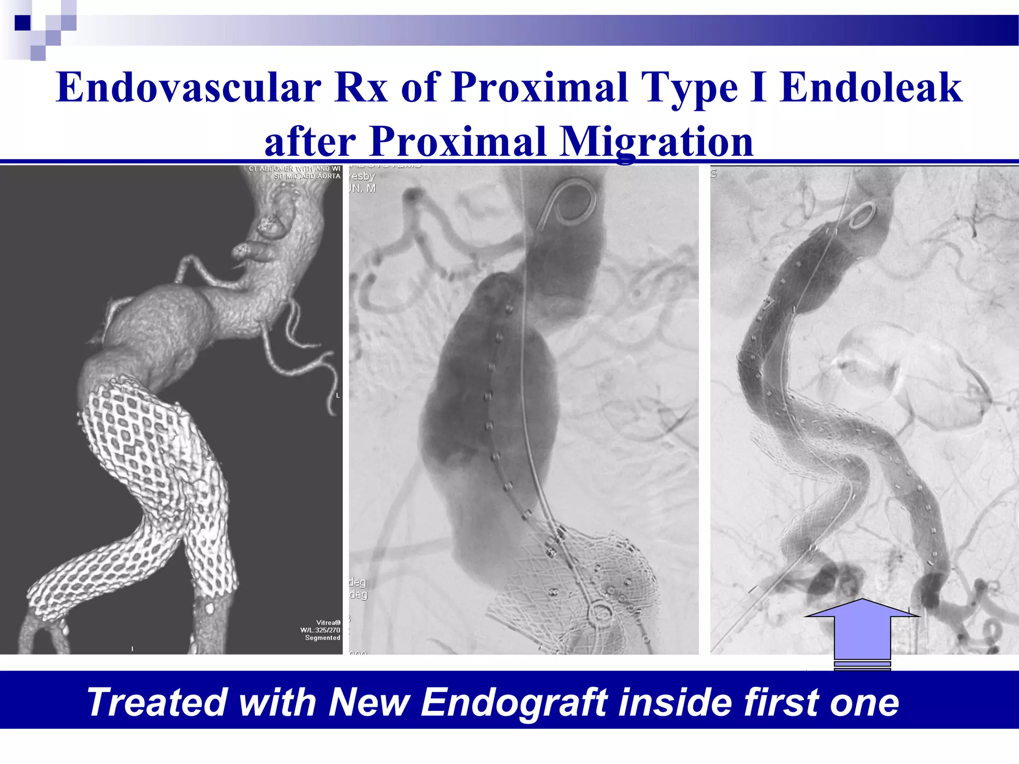
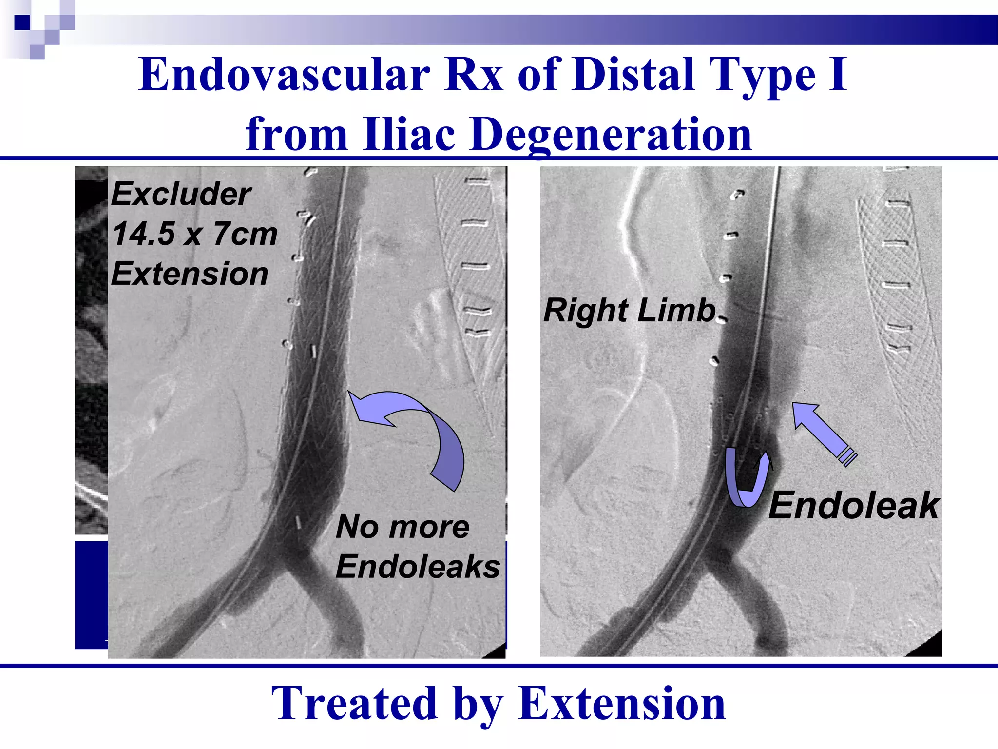
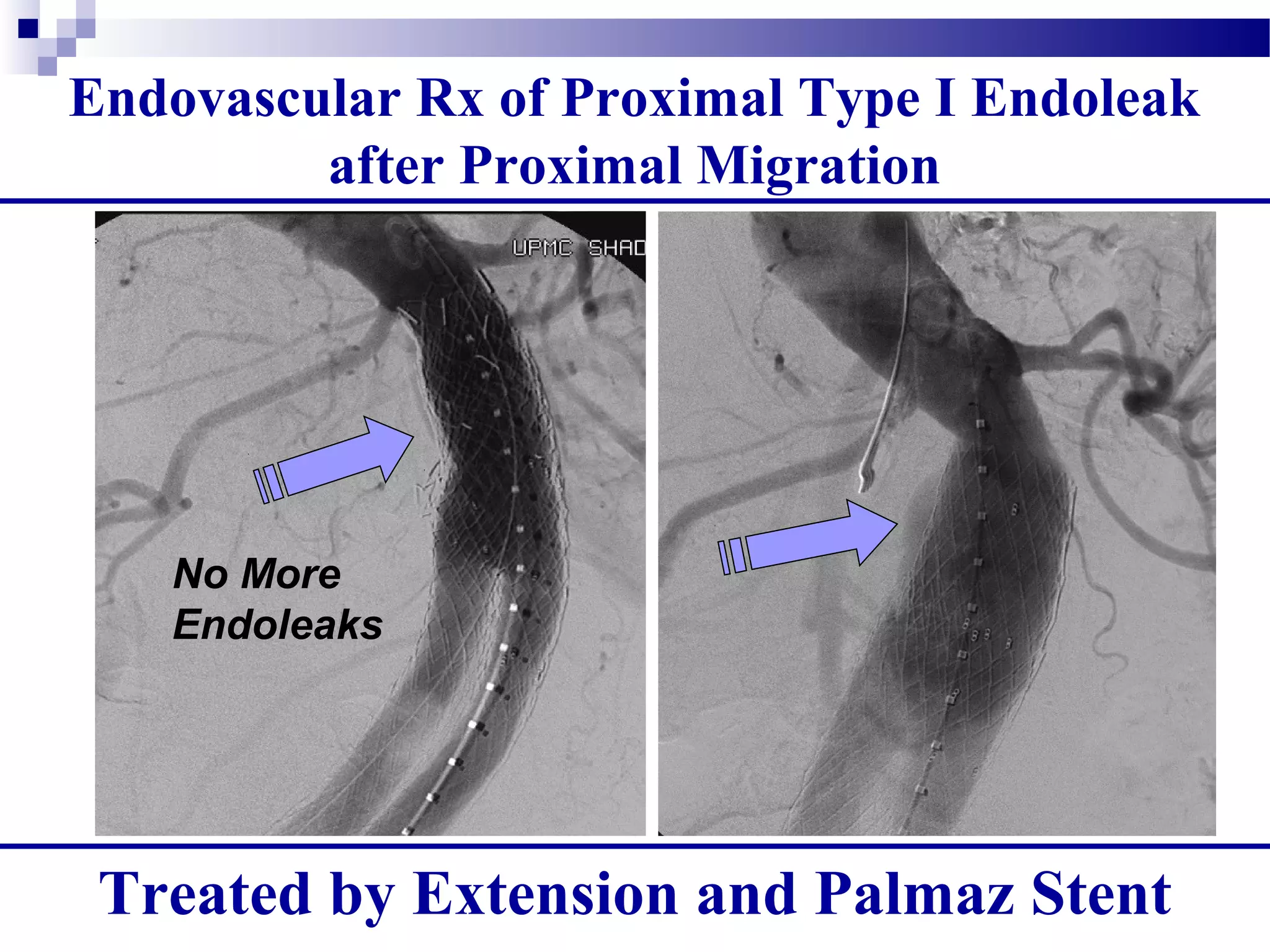
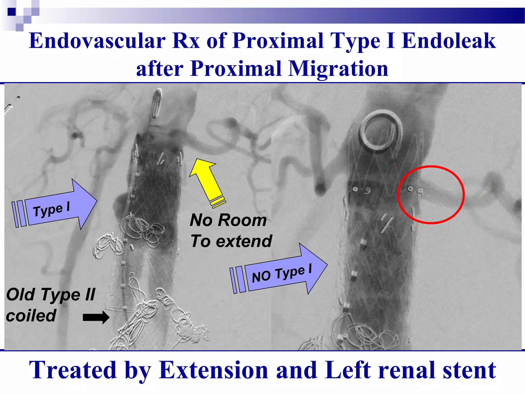
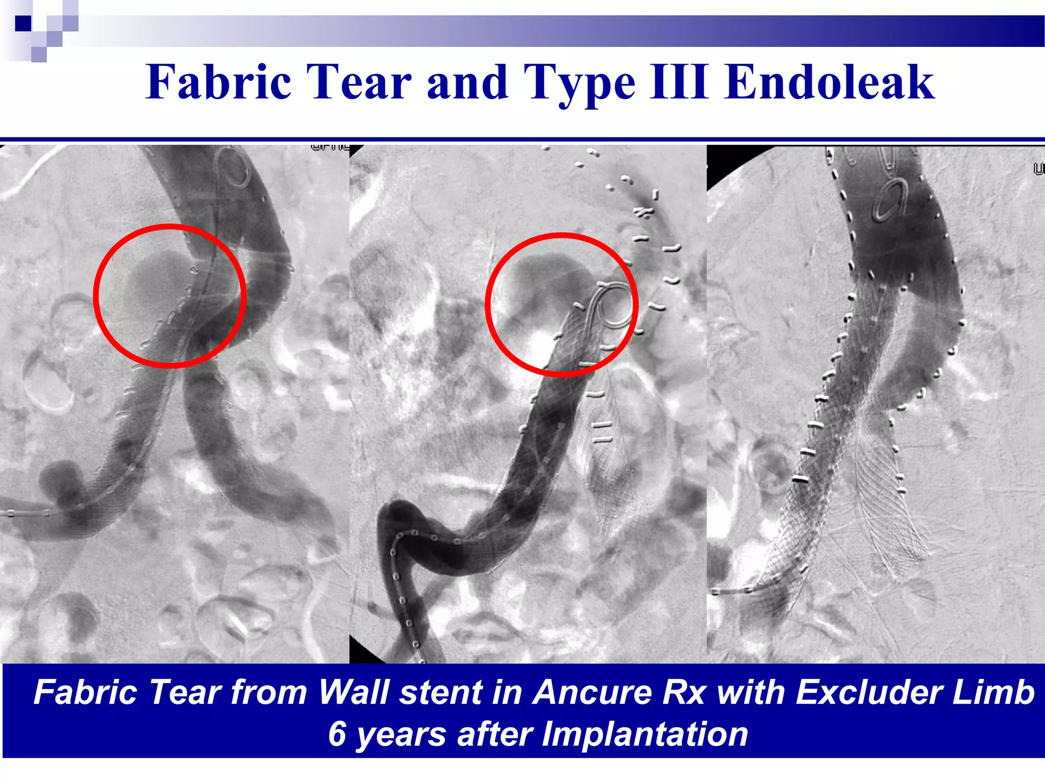
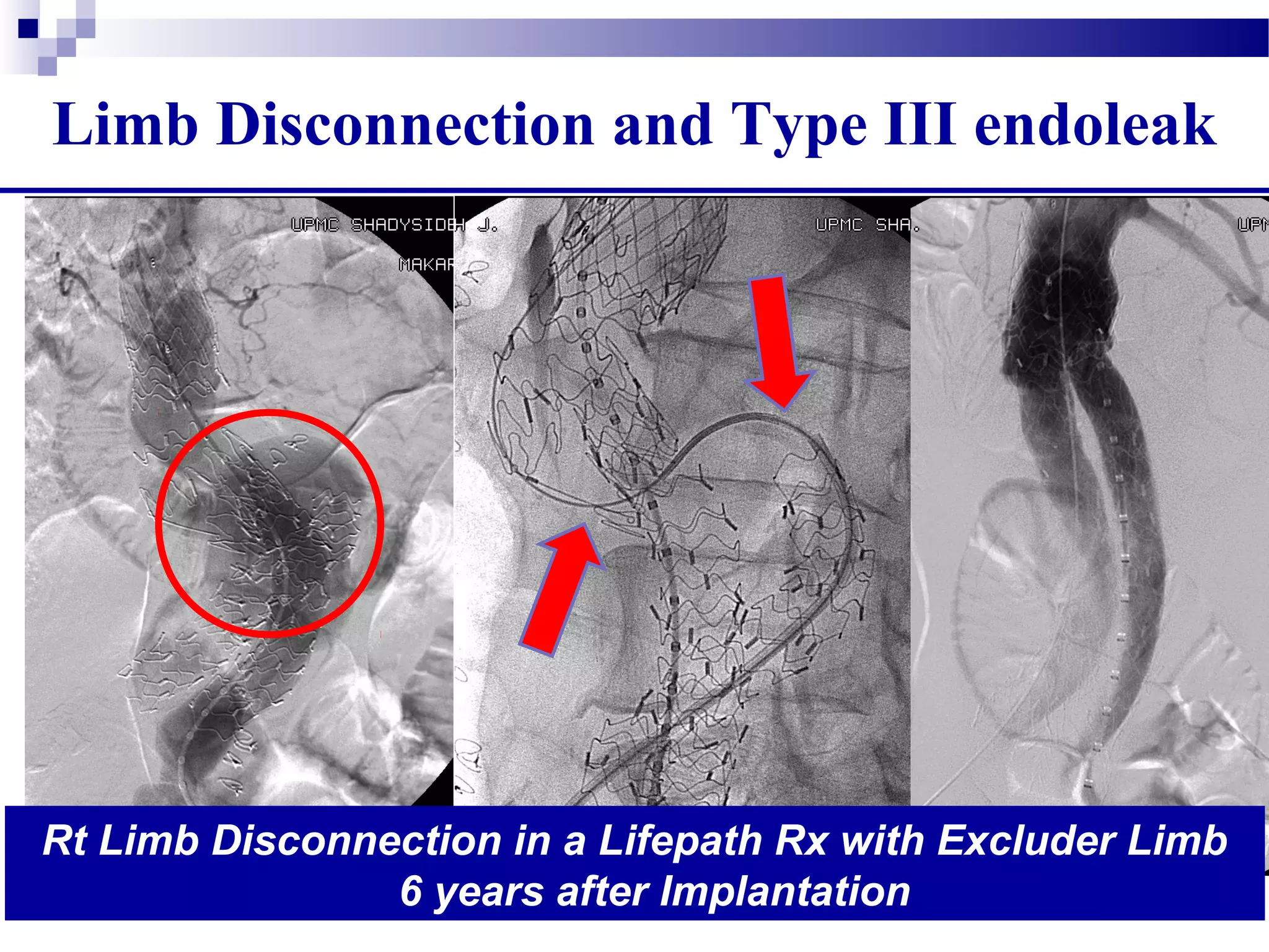
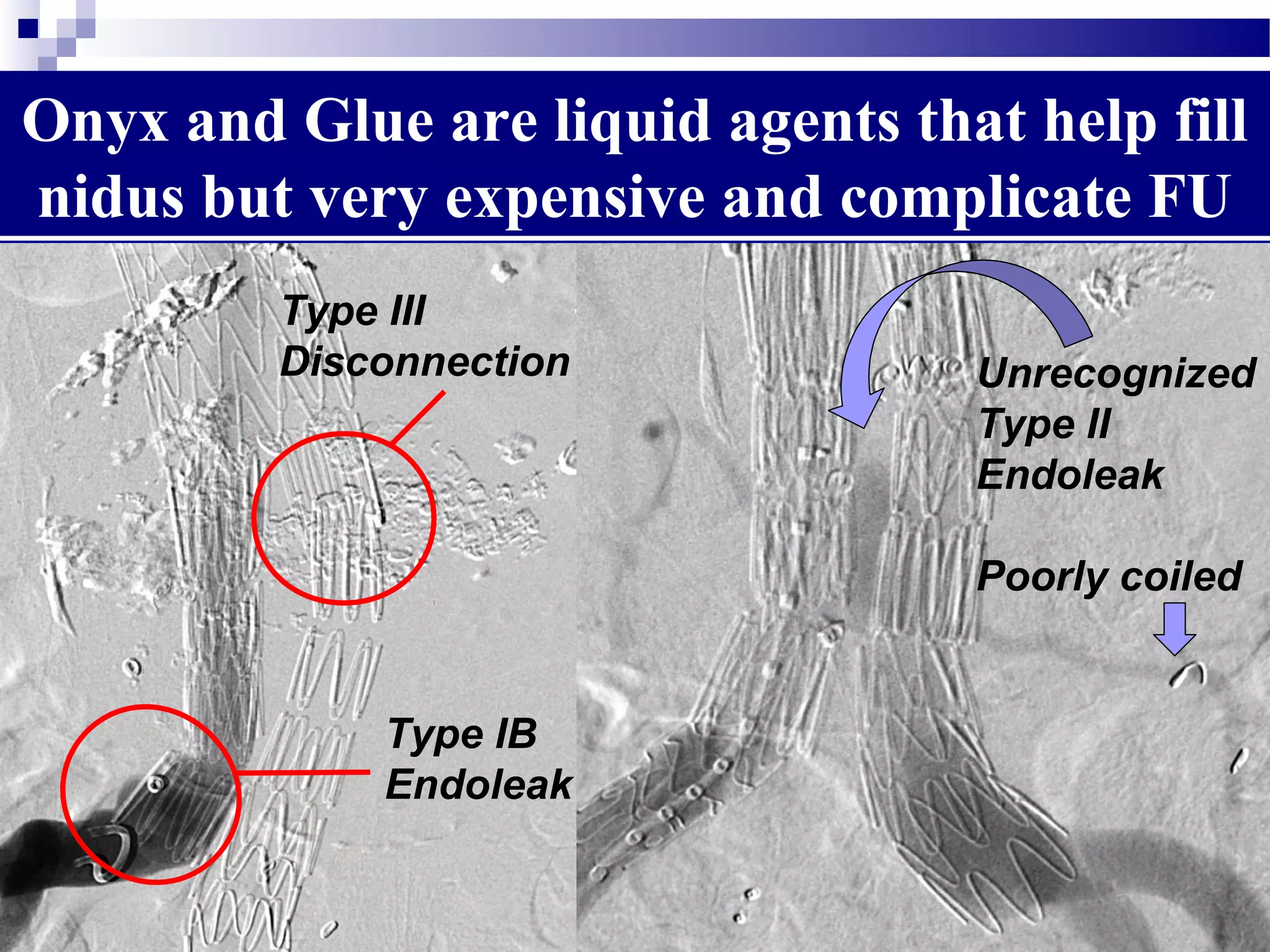
1. There are different types of endoleaks that can occur after EVAR, with Types I and III considered the most serious due to their risk of rupture if not treated.
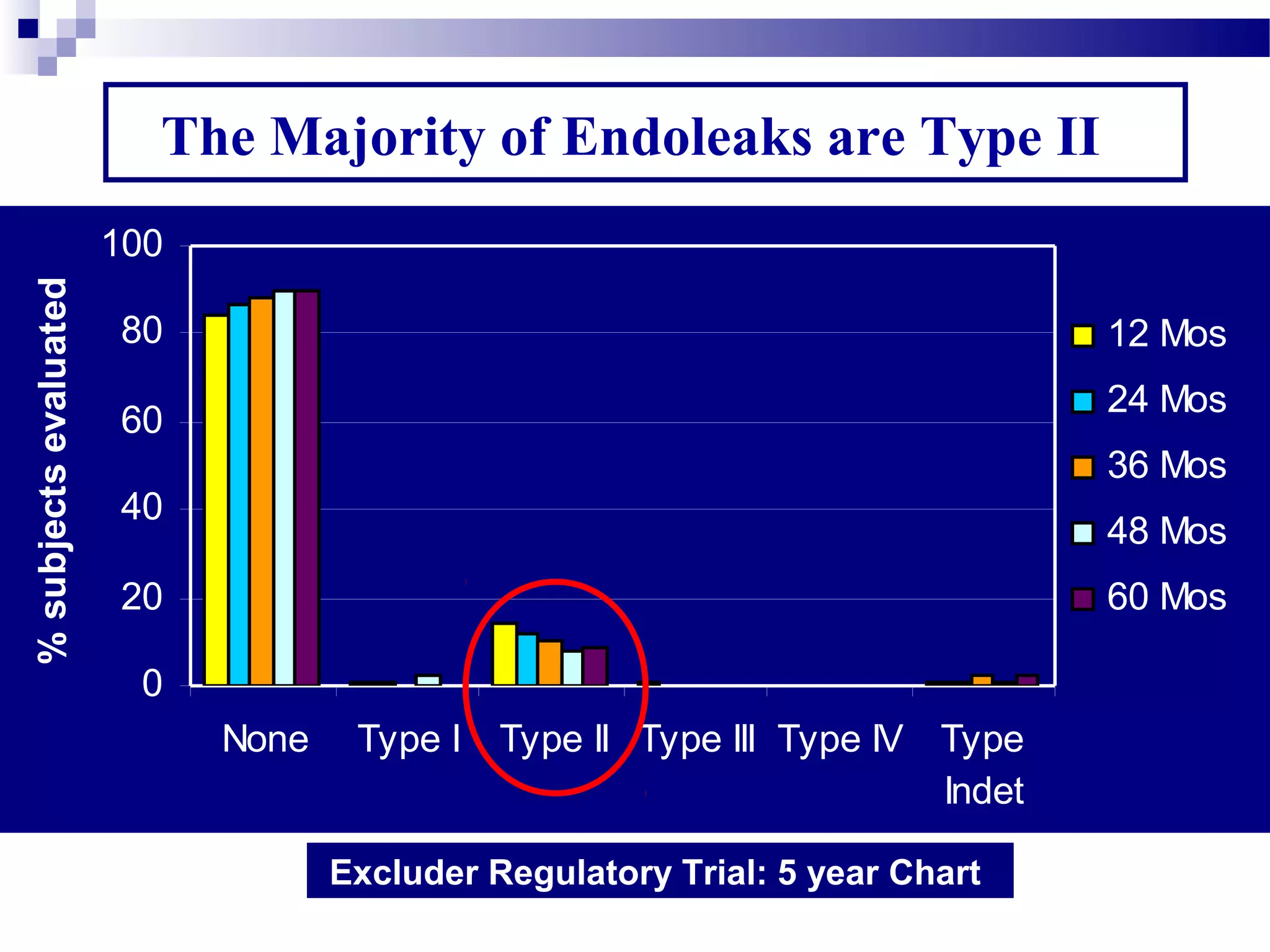
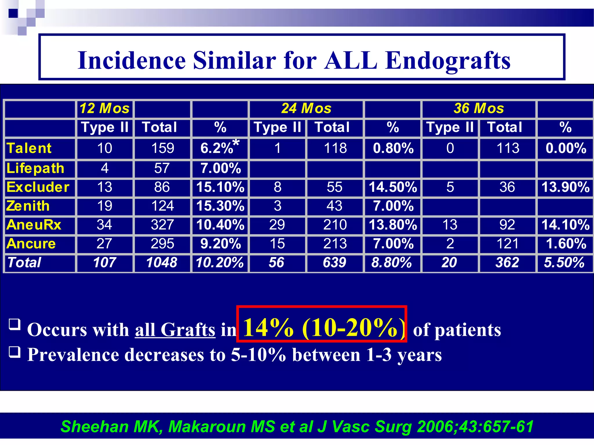
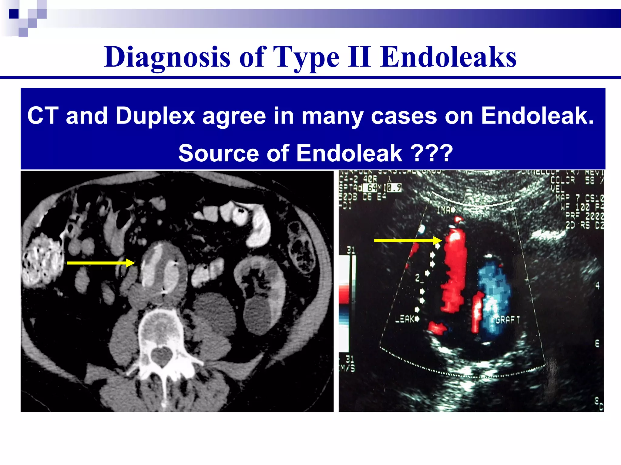
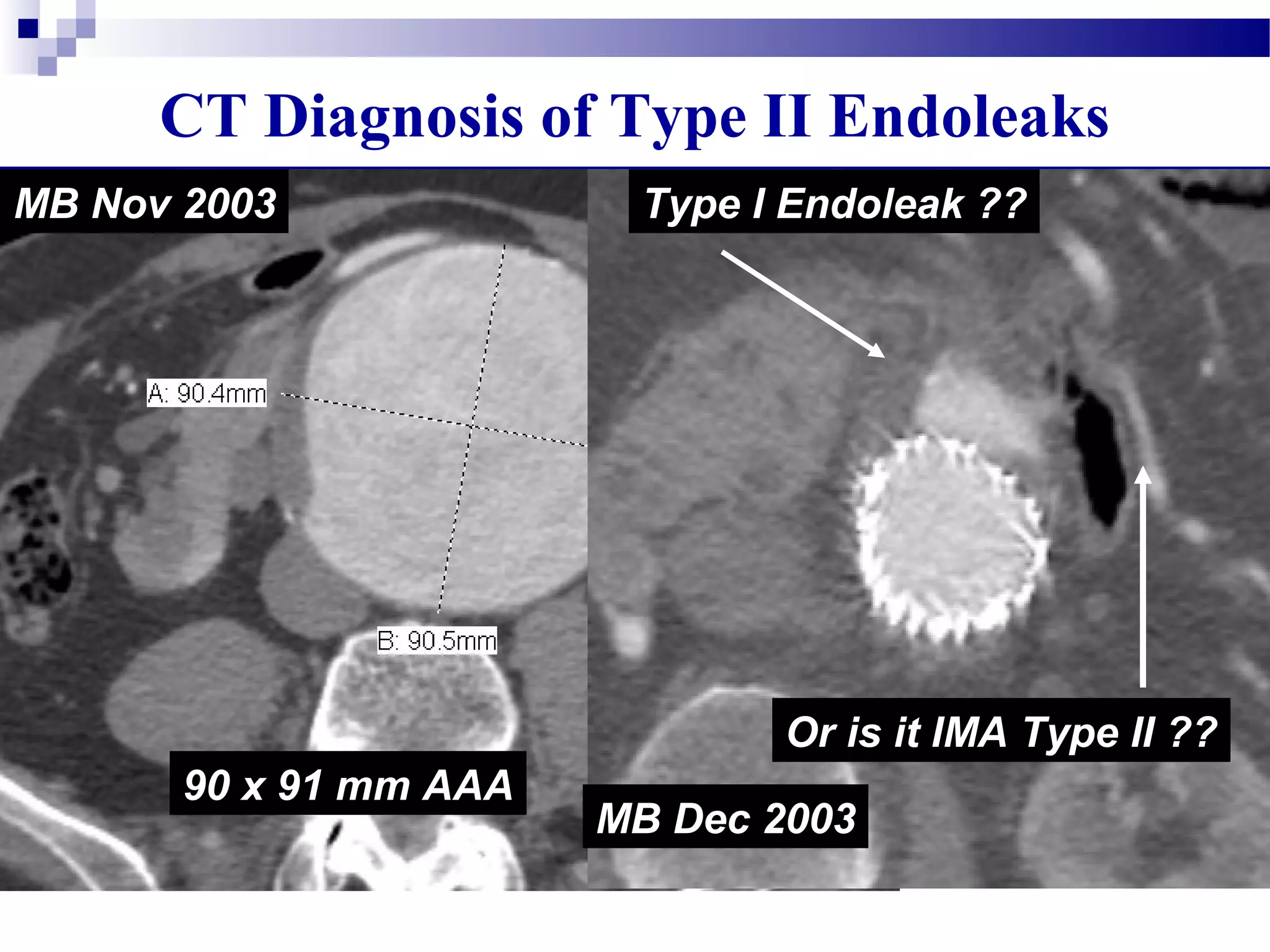
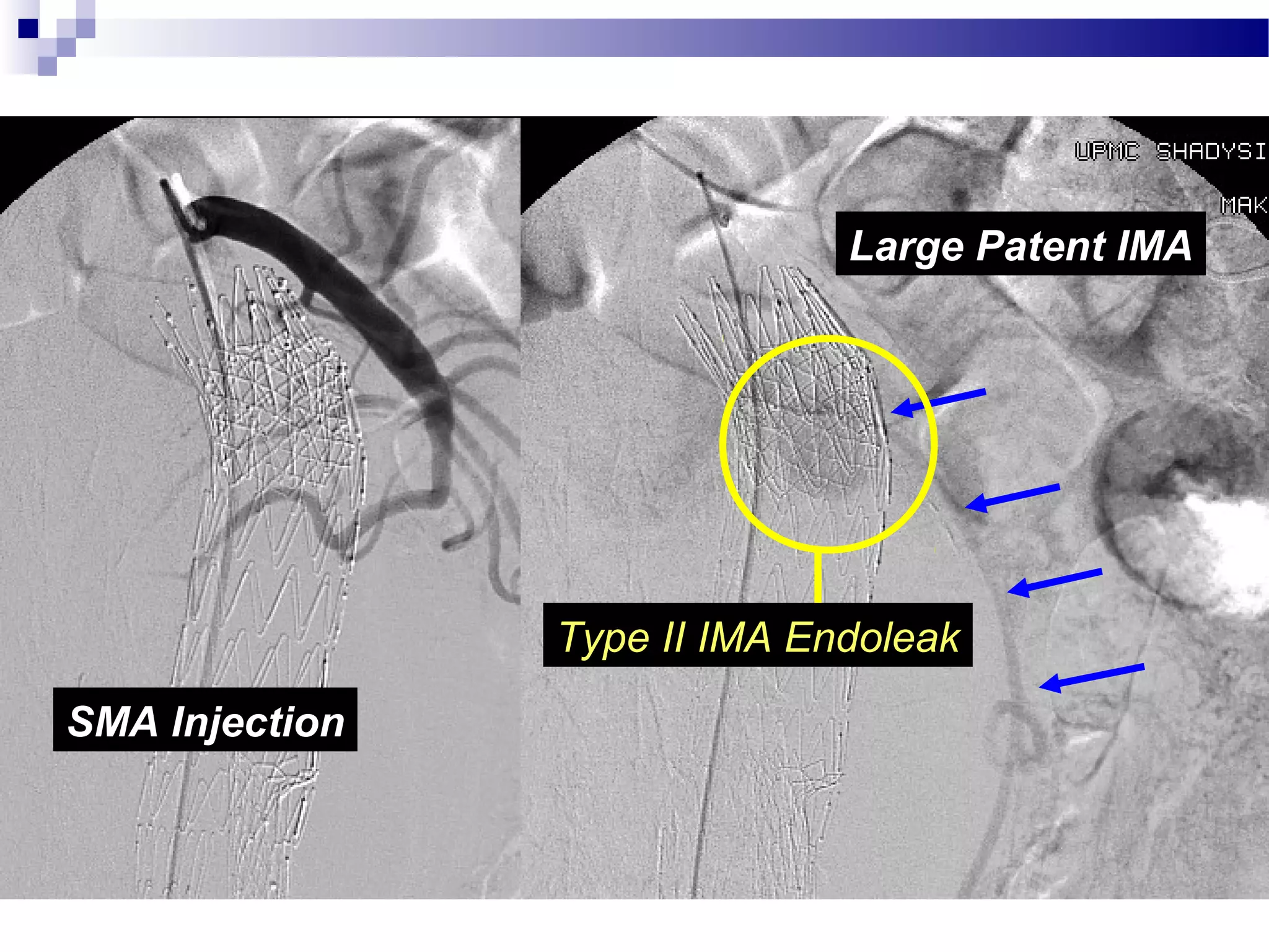
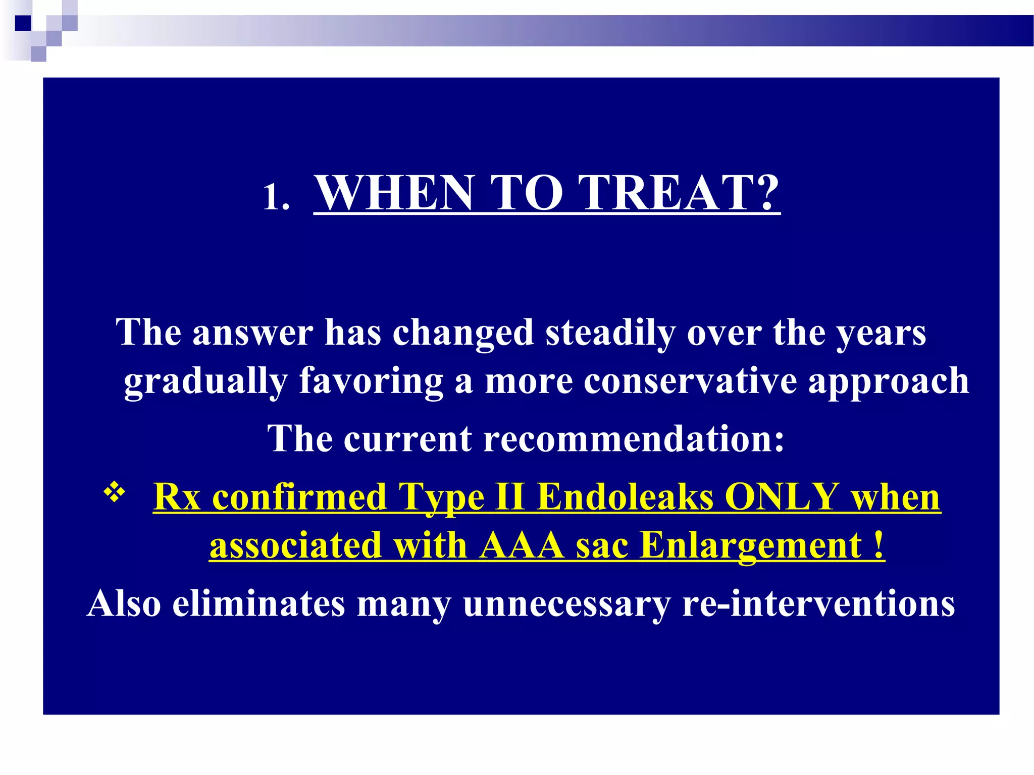
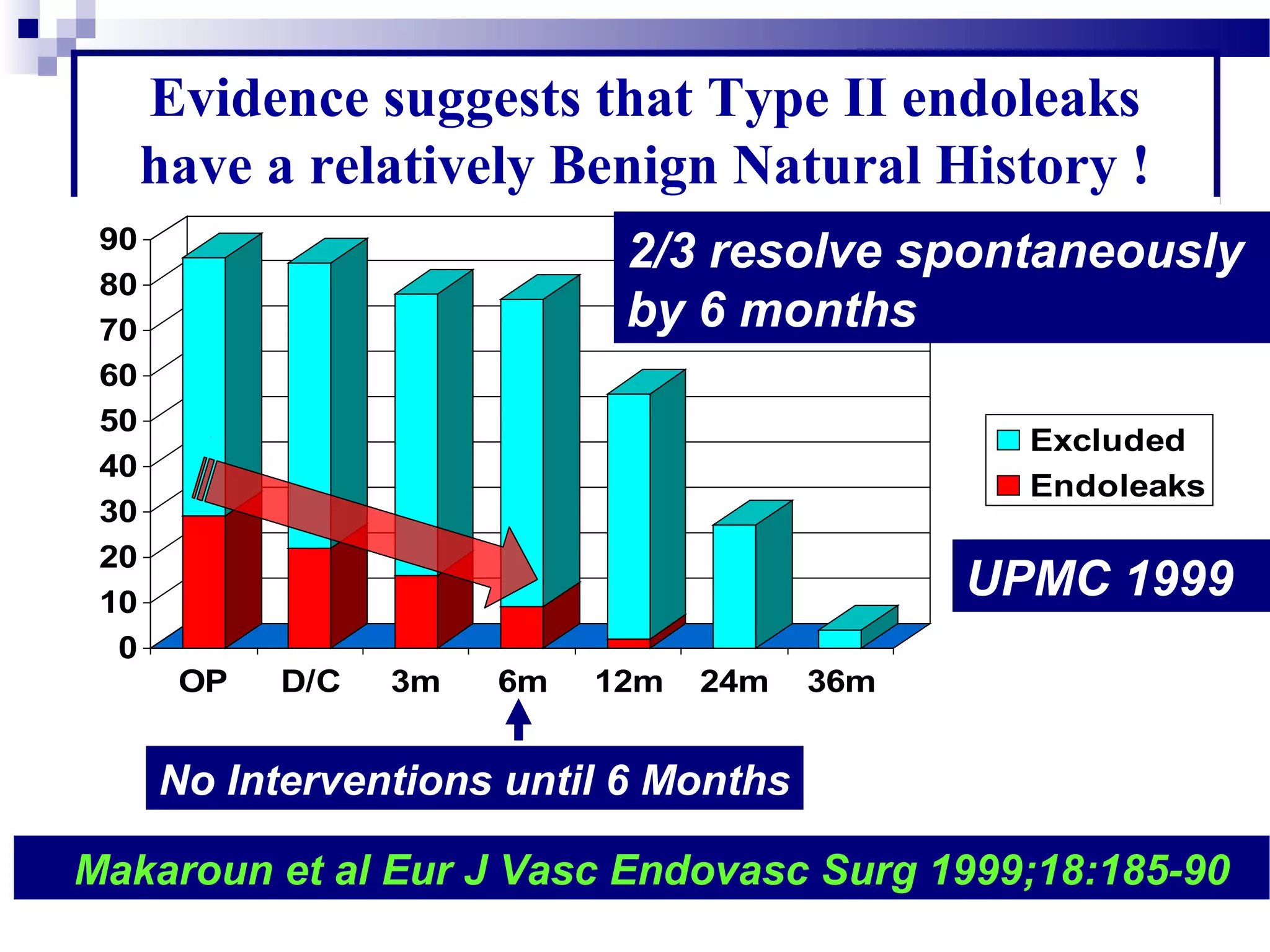
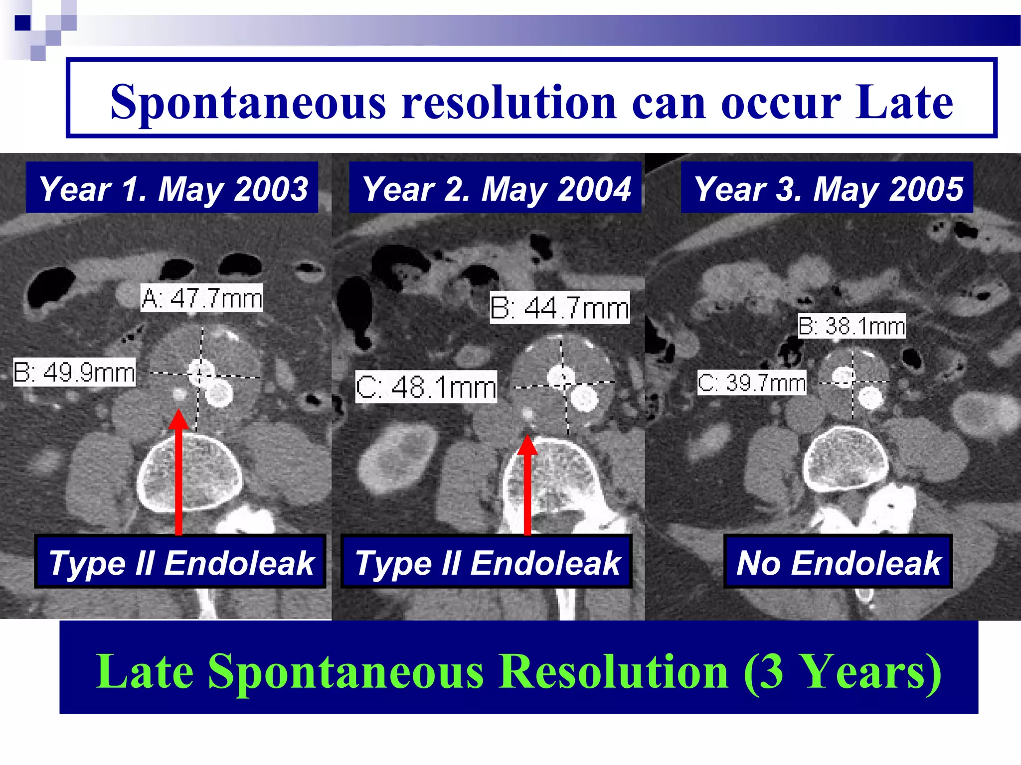
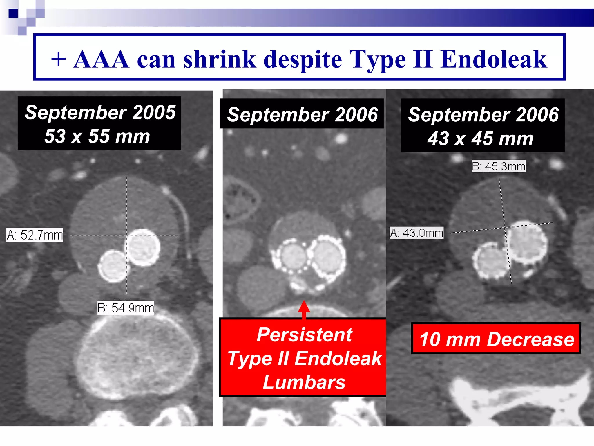
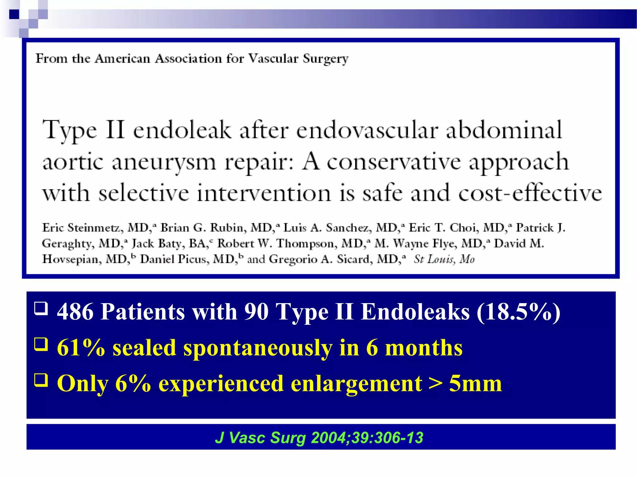
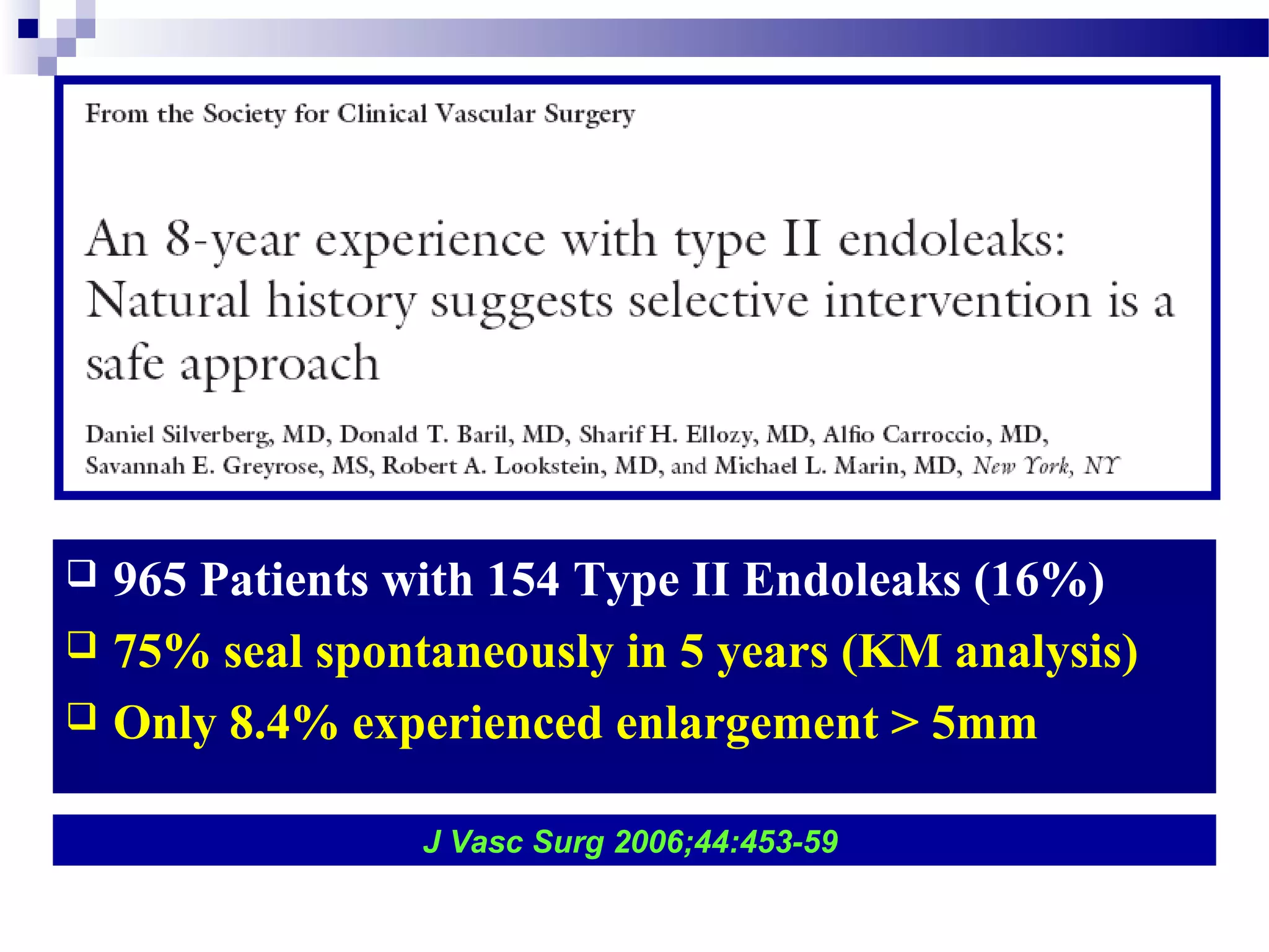
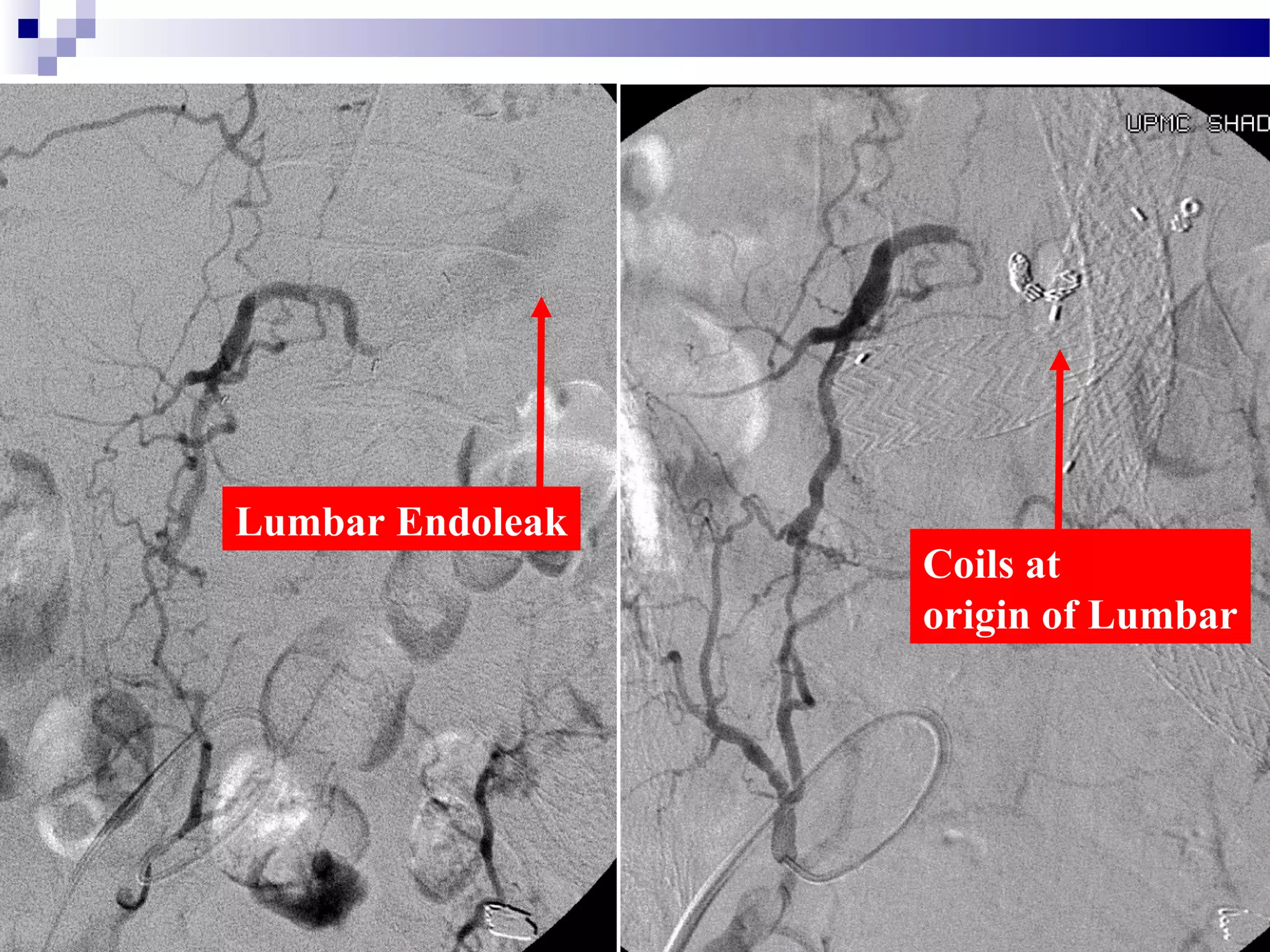
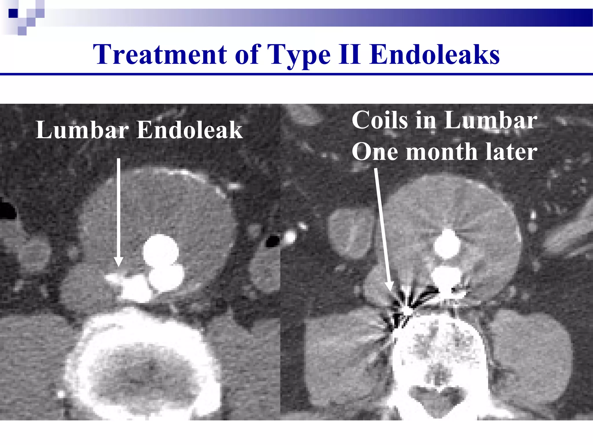
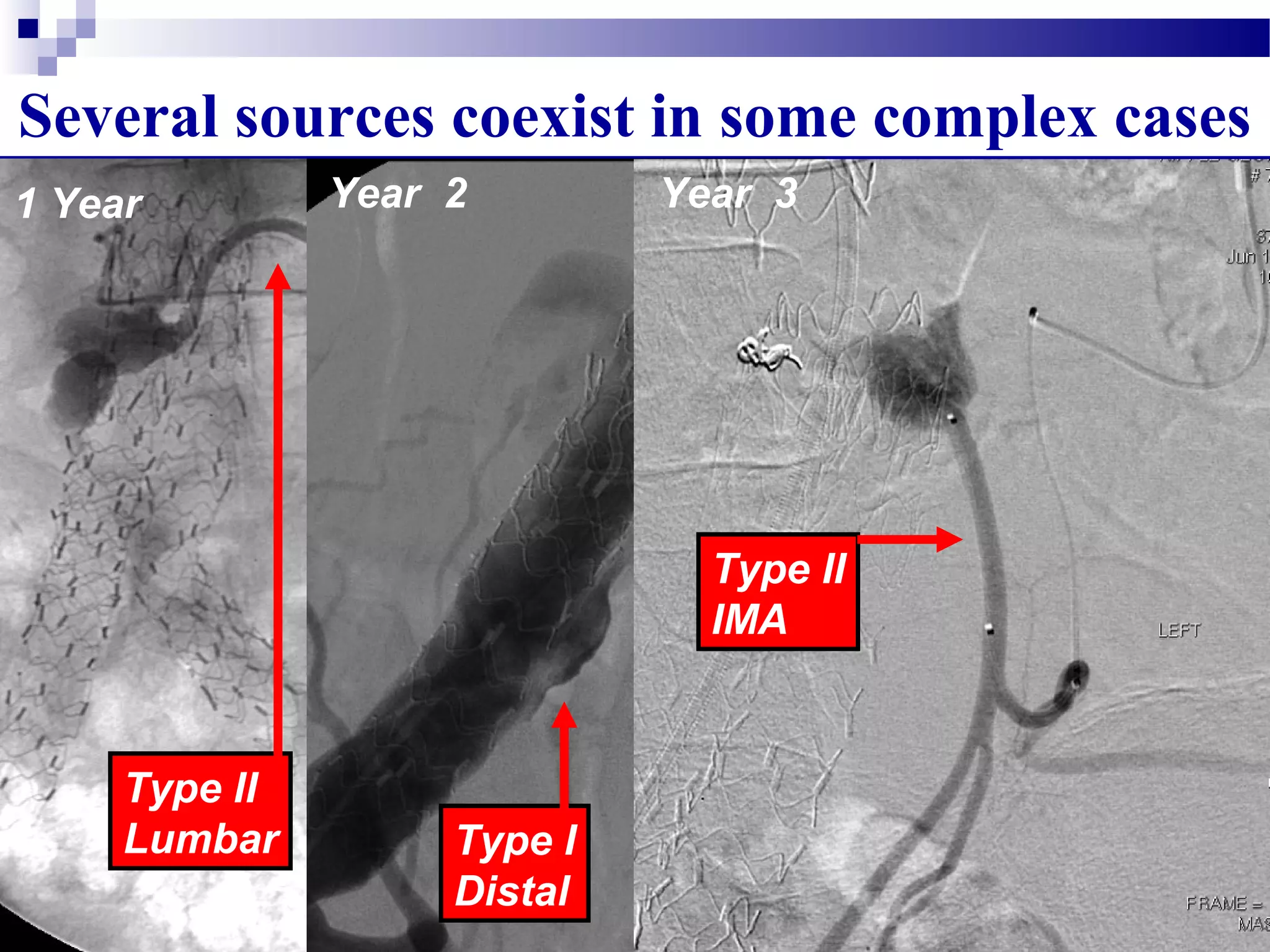
2. Type II endoleaks are more common but often have a benign natural history, with many resolving spontaneously. Treatment is only recommended if the aneurysm sac enlarges.
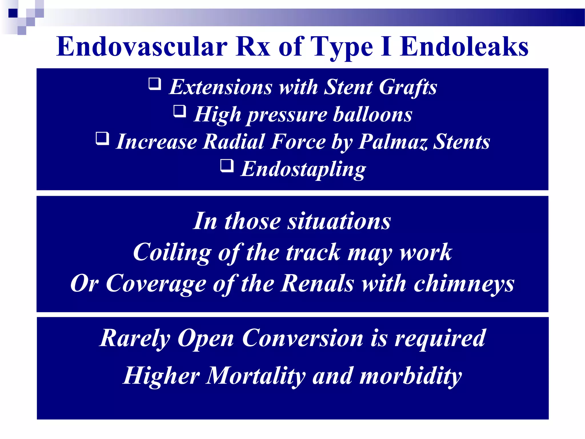
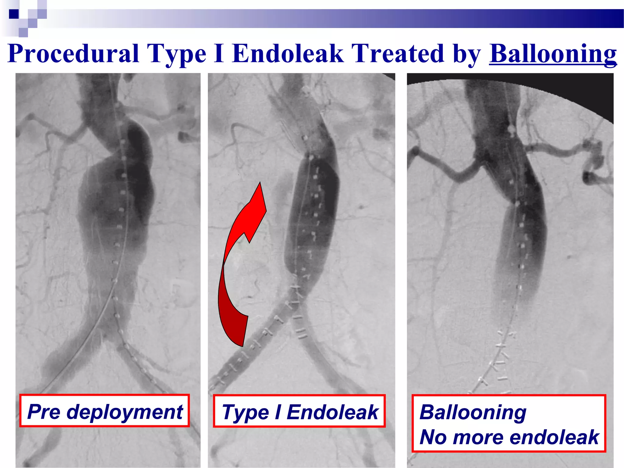
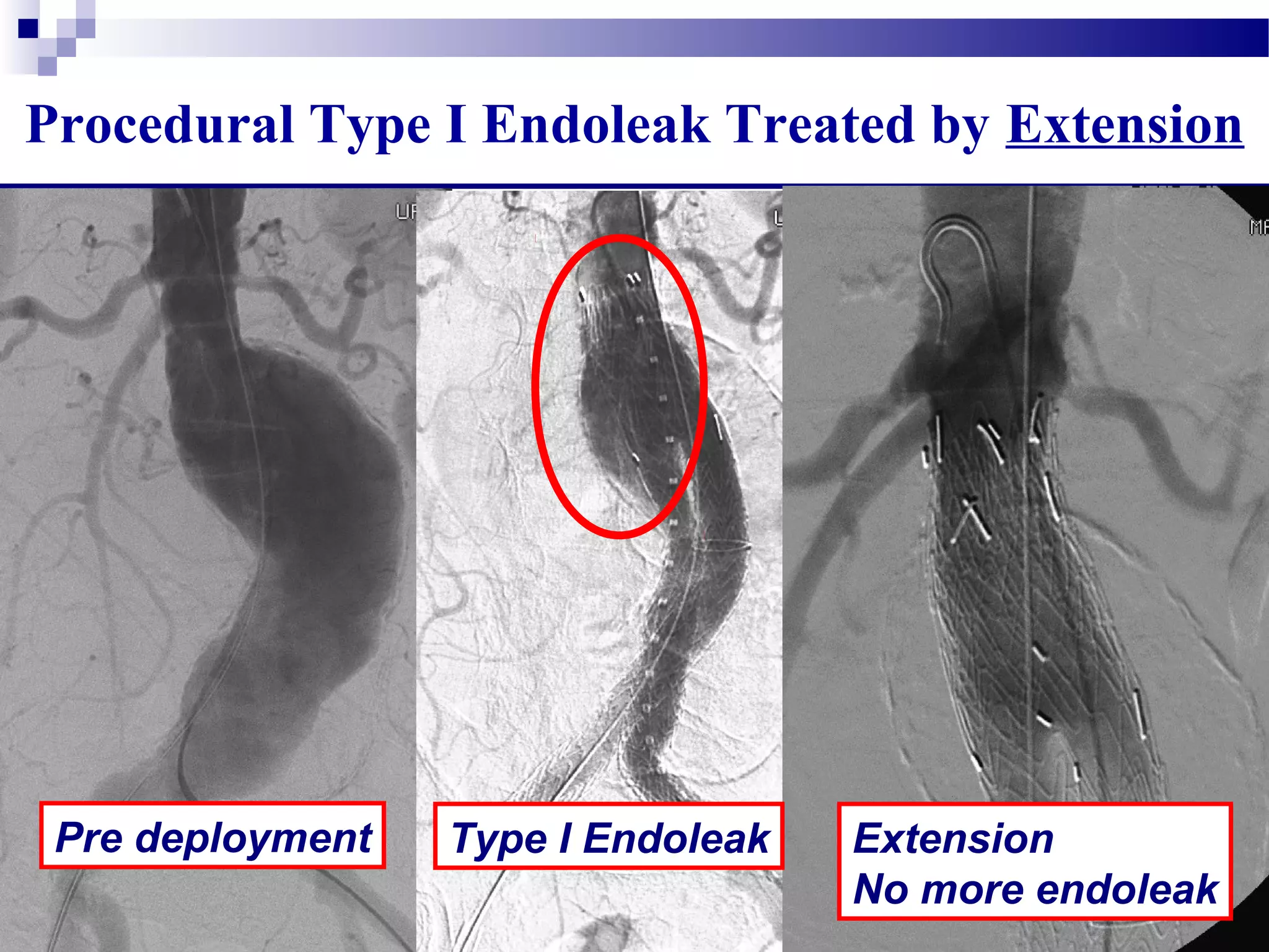
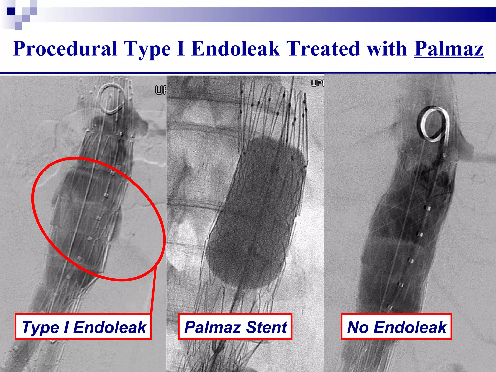
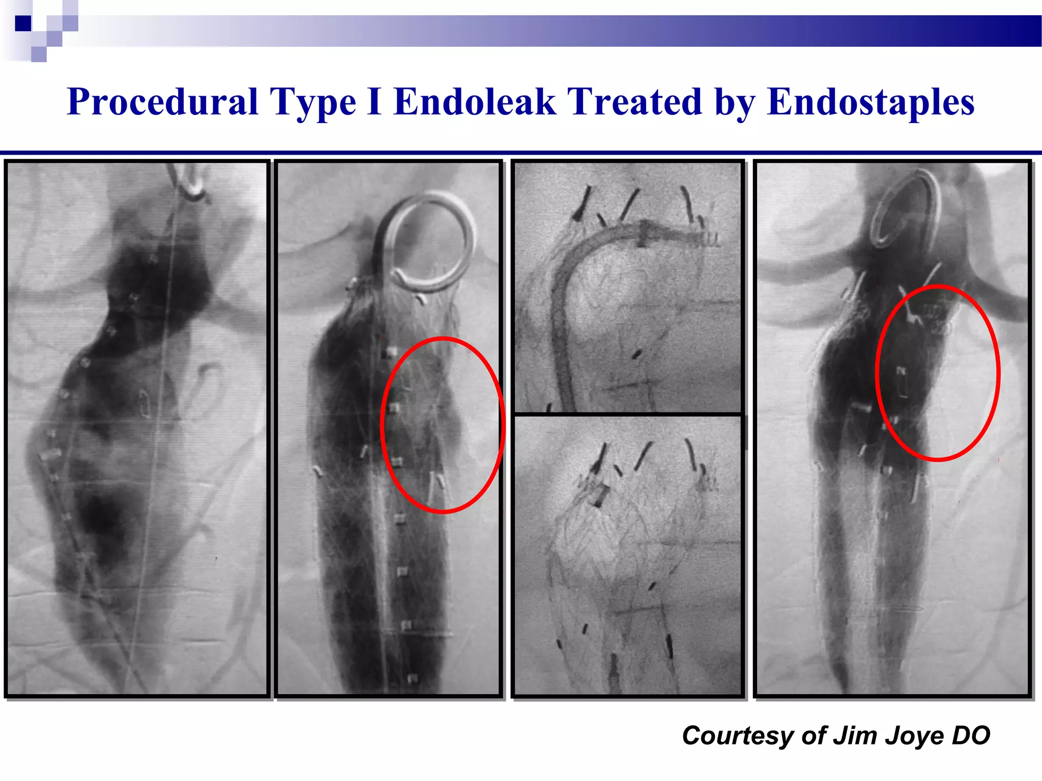
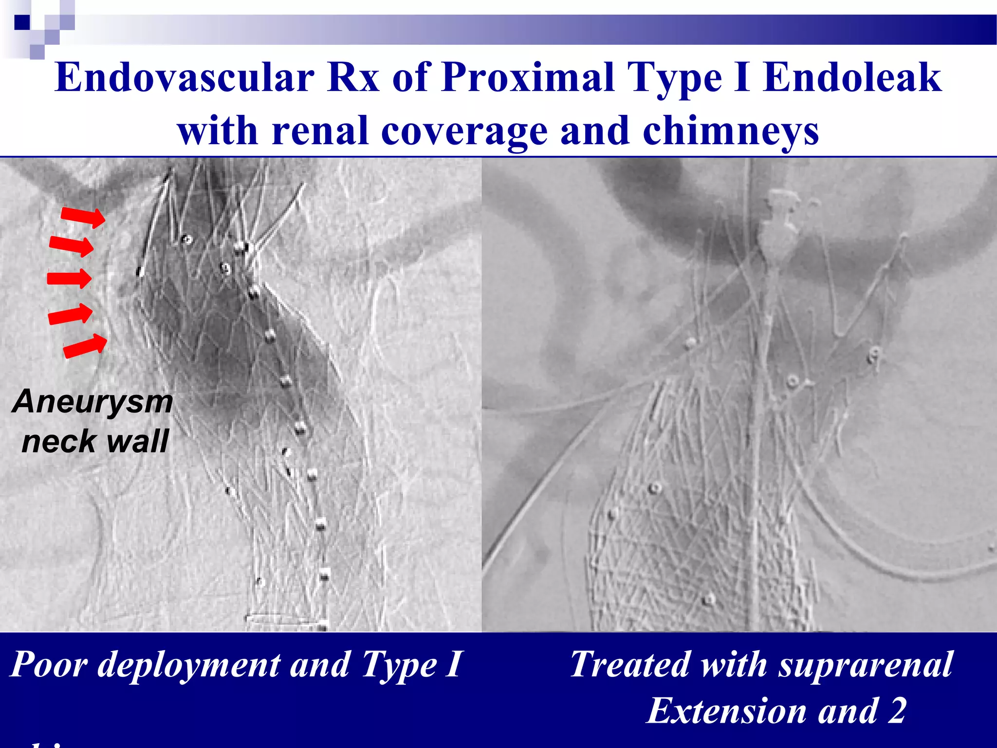
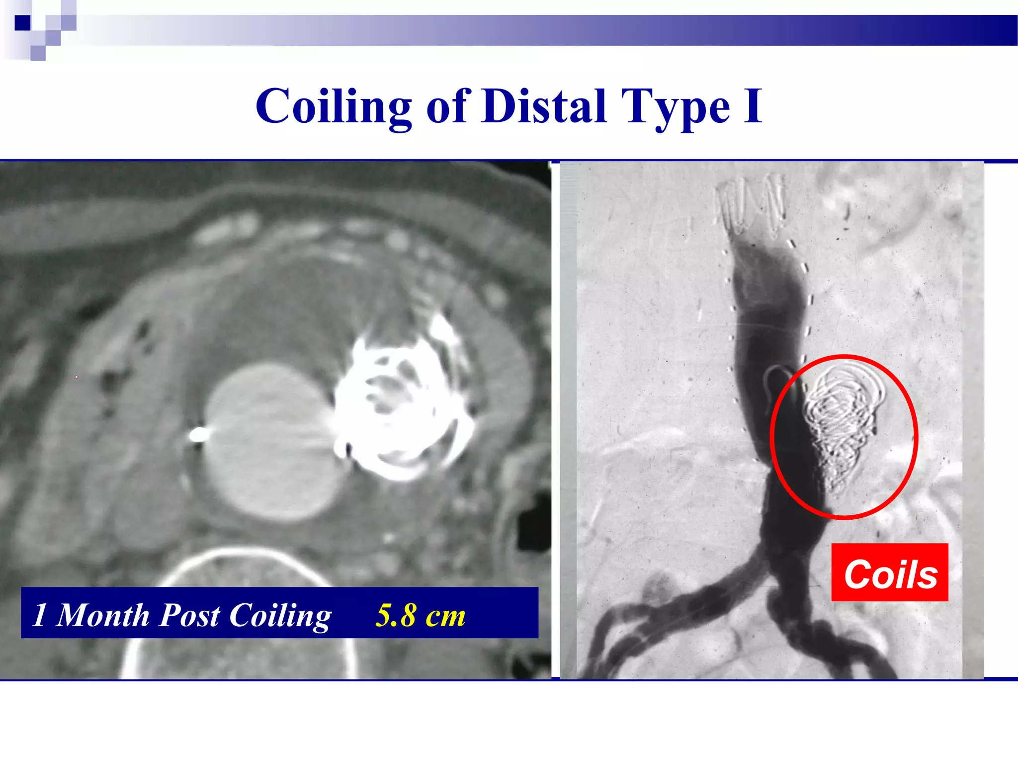
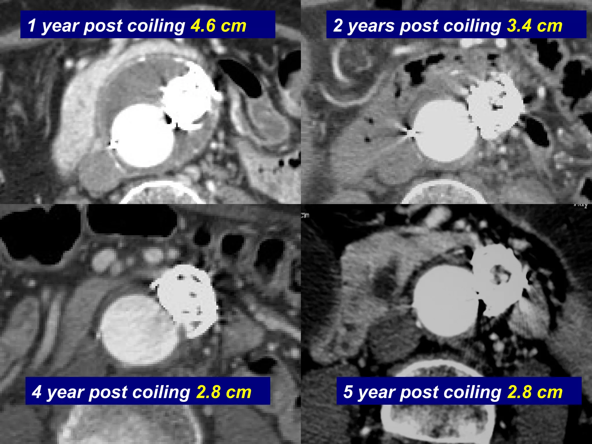
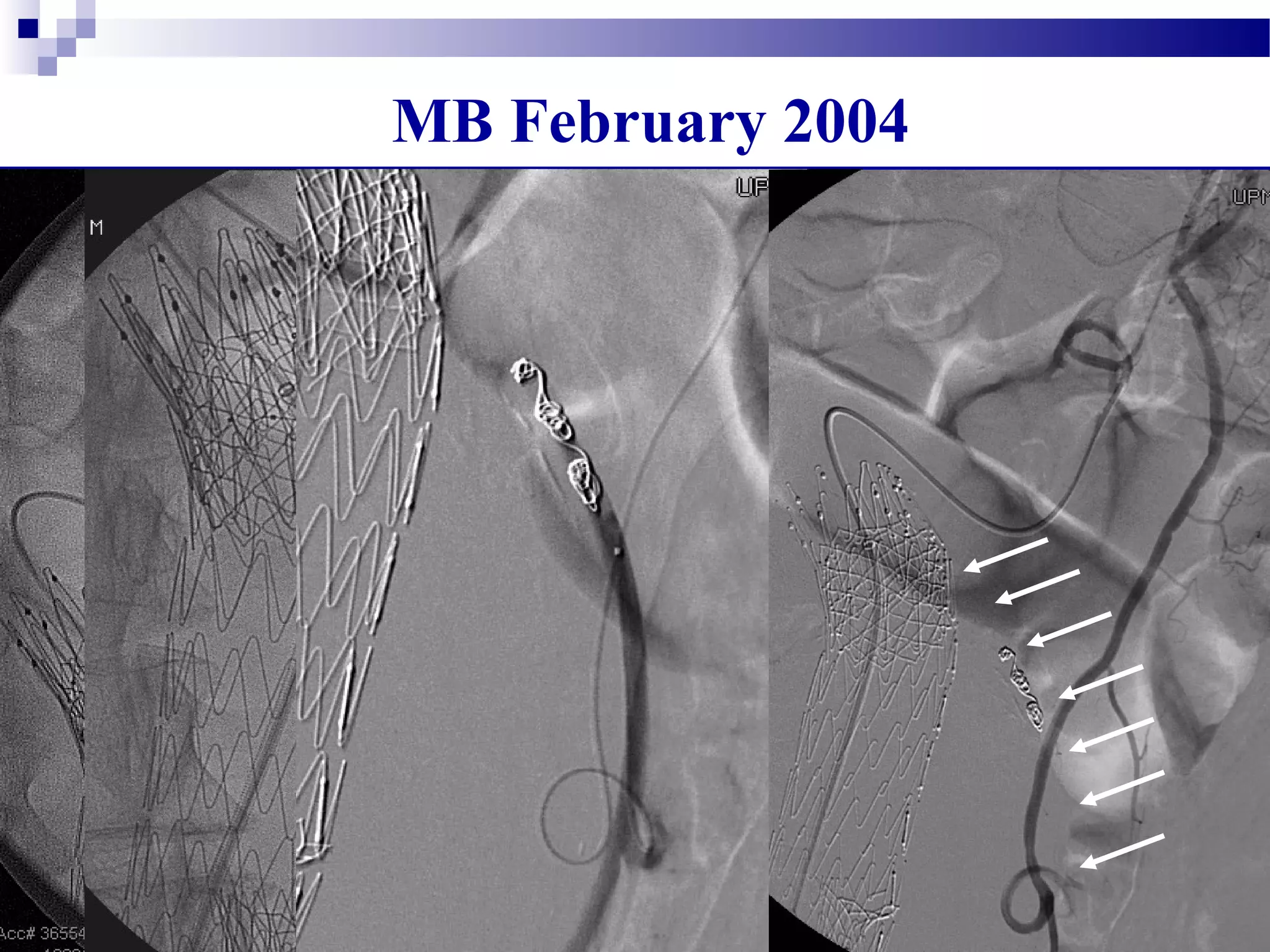
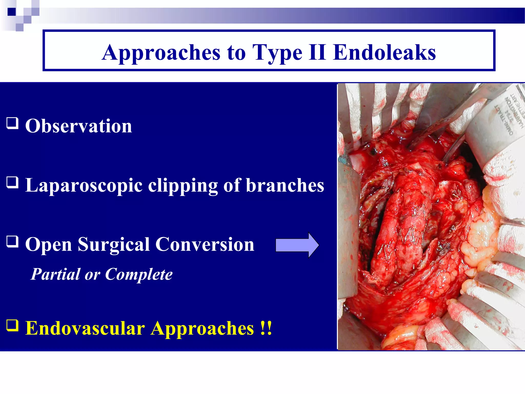
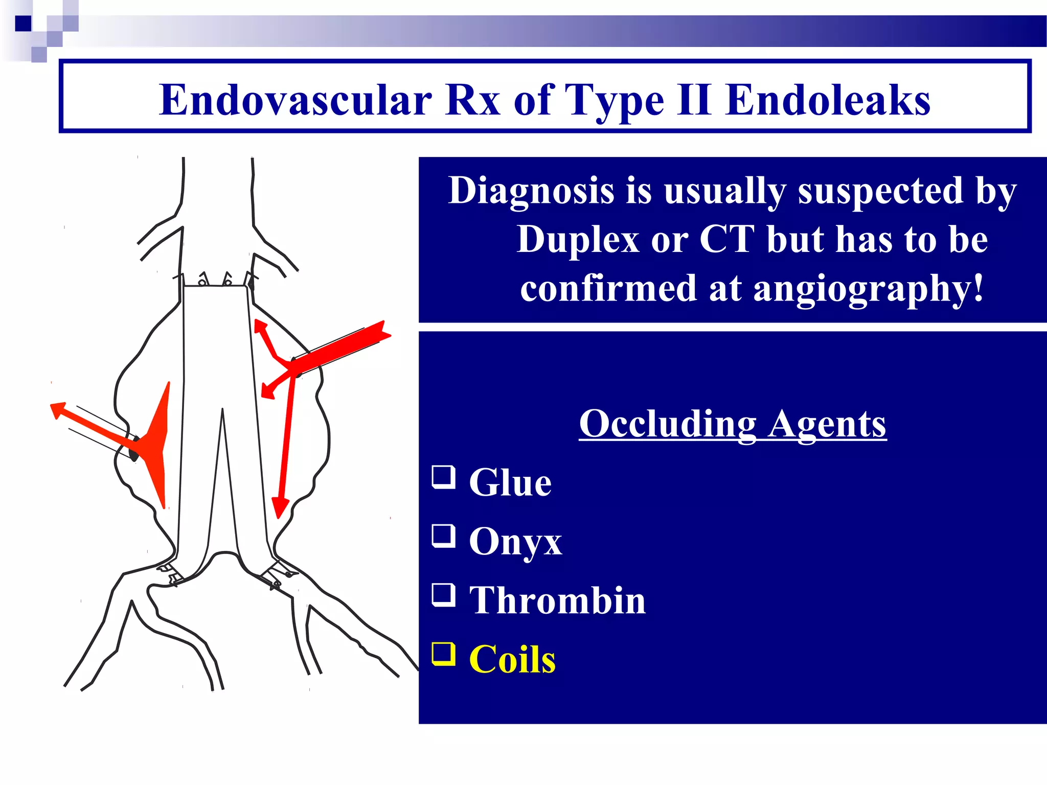
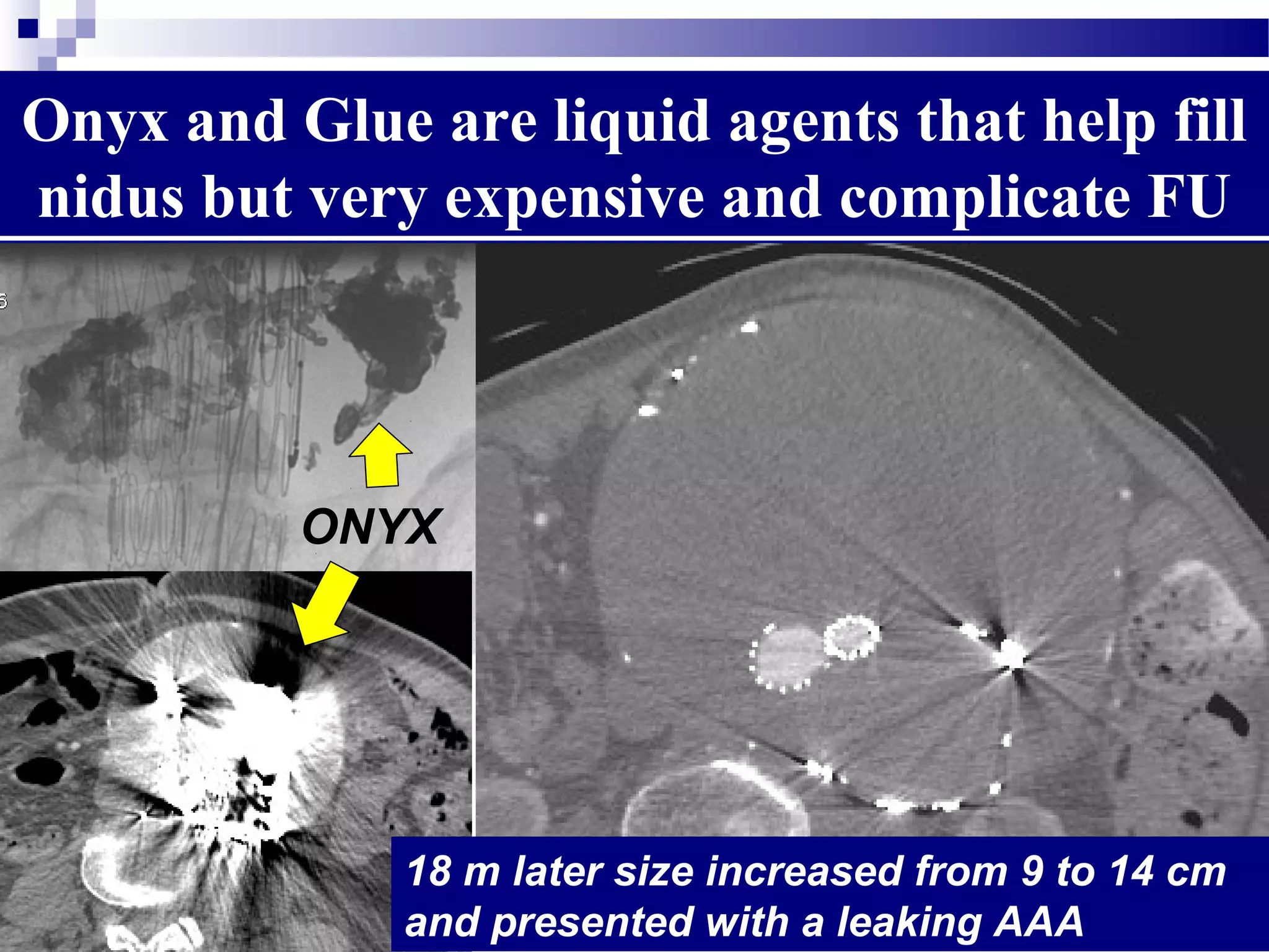
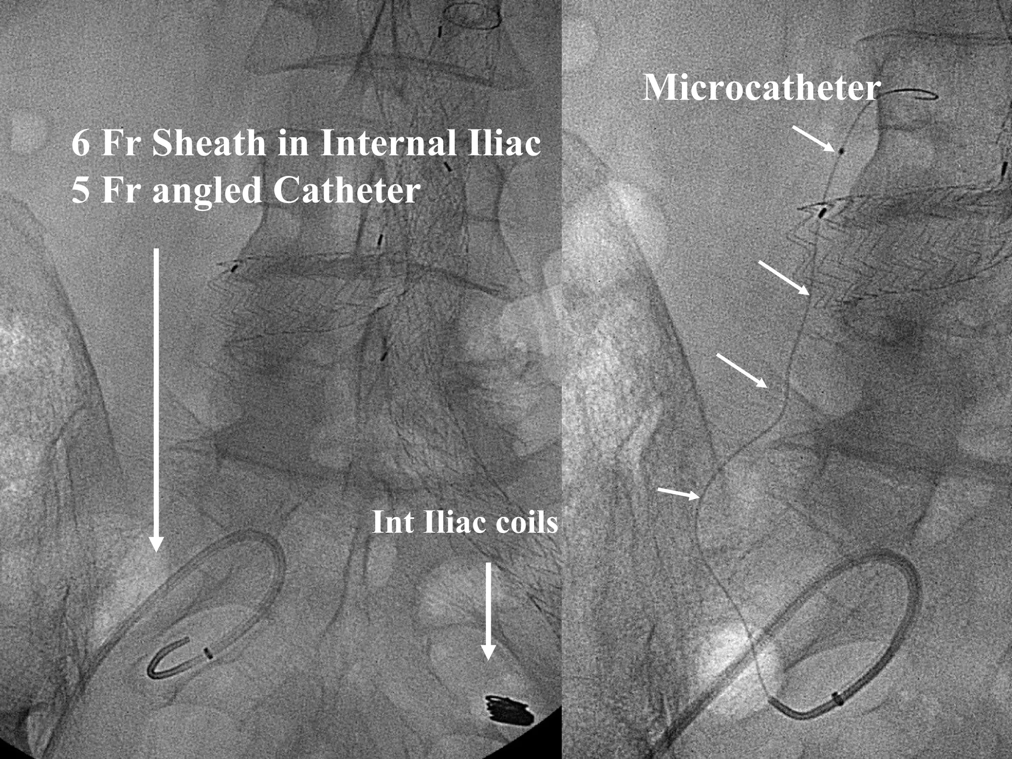
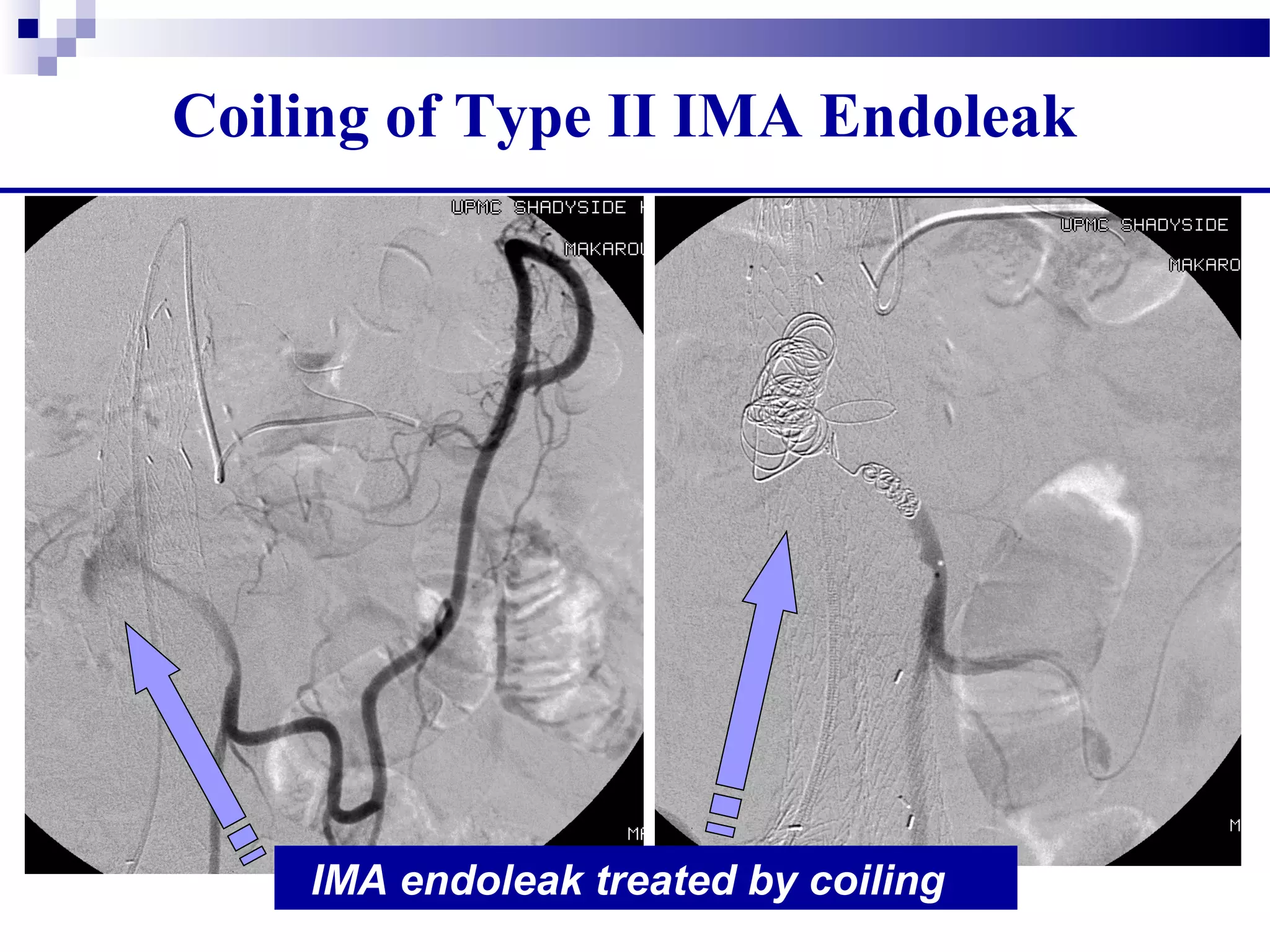
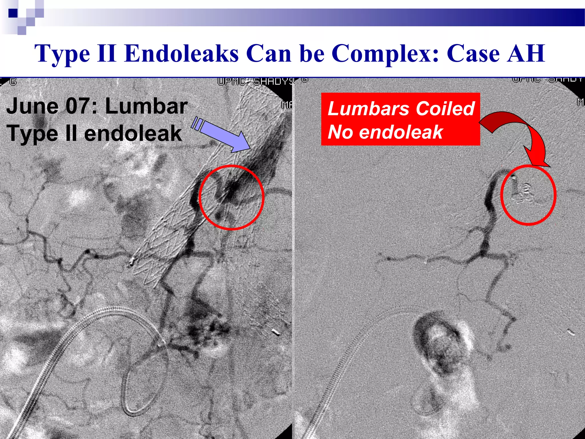
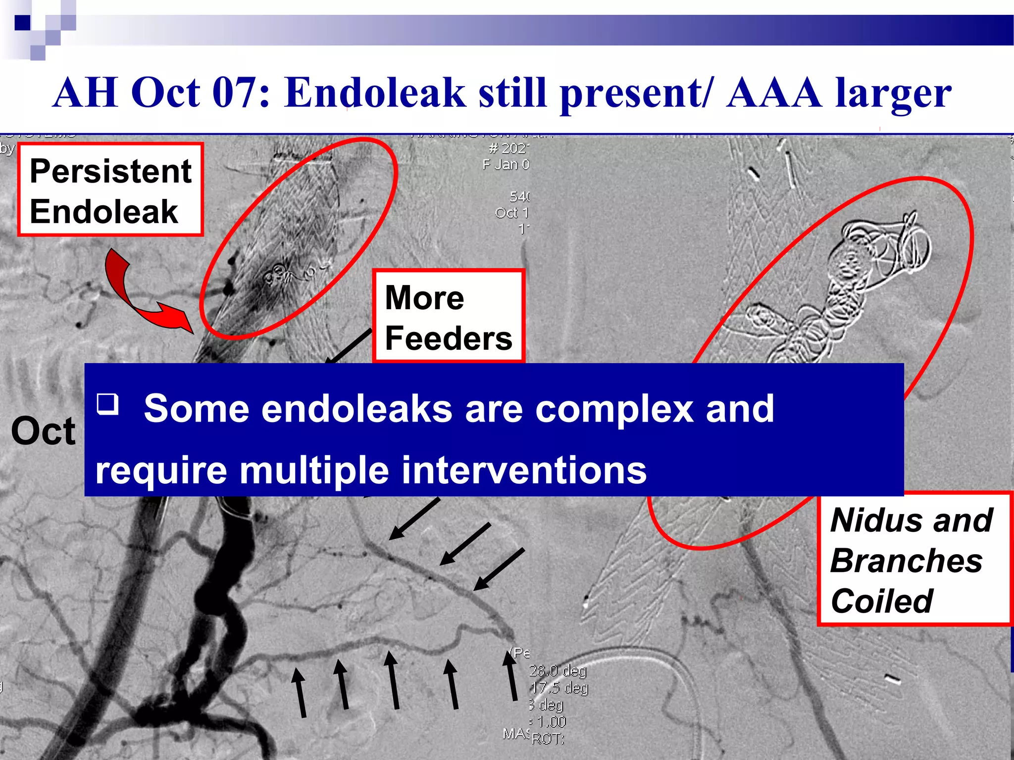
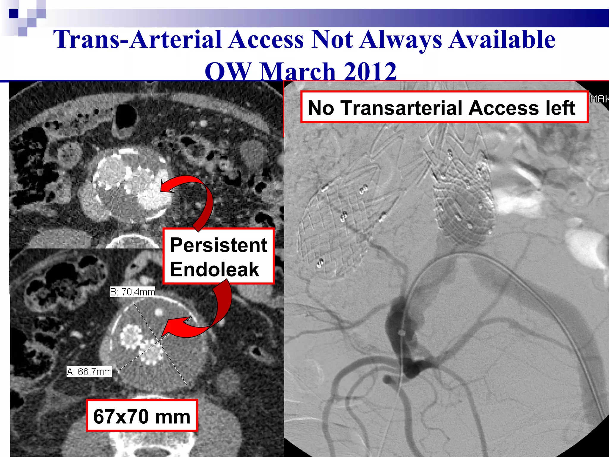
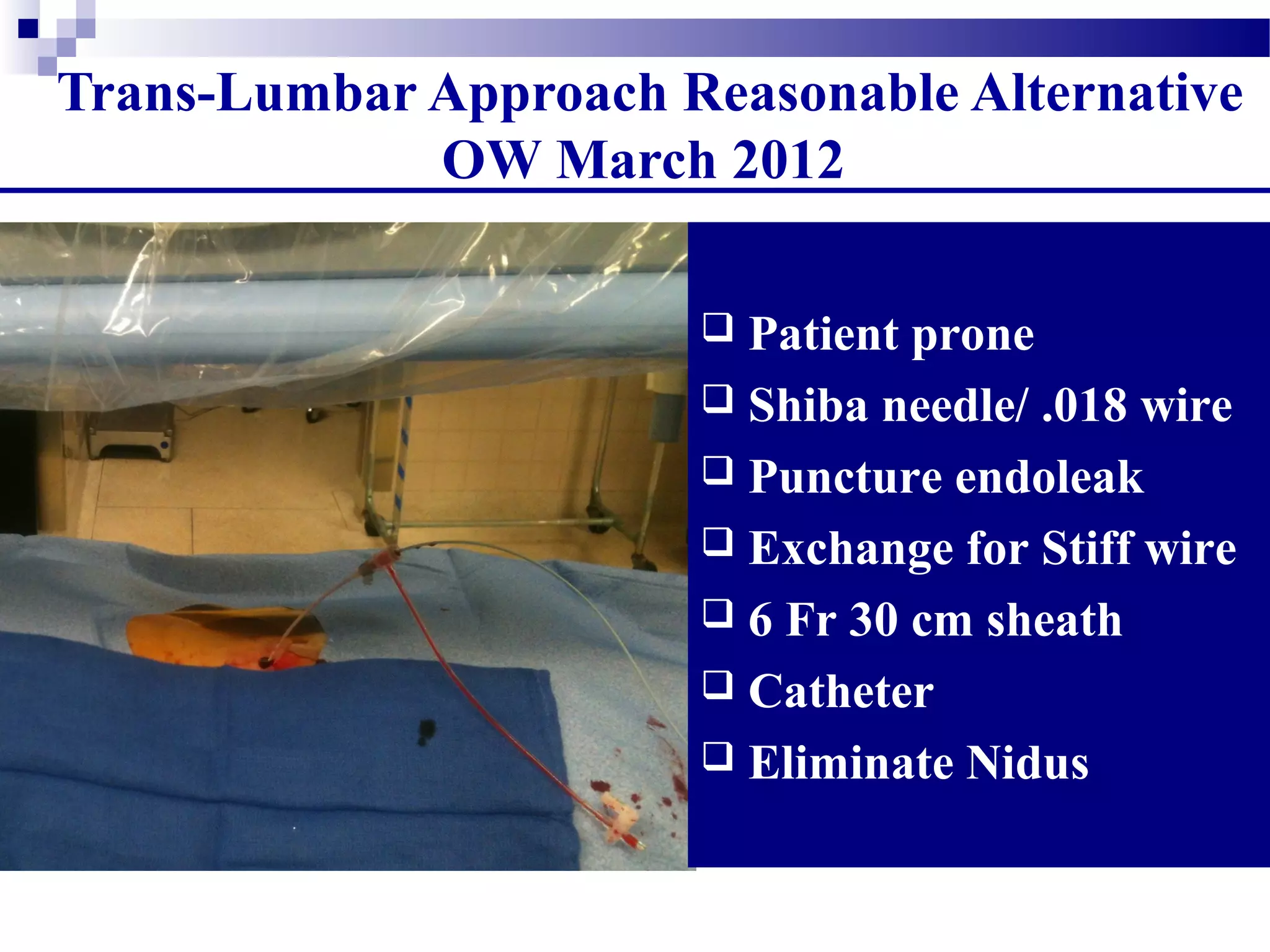
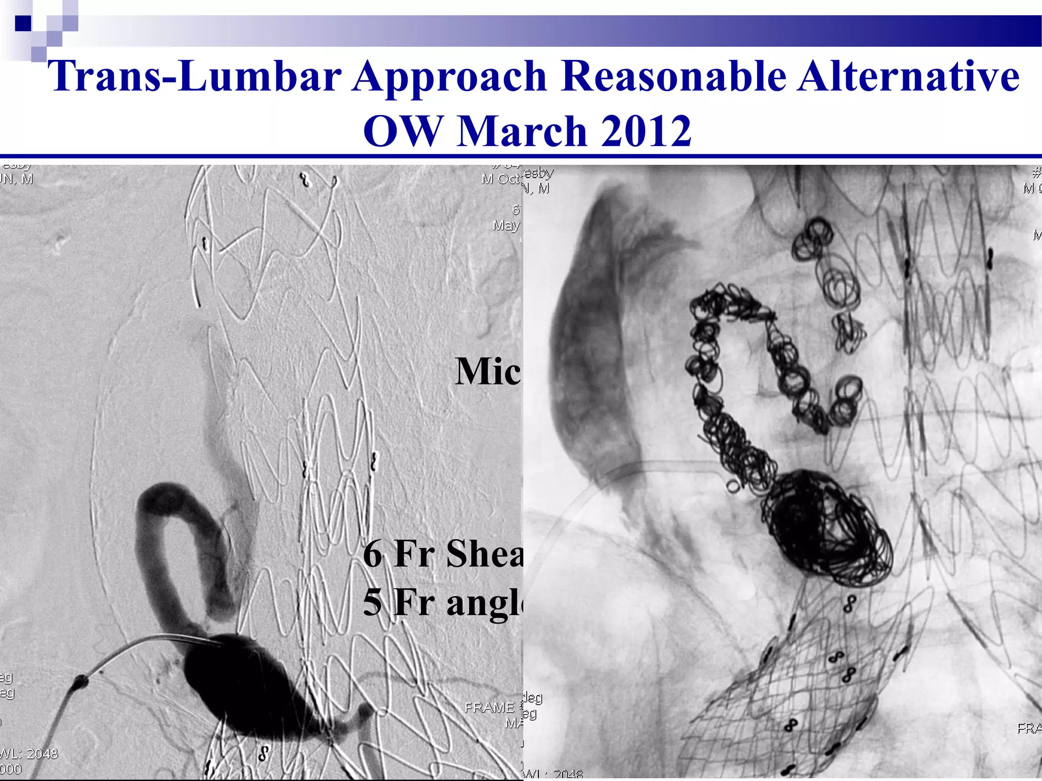
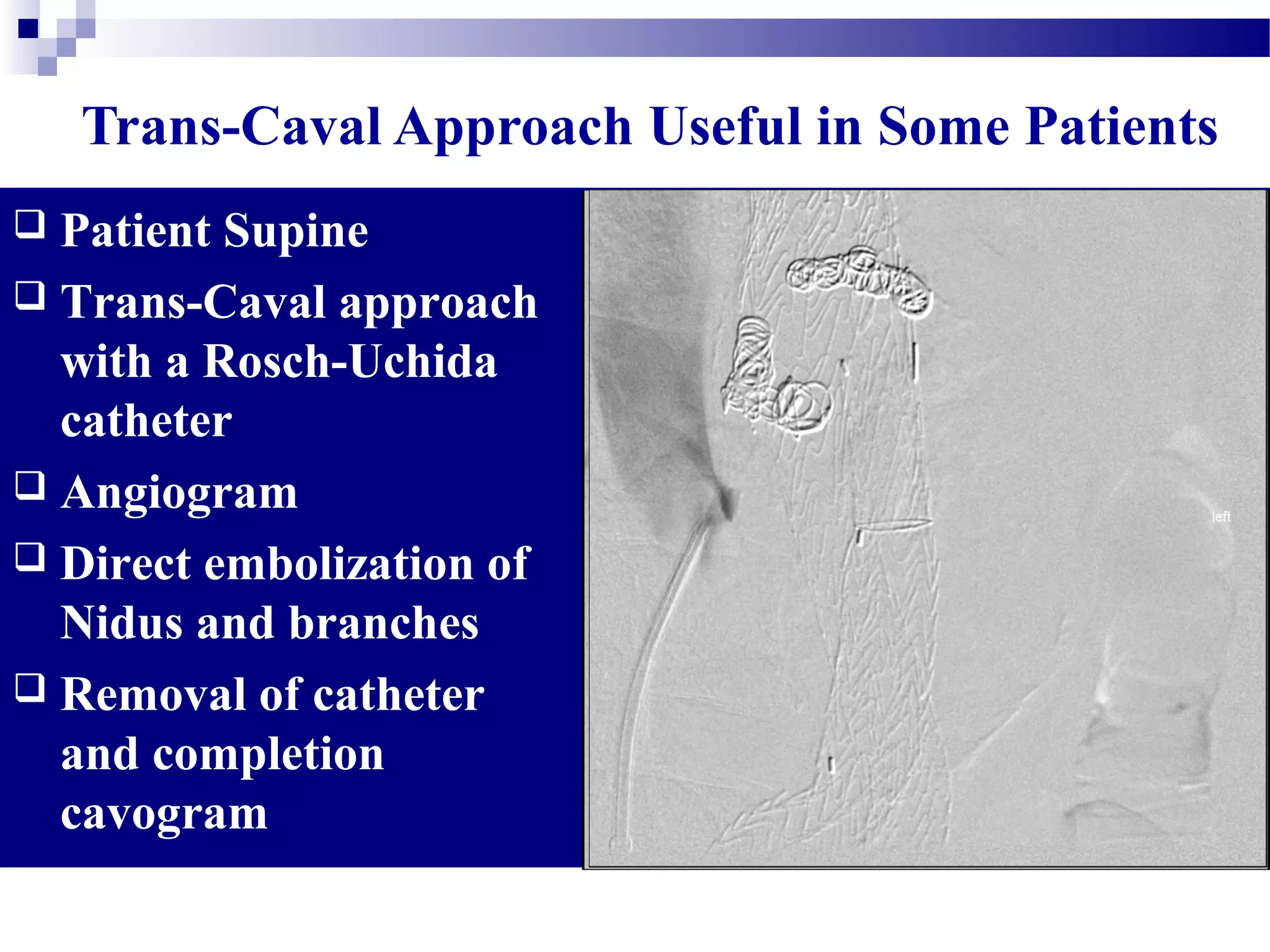
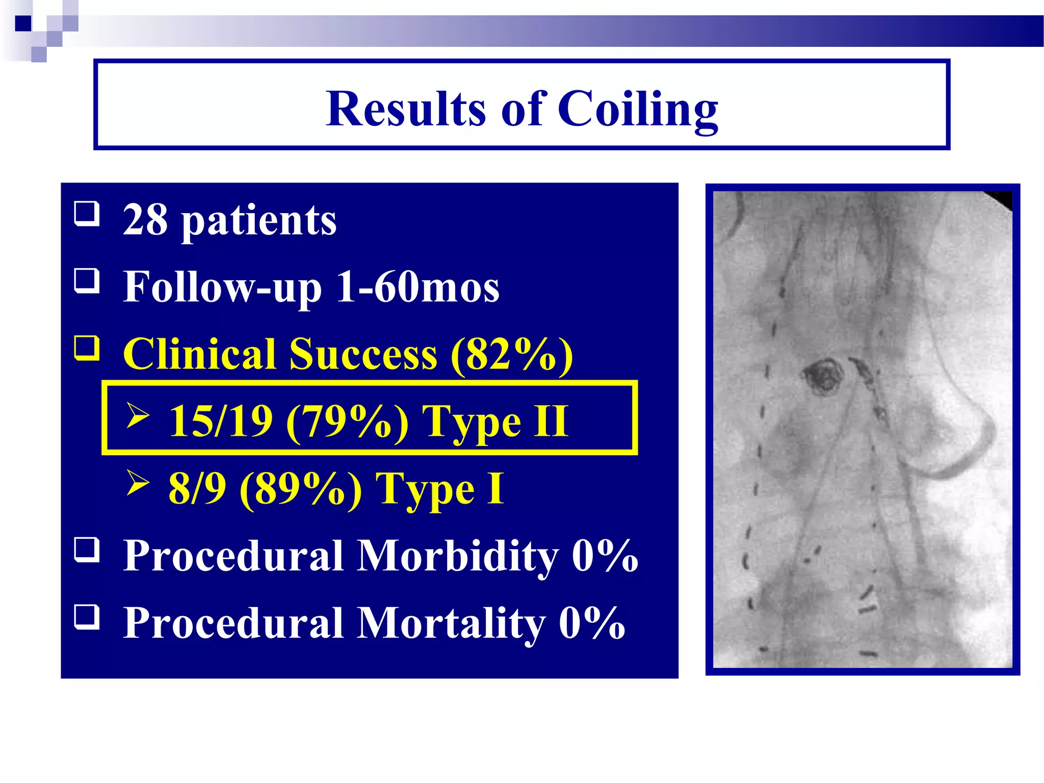
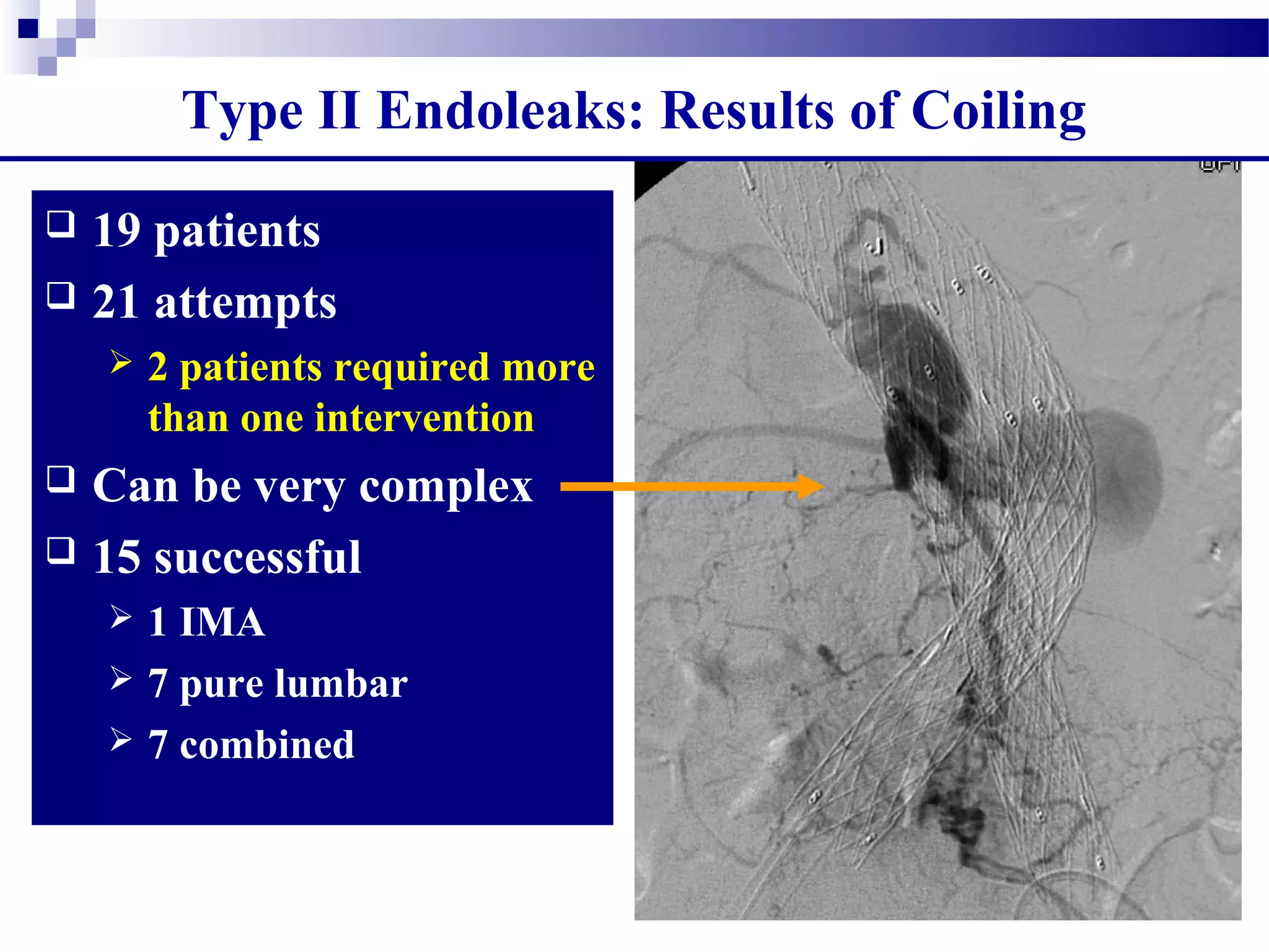
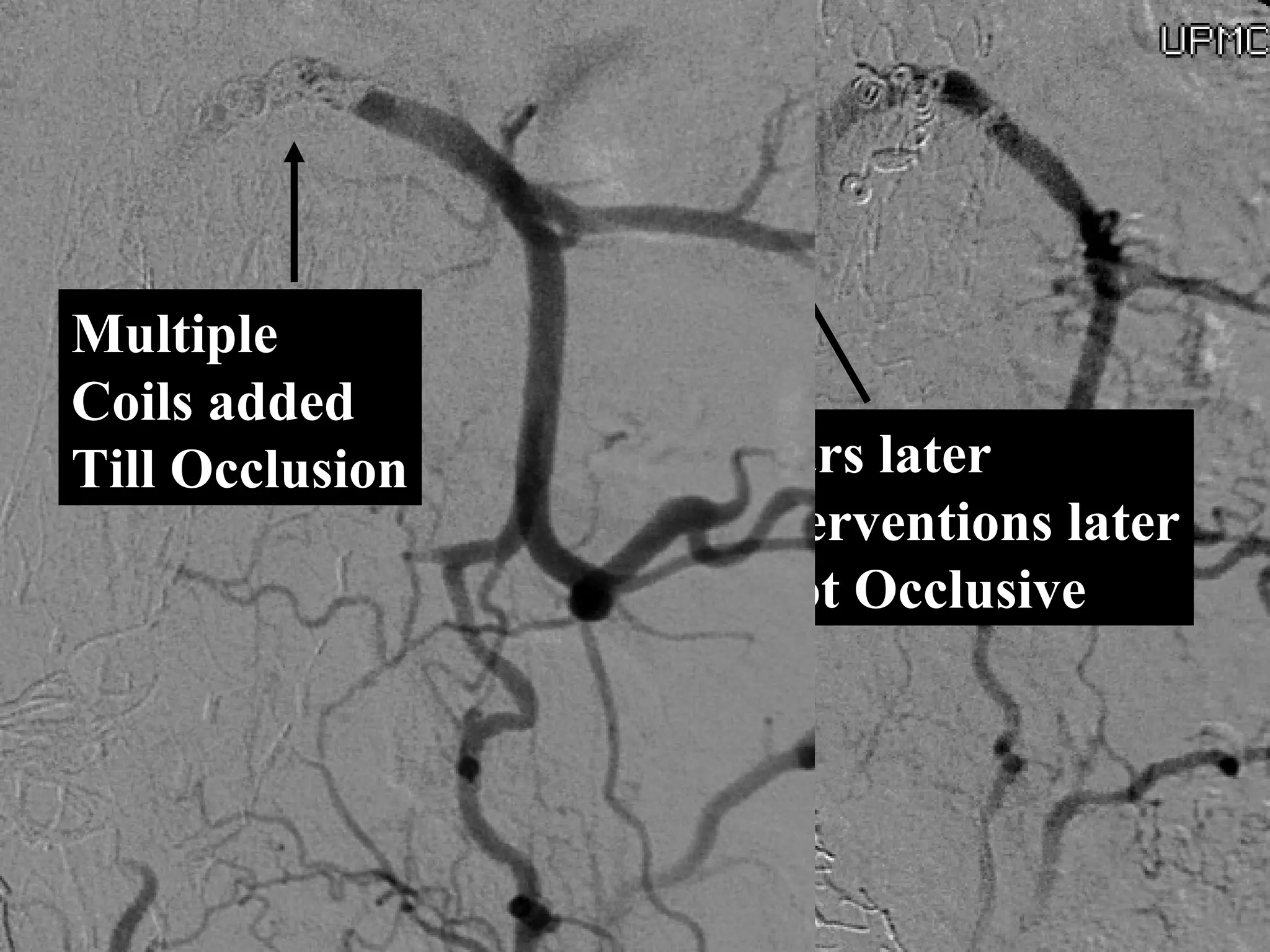
3. Endovascular techniques can be used to treat Type I, II, and III endoleaks via extensions, coils, glue, or onyx, with the goal of occluding the source of blood flow into the sac. Transarterial, translumbar, and transcaval approaches may be necessary depending on anatomy.
This document discusses endoleaks, which occur when blood flows outside the endoluminal graft used to treat an aneurysm but remains within the aneurysm sac. It classifies endoleaks into types 1-4 based on their cause. Type 1 endoleaks are graft-related due to issues at fixation sites, while type 2 is due to retrograde branch vessel flow. Type 3 results from graft integrity issues. The document outlines diagnosis and management strategies for different endoleak types, noting that types 1 and 3 can often be treated endovascularly. It also presents statistics on endoleak incidence and risk factors like proximal neck length, diameter and angulation.
CORONARY ENGAGEMENT
- Engaging the coronary artery ostia is one of the most essential steps of diagnostic angiography and PCI
- Using multiple catheters (Judkins, Amplatz) or single catheter (Tiger, Jacky)
- Requirements of an optimal catheter engagement: no pressure dampening, coaxial orientation, 2-3 mm engagement depth
- Pressure waveform monitor is of the utmost importance. Failure to recognize pressure damping/ventricularization followed by contrast injection can cause catastrophic complications.
FINAL MESSAGE
“Never take your eyes off the monitor and the pressure curve!”
“Serious complications in the cath lab often happen not out of ignorance or lack of expertise, but because of ignoring some basic principles and lack of cath lab discipline.”
Assessment of prosthetic valve functionSwapnil Garde
This document discusses the assessment of prosthetic valve function through various imaging modalities. It begins with an introduction to prosthetic valves and outlines topics to be covered, including classification of valve types. Evaluation methods like chest x-ray, fluoroscopy, echocardiography, and CT are described. Parameters assessed on each modality and guidelines for evaluation are provided. Complications of prosthetic valves and 3D imaging advances are also mentioned.
Aortic Valve Sparring Root Replacement David vs yacoubDicky A Wartono
The document discusses different techniques for aortic valve-sparing operations, including the remodeling technique developed by Yacoub and the reimplantation technique developed by David. It describes the technical steps for each procedure and compares their early mortality rates and long-term outcomes. While both techniques can successfully preserve native valve function, the reimplantation technique may be preferable to remodeling for certain patient anatomies or pathologies like bicuspid valves, Marfan syndrome, or acute aortic dissection. Intraoperative imaging is important for assessing valve competence after repair.
This document discusses the evaluation of severity of aortic stenosis. It covers clinical evaluation including symptoms and signs of severity. Echocardiographic assessment including Doppler assessment of peak transvalvular velocity and mean gradient is discussed. Classification of severity is described based on guidelines. The continuity equation for calculating aortic valve area is explained. Low-flow low-gradient aortic stenosis is addressed. The roles of cardiac catheterization, CT, and MRI in further evaluating severity are also summarized.
This document describes procedures for aortic valve replacement and repair. It discusses excising the native aortic valve and implanting a prosthetic valve using sutures placed around the annulus. For small annuli, the aortic root can be enlarged using techniques like the Nicks-Nunez or Konno-Rastan methods which involve patching the aortic wall. The document also outlines techniques for reconstructing valves, including patching leaflet perforations or tears.
The aortic root consists of the aortic annulus, sinuses of Valsalva, and sinotubular junction. It provides support for the aortic valve leaflets and connects the left ventricle to the ascending aorta. Abnormalities of the aortic root can cause aortic insufficiency. Surgical techniques for addressing aortic root pathology include replacement using a valve conduit or autograft, as well as techniques to enlarge the annulus such as the Nicks and Manouguian procedures. The choice of technique depends on factors like patient age and anatomy.
A 44-year old male presented with chest pain and was found to have acute prosthetic aortic valve failure due to abnormal pannus proliferation trapping the valve orifice. He underwent emergency aortic valve replacement. Pannus formation leading to valve obstruction is a known complication of prosthetic heart valves. Both mechanical and bioprosthetic valves carry risks of structural deterioration or valve obstruction. Transesophageal echocardiography is the gold standard for diagnosing prosthetic valve thrombosis, while treatment options include thrombolysis or surgery depending on the patient's clinical status and risks.
The Norwood procedure is the first of three surgeries required to treat single-ventricle conditions such as hypoplastic left heart syndrome (HLHS). Because the left side of the heart can’t be fixed, the series of surgeries rebuilds other parts of the heart.
The Norwood procedure is performed in the baby’s first or second week of life.to redirect the blood flow.
Three goals for the Norwood procedure:
1, Build a new aorta.
2, Direct blood from the right ventricle through the new aorta and on to the rest of the body.
3, Direct the right ventricle to pump blood to the lungs until the next surgery.
Intravascular ultrasonography (IVUS) provides images of coronary arteries and other blood vessels. It plays a critical role in understanding coronary disease and guiding interventional cardiology procedures. IVUS uses a catheter-mounted ultrasound transducer to create images. It can assess plaque, guide stent placement, detect complications, and characterize lesion morphology. IVUS provides detailed information to evaluate patients and optimize interventional strategies.
Prosthetic heart valves are used in 7-25% of infective endocarditis cases. The risk of infection is the same for mechanical and bioprosthetic valves at 5 years, but is higher for mechanical valves in the first 3 months. Infection risk cumulatively rises to 3.1% at 12 months and 5.7% at 60 months post-surgery. Infections typically present as low grade fever, new murmurs, or congestive heart failure. Echocardiography is used to evaluate prosthetic heart valves and detect complications.
1. The document describes the MitraClip procedure for percutaneous mitral valve repair.
2. The MitraClip procedure involves inserting a clip via the femoral vein to grasp and repair the mitral valve leaflets in a technique similar to the surgical Alfieri repair.
3. The key steps involve transseptal puncture, advancing the clip delivery system into the left atrium, positioning the clip below the mitral valve, grasping the leaflets with the clip, and releasing the clip to create a double orifice mitral valve.
This document provides an overview of acute aortic syndrome (AAS), including aortic dissection, intramural hematoma, penetrating aortic ulcer, and ruptured aortic aneurysm. It discusses the pathophysiology, imaging appearance and protocols, classification systems, and reporting considerations for each condition. CT and MRI are highlighted as the primary imaging modalities. The radiologist plays an important role in diagnosing AAS, determining treatment approaches such as endovascular intervention, and evaluating outcomes through follow up imaging. Proper technique and systematic reporting of anatomic details are essential for clinical management of these life-threatening aortic emergencies.
Peripheral Angioplasty / Endovascular Management of PVD - PrinciplesSaurabh Joshi
This presentation covers the principles of peripheral angioplasty with and explanation of the TASC stratification and selection of appropriate management according to current guidelines. Endovascular management of peripheral vascular disease.
in endo era. aortic is one of the industrial driven medical proedure & one of the most expensive. Its morbidity are quoet acceptable, but......... surgeon if the best deal behind this awsome techno
Treatment of thoracoabdominal aortic aneurysms. surgery alone or hybriduvcd
This document summarizes treatment approaches for thoracoabdominal aortic aneurysms including open surgical repair and hybrid repair techniques. It presents data on outcomes from open surgical repair at Lenox Hill Hospital including mortality rates of 6%, paraplegia rates of 2%, and risk factors for mortality such as need for hemodialysis or ventilation over 48 hours. It also compares outcomes from open repair to endovascular and hybrid techniques reported in other studies. Both open and hybrid repair are described as extremely complex operations, with open repair remaining the standard approach for appropriately selected patients.
The document discusses ruptured aneurysms of the aorta, specifically focusing on ruptured abdominal aortic aneurysms (RAAAs). It describes the typical presentation of RAAAs, which includes abdominal or back pain, hypotension, and the potential presence of a pulsatile abdominal mass. It notes that RAAAs have a high mortality rate if not treated emergently through open repair or potentially endovascular aneurysm repair (EVAR). Unusual presentations of RAAAs are also discussed, which can include symptoms like leg paralysis or groin/testicular pain that mimic other conditions and delay diagnosis.
Technique of peripheral angiogram and complicationMai Parachy
The document discusses techniques for peripheral angiograms and potential complications. It covers operating room preparation including equipment such as needles, guide wires, sheaths, and catheters. Access site selection is discussed including the common femoral, popliteal, tibial, brachial, subclavian, and radial arteries. The angiogram procedure is outlined including artery puncture, sheath placement, guidewire insertion, catheter selection, contrast injection, and closure techniques such as manual compression or closure devices. Complications from the procedure are also mentioned.
Segmental approach and evaluation of cardiac morphologyNizam Uddin
This document discusses the segmental approach to evaluating cardiac morphology in congenital heart disease. It describes the three major cardiac segments - atria, ventricles, and great arteries - and how any combination of anomalies in these segments can occur. It emphasizes using a step-by-step approach to characterize each segment anatomically, describe the connections between segments, and identify any associated anomalies. MRI can help clarify complex intracardiac anatomy, characterize atrial and ventricular septal defects, and evaluate postoperative patients - complementing echocardiography in the evaluation, diagnosis and management of congenital heart disease.
1) Transthoracic and transesophageal echocardiography are important modalities for assessing atrial septal defects (ASDs). TTE can identify RV volume overload and septal flattening, while TEE precisely measures defect size and evaluates rim morphology.
2) The four main types of ASDs - ostium secundum, ostium primum, sinus venosus, and coronary sinus defects - have distinguishing echo features. Doppler can demonstrate shunt direction and magnitude.
3) Echocardiography guides percutaneous ASD closure by assessing defect and rim anatomy, device sizing, and post-procedure result. Understanding echo features is key to ensuring procedure success.
The document discusses various aortic arch anomalies including their embryology and imaging appearance. The normal left aortic arch anatomy is described along with common variants such as bovine arch. Congenital anomalies including left aortic arch with aberrant right subclavian artery and right aortic arch are explained in detail with their embryological basis and imaging features. Rare anomalies such as cervical aortic arch are also briefly covered.
This document provides information on transposition of the great arteries (TGA), including its definition, theories of development, morphology, clinical features, diagnosis, and management. Some key points:
- TGA is a congenital heart defect where the aorta arises from the right ventricle and the pulmonary artery arises from the left ventricle, rather than their normal positions.
- There are several theories for its development during embryogenesis. Its morphology involves abnormalities in the ventricles, arteries, valves, and other structures compared to normal.
- Presentation depends on mixing between circulations. Poor mixing in infants with intact septum leads to severe cyanosis. Better mixing with a VSD or P
This document discusses acute aortic syndrome, including a case presentation of a 55-year-old female with chest pain. Key details include:
1. The patient presented with sudden onset chest pain and was found to have hypertension on examination.
2. Initial workup including ECG, labs and chest x-ray were non-diagnostic but showed a widened mediastinum.
3. Echocardiogram and CT angiogram revealed an aortic dissection involving the aortic arch and descending thoracic aorta.
4. She was referred urgently for cardiovascular surgery to treat this life-threatening condition within 24 hours of presentation.
- L-TGA, also known as corrected transposition of the great arteries, is a rare congenital heart defect where the ventricles are transposed and the atrioventricular valves are discordant.
- The embryological cause is abnormal leftward looping of the heart during development, resulting in the morphologic right ventricle being on the left side and pumping blood to the lungs, while the morphologic left ventricle is on the right side and pumps blood to the body.
- Associated abnormalities are common, including ventricular septal defects, pulmonary stenosis, tricuspid valve anomalies, and conduction system abnormalities. Long term, the right ventricle is poorly suited to function as the systemic
This document discusses aortic aneurysms, including their anatomy, physiology, risk factors, diagnosis, and management. It provides details on:
1) The layers of the aortic wall and how they give the aorta elasticity and strength.
2) Factors that cause the aortic wall to stiffen with age like increases in collagen and calcification of elastic fibers.
3) Definitions of aortic aneurysm and classifications based on location and shape. Thoracic aortic aneurysms involve the ascending aorta while abdominal aortic aneurysms are infrarenal.
4) Screening recommendations, diagnosis using imaging like ultrasound, CT and echocardiography, and considerations for open surgical repair
- Koch's triangle delineates the location of the atrioventricular node. It is bounded posteriorly by the tendon of Todaro, anteriorly by the tricuspid valve septal leaflet, and inferiorly by the coronary sinus ostium.
- The atrioventricular node and His bundle are located near the apex of the triangle where the His bundle penetrates the central fibrous body. Catheter ablation for atrioventrial nodal reentrant tachycardia often targets the slow pathway region within the triangle.
- The dimensions and structures within Koch's triangle can vary between individuals, which is clinically relevant for catheter ablation procedures guided by anatomic landmarks in this region.
Eversion or standard carotid endarterectomy local or general anesthesia does ...uvcd
1) This document discusses different techniques for carotid endarterectomy (CEA), including eversion endarterectomy (EEA) versus standard CEA with patchplasty, and whether general (GA) or local anesthesia (LA) makes a difference.
2) Meta-analyses and randomized trials like the GALA trial found no significant differences in stroke or death rates between EEA versus CEA, or between GA versus LA.
3) The conclusions are that the choice of surgical technique and anesthesia method depends on surgeon and patient factors, with completion imaging advisable, but overall the scientific evidence shows no difference in outcomes between the various options.
TYPE II ENDOLEAK: FROM TREATMENT OF COMPLICATION TO PREVENTIONSalvatore Ronsivalle
Congress presentation in 10°S.Paulo 2010 Vascular Surgery Meeting
Presentazione al 10 congresso di Chirurgia Vascolare di S.Paulo 2010
(Chirurgia Vascolare-ULSS 15 Alta Padovana)
(Vascular Surgery -ULSS 15 Alta Padovana)
J ENDOVASC THER 2012;19:128–130-Letters to he Editors-Type II Endoleak: From Treatment of a Complication to Prevention
(Chirurgia Vascolare-ULSS 15 Alta Padovana)
(Vascular Surgery -ULSS 15 Alta Padovana)
This document discusses the case of a patient who presented to the emergency room with an odontoid fracture. It provides details on the patient's management, which included placement in a Philadelphia collar and imaging of the cervical spine. The fracture was diagnosed as a Type II odontoid fracture without neurologic deficit. Management included CT of the cervical spine, placement in skull traction, pain control, and bed rest. The rest of the document reviews odontoid fractures, including classification, associated conditions, symptoms, imaging, and treatment options both non-operative and operative.
A New Management For Hypogastric Flow Exclusion In Evar Using An Extension Of...Salvatore Ronsivalle
OUR EXPERIENCE:A NEW MANAGEMENT FOR HYPOGASTRIC FLOW EXCLUSION USING AN EXTENSION OF THE SAC THROMBIZATION PROCEDURE
NOSTRA ESPERIENZA: UN NUOVO MODO DI ESCLUDERE L’ARTERIA IPOGASTRICA USANDO UN' ESTENSIONE DELLA PROCEDURA DI TROMBIZZAZIONE DELLA SACCA (Chirurgia Vascolare-ULSS 15 Alta Padovana)
(Vascular Surgery -ULSS 15 Alta Padovana)
Fractures of the femoral neck in children can have high complication rates. Type II fractures have the highest risk of avascular necrosis (AVN) at 50%. Internal fixation is now preferred over casting for displaced fractures to reduce complications like coxa vara and nonunion. AVN is the most common complication and is associated with fracture displacement and hematoma formation. Urgent surgical decompression may reduce AVN risks. Hip dislocations also have risks of AVN and are treated with prompt closed reduction or open reduction if closed fails.
This document discusses type 2 endoleaks, which occur in about 60% of endovascular aneurysm repair (EVAR) complications. While type 2 endoleaks are the most common type of endoleak, their clinical significance is debated. The document reviews evidence that type 2 endoleaks can cause sac expansion and rupture over time. It also discusses various treatment options for type 2 endoleaks including transarterial embolization, with limited long-term success rates, and concludes that current techniques are probably not effective in the long term management of type 2 endoleaks.
This document discusses the history and techniques of mitral valve surgery. It begins with a brief history of mitral valve repair surgery from 1902 to present. It then describes various techniques for mitral valve repair including leaflet resection, sliding plasty, chordal replacement, and annuloplasty. Indications for mitral valve surgery include symptomatic patients with severe mitral regurgitation or asymptomatic patients with reduced left ventricular function. Mitral valve repair is generally preferred over replacement when possible. Surgical outcomes are improved with repair compared to replacement.
Dr. Abhishek presented on coronary artery perforation during PCI. Key points included:
- Incidence ranges from 0.19-3% with increased mortality risk. Risk factors include complex lesions and older age.
- Perforations are classified anatomically and by severity (Ellis classification). Large vessel perforations are highest risk.
- Management involves balloon inflation, covered stents, or catheter techniques to seal the perforation. Distal perforations can be managed with balloon occlusion or embolization.
- Outcomes depend on severity but type III perforations have high mortality. Monitoring for delayed tamponade is important.
Fracture of shaft and distal part of Femoral bone by Dr. Ammar AlsabaeAmmar Alsbae
This ppt show the fracture of shaft and distal part ( condylar and supracondylar ) of femuarl bone which include anatomy , classification , clinical picture , diagnosis , treatment and complications .
This PPT prepared by Ammar Alsabae , A medical student , faculity of medicine , Taiz university . Yemen .
J ENDOVASC THER 2010;17:517–524-Clinical Investigation- Aneurysm Sac ‘‘Thrombization’’ and Stabilization
in EVAR: A Technique to Reduce the Risk of Type II Endoleak
(Chirurgia Vascolare-ULSS 15 Alta Padovana)
(Vascular Surgery -ULSS 15 Alta Padovana)
J ENDOVASC THER 2005;12:579–582- Tecnical Note-Fibrin Glue Aneurysm Sac Embolization
at the Time of Endografting
(Chirurgia Vascolare-ULSS 15 Alta Padovana)
(Vascular Surgery -ULSS 15 Alta Padovana)
1) Peripheral artery disease (PAD) is common in patients with diabetes and a leading cause of lower limb amputation.
2) Endovascular therapy is now the preferred initial approach for revascularization in diabetic foot patients with PAD, as it is less invasive and risky compared to surgery.
3) Techniques like balloon angioplasty and stenting have high success rates for revascularization and allow salvaging over 90% of threatened limbs, even in high-risk patients, when combined with good diabetic control, wound debridement, and antibiotics.
A v presentations /certified fixed orthodontic courses by Indian dental academy Indian dental academy
The Indian Dental Academy is the Leader in continuing dental education , training dentists in all aspects of dentistry and offering a wide range of dental certified courses in different formats.
Indian dental academy provides dental crown & Bridge,rotary endodontics,fixed orthodontics,
Dental implants courses.for details pls visit www.indiandentalacademy.com ,or call
0091-9248678078
Total knee arthroplasty (TKA) can have various complications that are grouped into intraoperative, early postoperative, and late postoperative categories. Intraoperative complications include vascular injuries, neurological injuries, and injuries to the extensor mechanism. Early postoperative complications include bleeding, infections, deep vein thrombosis, and pulmonary embolism. Late complications involve issues like instability, stiffness, fractures around the prosthesis, loosening of the prosthesis, and patellofemoral joint problems. Careful surgical technique and appropriate preventative measures can help reduce the risk of complications from TKA.
Therapeutic endoscopy is used in GI surgery to directly examine and treat problems in the digestive tract. It allows diagnosis and treatment without invasive surgery. Common therapeutic endoscopic procedures described include hemostasis for bleeding ulcers or varices, polypectomy, stricture dilation, stent placement, and debridement for conditions like achalasia. New techniques under development include natural orifice transluminal endoscopic surgery (NOTES) to perform surgical procedures without external incisions by entering through natural openings. Therapeutic endoscopy provides minimally invasive options for many GI conditions.
This document discusses fractures around the elbow joint that commonly occur in children, including supracondylar fractures of the humerus, lateral condyle fractures of the humerus, and distal radial fractures. It provides details on the classification, epidemiology, clinical presentation, complications, and treatment options for these pediatric elbow injuries. Common fracture types are described along with approaches to nonoperative and operative management depending on the degree of displacement and integrity of the articular surface. Complications addressed include malunion, growth disturbances, and avascular necrosis.
Emmanouil S. Brilakis - Complications – how to manageEuro CTO Club
The document discusses complications that can occur during percutaneous coronary interventions and their management. It covers different types of perforations including main vessel, distal vessel, and collateral perforations. It provides guidance on managing large vessel perforations with contralateral access and covered stents, and distal vessel perforations using the "block and deliver" technique with fat or coils. Other complications reviewed include dissections, equipment loss, and stent retrieval techniques. The importance of promptly detecting and treating complications to prevent hemodynamic instability is emphasized.
Current role of tever in acute and chronic dissection results in chinauvcd
Current Role of TEVER in Acute and Chronic Dissection: Results in China discusses the increasing rates of acute and chronic type B aortic dissection in China, with over 15,000 new cases annually. While there are no standardized treatment guidelines in China, TEVAR is commonly used to treat over 70% of cases, especially those with complications. The use of TEVAR has grown significantly in China since the first case in 1999, with over 12,000 cases treated in 2012. The summary discusses developments in TEVAR techniques and management strategies used in China for various aortic dissection cases and complications.
This document discusses condylar fractures of the mandible. It begins with an introduction and overview of condylar fracture classification systems. It then covers the etiology, clinical examination, principles of treatment, and treatment options for condylar fractures, including closed and open reduction techniques. Complications of treatment are also outlined. The document emphasizes that the treatment approach depends on factors like the patient's age, fracture characteristics, and whether other injuries are present. The goal of treatment is to achieve a stable occlusion and restore function through both surgical and non-surgical means.
the slide describes femoral fracture with case presentations as well as rediological diagnosis ,when opened and closed .the management from emergency period and through to stabilization
Similar to Management of endoleaks after evar asvs 2013 (20)
XP 2024 presentation: A New Look to Leadershipsamililja
Presentation slides from XP2024 conference, Bolzano IT. The slides describe a new view to leadership and combines it with anthro-complexity (aka cynefin).
This presentation by Professor Alex Robson, Deputy Chair of Australia’s Productivity Commission, was made during the discussion “Competition and Regulation in Professions and Occupations” held at the 77th meeting of the OECD Working Party No. 2 on Competition and Regulation on 10 June 2024. More papers and presentations on the topic can be found at oe.cd/crps.
This presentation was uploaded with the author’s consent.
This presentation by OECD, OECD Secretariat, was made during the discussion “Competition and Regulation in Professions and Occupations” held at the 77th meeting of the OECD Working Party No. 2 on Competition and Regulation on 10 June 2024. More papers and presentations on the topic can be found at oe.cd/crps.
This presentation was uploaded with the author’s consent.
Mastering the Concepts Tested in the Databricks Certified Data Engineer Assoc...SkillCertProExams
• For a full set of 760+ questions. Go to
https://skillcertpro.com/product/databricks-certified-data-engineer-associate-exam-questions/
• SkillCertPro offers detailed explanations to each question which helps to understand the concepts better.
• It is recommended to score above 85% in SkillCertPro exams before attempting a real exam.
• SkillCertPro updates exam questions every 2 weeks.
• You will get life time access and life time free updates
• SkillCertPro assures 100% pass guarantee in first attempt.
Collapsing Narratives: Exploring Non-Linearity • a micro report by Rosie WellsRosie Wells
Insight: In a landscape where traditional narrative structures are giving way to fragmented and non-linear forms of storytelling, there lies immense potential for creativity and exploration.
'Collapsing Narratives: Exploring Non-Linearity' is a micro report from Rosie Wells.
Rosie Wells is an Arts & Cultural Strategist uniquely positioned at the intersection of grassroots and mainstream storytelling.
Their work is focused on developing meaningful and lasting connections that can drive social change.
Please download this presentation to enjoy the hyperlinks!
Suzanne Lagerweij - Influence Without Power - Why Empathy is Your Best Friend...Suzanne Lagerweij
This is a workshop about communication and collaboration. We will experience how we can analyze the reasons for resistance to change (exercise 1) and practice how to improve our conversation style and be more in control and effective in the way we communicate (exercise 2).
This session will use Dave Gray’s Empathy Mapping, Argyris’ Ladder of Inference and The Four Rs from Agile Conversations (Squirrel and Fredrick).
Abstract:
Let’s talk about powerful conversations! We all know how to lead a constructive conversation, right? Then why is it so difficult to have those conversations with people at work, especially those in powerful positions that show resistance to change?
Learning to control and direct conversations takes understanding and practice.
We can combine our innate empathy with our analytical skills to gain a deeper understanding of complex situations at work. Join this session to learn how to prepare for difficult conversations and how to improve our agile conversations in order to be more influential without power. We will use Dave Gray’s Empathy Mapping, Argyris’ Ladder of Inference and The Four Rs from Agile Conversations (Squirrel and Fredrick).
In the session you will experience how preparing and reflecting on your conversation can help you be more influential at work. You will learn how to communicate more effectively with the people needed to achieve positive change. You will leave with a self-revised version of a difficult conversation and a practical model to use when you get back to work.
Come learn more on how to become a real influencer!
Carrer goals.pptx and their importance in real lifeartemacademy2
Career goals serve as a roadmap for individuals, guiding them toward achieving long-term professional aspirations and personal fulfillment. Establishing clear career goals enables professionals to focus their efforts on developing specific skills, gaining relevant experience, and making strategic decisions that align with their desired career trajectory. By setting both short-term and long-term objectives, individuals can systematically track their progress, make necessary adjustments, and stay motivated. Short-term goals often include acquiring new qualifications, mastering particular competencies, or securing a specific role, while long-term goals might encompass reaching executive positions, becoming industry experts, or launching entrepreneurial ventures.
Moreover, having well-defined career goals fosters a sense of purpose and direction, enhancing job satisfaction and overall productivity. It encourages continuous learning and adaptation, as professionals remain attuned to industry trends and evolving job market demands. Career goals also facilitate better time management and resource allocation, as individuals prioritize tasks and opportunities that advance their professional growth. In addition, articulating career goals can aid in networking and mentorship, as it allows individuals to communicate their aspirations clearly to potential mentors, colleagues, and employers, thereby opening doors to valuable guidance and support. Ultimately, career goals are integral to personal and professional development, driving individuals toward sustained success and fulfillment in their chosen fields.
This presentation, created by Syed Faiz ul Hassan, explores the profound influence of media on public perception and behavior. It delves into the evolution of media from oral traditions to modern digital and social media platforms. Key topics include the role of media in information propagation, socialization, crisis awareness, globalization, and education. The presentation also examines media influence through agenda setting, propaganda, and manipulative techniques used by advertisers and marketers. Furthermore, it highlights the impact of surveillance enabled by media technologies on personal behavior and preferences. Through this comprehensive overview, the presentation aims to shed light on how media shapes collective consciousness and public opinion.
1. Management of Endoleaks after EVAR
Michel Makaroun MD
Co-Director UPMC Heart and Vascular Institute
Professor and Chief, Division of Vascular Surgery
University of Pittsburgh School of Medicine
3. Type I
Attachment leak
Type II
Branch flow
Type III
Defect in graft or
modular disconnection
Type IV
Fabric porosity
The Different Types of Endoleaks
4. There is almost uniform consensus about
Type I and III Endoleaks
They are serious and associated with a
significant risk of rupture!
Should be treated whenever feasible:
either with
Endovascular Salvage or
Open Conversion
8. World Review of Ruptures after EVAR
55% (129/235) of All Ruptures
are due to Type I endoleaks
38 of the ruptures in the first 30 days
9. Intrasac Pressure Measurements
Before Exclusion
Mean Pressure: 75 mmHg
After EVAR with Type I
Mean Pressure: 111 mmHgBefore Implantation Type I endoleak
10. Earliest EVAR Tube Experience
Parodi first 50 patients (1995)
5 Type I endoleaks (10%) : 3 proximal 2 distal
4 died by 8 months, one from Rupture @2 months
20% Mortality from Rupture 1st
year !
11. Earliest EVAR Bifurcated Experience
Chuter first 41 patients (1996)
9 Type I endoleaks (22%)
2 Type I died within 3 days from rupture
22% Mortality from Rupture!
12. Early Experience proved Type I Endoleaks to be serious.
ALL Type I Endoleaks have since been treated when feasible
at original procedure or when discovered!!
1. Incidence has decreased significantly
2. Very few type I endoleaks are monitored conservatively
Small endoleaks missed at completion angiography
Endoleaks difficult to manage by endovascular means
in sick patients with limited life expectancy
13. Endovascular Rx of Type I Endoleaks
Extensions with Stent Grafts
High pressure balloons
Increase Radial Force by Palmaz Stents
Endostapling
Extension Simple and effective but can be limited by
1. Renals close to the proximal end
2. Essential internal iliac artery
In those situations
Coiling of the track may work
Or Coverage of the Renals with chimneys
Rarely Open Conversion is required
Higher Mortality and morbidity
14. Procedural Type I Endoleak Treated by Ballooning
Pre deployment Type I Endoleak Ballooning
No more endoleak
15. Procedural Type I Endoleak Treated by Extension
Pre deployment Type I Endoleak Extension
No more endoleak
16. Procedural Type I Endoleak Treated with Palmaz
Type I Endoleak Palmaz Stent No Endoleak
17. Procedural Type I Endoleak Treated by Endostaples
Courtesy of Jim Joye DO
18. Late Type I Endoleaks
Can be due to Migration
Aneurysmal degeneration of neck
Enlargement of Iliac arteries
Angulation
19. Treated with New Endograft inside first one
Endovascular Rx of Proximal Type I Endoleak
after Proximal Migration
20. Endovascular Rx of Distal Type I
from Iliac Degeneration
7 years post Ancure:
Distal Type I Endoleak
Right Limb
Endoleak
Excluder
14.5 x 7cm
Extension
No more
Endoleaks
Treated by Extension
21. Endovascular Rx of Proximal Type I Endoleak
after Proximal Migration
3 years post AneuRx:
Migration and Proximal
Type I
No More
Endoleaks
Treated by Extension and Palmaz Stent
22. Endovascular Rx of Proximal Type I Endoleak
after Proximal Migration
Treated by Extension and Left renal stent
Type I
Old Type II
coiled
NO Type I
No Room
To extend
23. Endovascular Rx of Proximal Type I Endoleak
with renal coverage and chimneys
Aneurysm
neck wall
Poor deployment and Type I Treated with suprarenal
Extension and 2
Failed
Extension
Palmaz
Staples
24. Coiling of Distal Type I
6 months post Tube Ancure
Distal Type I
Graft
Endoleak
Coils
1 Month Post Coiling 5.8 cm
25. Coiling of Distal Type I
1 year post coiling 4.6 cm 2 years post coiling 3.4 cm
5 year post coiling 2.8 cm4 year post coiling 2.8 cm
26. Type I
Open Conversion
Does not always require complete Explantation
Operative Mortality: 5-10%
High Morbididty
Conversion To Open Repair
28. Fabric Tear and Type III Endoleak
Fabric Tear from Wall stent in Ancure Rx with Excluder Limb
6 years after Implantation
29. Limb Disconnection and Type III endoleak
Rt Limb Disconnection in a Lifepath Rx with Excluder Limb
6 years after Implantation
30. How about Type II Endoleaks?
The opinions here are much more divided !
31. The Majority of Endoleaks are Type II
0
20
40
60
80
100
None Type I Type II Type III Type IV Type
Indet
%subjectsevaluated
12 Mos
24 Mos
36 Mos
48 Mos
60 Mos
Excluder Regulatory Trial: 5 year Chart
32. 12 Mos
Type II Total % Type II Total % Type II Total %
Talent 10 159 6.2%* 1 118 0.80% 0 113 0.00%
Lifepath 4 57 7.00%
Excluder 13 86 15.10% 8 55 14.50% 5 36 13.90%
Zenith 19 124 15.30% 3 43 7.00%
AneuRx 34 327 10.40% 29 210 13.80% 13 92 14.10%
Ancure 27 295 9.20% 15 213 7.00% 2 121 1.60%
Total 107 1048 10.20% 56 639 8.80% 20 362 5.50%
24 Mos 36 Mos
Occurs with all Grafts in 14% (10-20%) of patients
Prevalence decreases to 5-10% between 1-3 years
Sheehan MK, Makaroun MS et al J Vasc Surg 2006;43:657-61
Incidence Similar for ALL Endografts
33. Diagnosis of Type II Endoleaks
CT and Duplex agree in many cases on Endoleak.
Source of Endoleak ???
35. Source can be difficult to determine
Some endoleaks are very complex
90 x 91 mm AAA
MB Nov 2003
MB Dec 2003
Type I Endoleak ??
Or is it IMA Type II ??
CT Diagnosis of Type II Endoleaks
38. 1. WHEN TO TREAT?
The answer has changed steadily over the years
gradually favoring a more conservative approach
The current recommendation:
Rx confirmed Type II Endoleaks ONLY when
associated with AAA sac Enlargement !
Also eliminates many unnecessary re-interventions
39. Evidence suggests that Type II endoleaks
have a relatively Benign Natural History !
0
10
20
30
40
50
60
70
80
90
OP D/C 3m 6m 12m 24m 36m
Excluded
Endoleaks
No Interventions until 6 Months
2/3 resolve spontaneously
by 6 months
Makaroun et al Eur J Vasc Endovasc Surg 1999;18:185-90
UPMC 1999
40. Spontaneous resolution can occur Late
Year 1. May 2003
Type II Endoleak
Year 2. May 2004
Type II Endoleak
Year 3. May 2005
No Endoleak
Late Spontaneous Resolution (3 Years)
42. 486 Patients with 90 Type II Endoleaks (18.5%)
61% sealed spontaneously in 6 months
Only 6% experienced enlargement > 5mm
J Vasc Surg 2004;39:306-13
43. 965 Patients with 154 Type II Endoleaks (16%)
75% seal spontaneously in 5 years (KM analysis)
Only 8.4% experienced enlargement > 5mm
J Vasc Surg 2006;44:453-59
44. So Should we Ignore Type II Endoleaks?
Probably not!
45. Review of 270 Aneurysm Ruptures after EVAR
Endoleaks the cause of rupture in 160 patients
Type I or III in 114 Patients
Type II in 23 Patients
Eur J Vasc Endovasc Surg 2009;37:15-22
46. Type II Endoleaks
Usually run a benign course
But can rarely result in rupture
Should ONLY be treated when associated with
AAA enlargement!
Caveat: Increasing Sac Size is an unproven surrogate
for the potential of future rupture but quite likely
47. 2. How to do it?
There is no consensus as to the best way to treat
Type II Endoleaks, as they can be very different
from each other and can be very complex to treat.
48. Approaches to Type II Endoleaks
Observation
Laparoscopic clipping of branches
Open Surgical Conversion
Partial or Complete
Endovascular Approaches !!
49. Endovascular Rx of Type II Endoleaks
Multiple Branch Vessels involved
IMA
Multiple sets of Lumbars
Other branches
Large Nidus
Diagnosis is usually suspected by
Duplex or CT but has to be
confirmed at angiography!
Principle of Endo RX
Obliterate the feeding vessels
and if possible the nidus
Three Different Approaches
Trans-Arterial catheterization:
More technically demanding but
potentially more effective
Translumbar puncture
Transcaval direct access
Rx Nidus. Difficult to get vessels
Occluding Agents
Glue
Onyx
Thrombin
Coils
50. Onyx and Glue are liquid agents that help fill
nidus but very expensive and complicate FU
ONYX
18 m later size increased from 9 to 14 cm
and presented with a leaking AAA
51. Onyx and Glue are liquid agents that help fill
nidus but very expensive and complicate FU
Type III
Disconnection
Type IB
Endoleak
Unrecognized
Type II
Endoleak
Poorly coiled
52. 2. How I do it
Technical Notes
Trans-arterial Coaxial System
Micro-catheters
Coils
Can deliver very long coils if needed (Interlocks)
Use Saline flush for short ones instead of coil pushers
Make sure it is occluded
Proximal lumbars (L1-L3) near impossible to reach
53. Int Iliac coils
6 Fr Sheath in Internal Iliac
5 Fr angled Catheter
Microcatheter
56. Coiling of Type II IMA Endoleak
IMA endoleak treated by coiling
57. Type II Endoleaks Can be Complex: Case AH
June 07: Lumbar
Type II endoleak
Microcatheter
Access
Lumbars Coiled
No endoleak
58. AH Oct 07: Endoleak still present/ AAA larger
Oct 07
Persistent
Endoleak
More
Feeders
Renegade
Micro
Catheter
Access to
AAA Sac
Complex Endoleak
Nidus and
Branches
Coiled
Some endoleaks are complex and
require multiple interventions
59. Trans-Arterial Access Not Always Available
OW March 2012
Persistent
Endoleak
67x70 mm
Type II Endoleak
No Transarterial Access Right
No Transarterial Access left
60. Trans-Lumbar Approach Reasonable Alternative
OW March 2012
Patient prone
Shiba needle/ .018 wire
Puncture endoleak
Exchange for Stiff wire
6 Fr 30 cm sheath
Catheter
Eliminate Nidus
62. Trans-Caval Approach Useful in Some Patients
Patient Supine
Trans-Caval approach
with a Rosch-Uchida
catheter
Angiogram
Direct embolization of
Nidus and branches
Removal of catheter
and completion
cavogram
63. 3. Does it Work?
A qualified YES!
Of course conversions (both partial and complete) do
work but associated morbidity is high
Endovascular interventions are tedious and will work
in most, if operator is experienced and persistent
64. 3. Does it Work?
Unfortunately, Very little long term data exists!
It is easy to make claims of effectiveness since:
a) Many interventions were carried too early when
most endoleaks would have resolved spontaneously
b) Many techniques obstruct future imaging
c) No clear endpoint of effectiveness: Size of AAA
65. UPMC experience 1995- 2003
All Trans-Arterial coiling
Endoleaks only treated if persistent > 6 months
Success: No leaks and stable or shrinking AAA sac
FU: Mean 18 months
J Vasc Surg 2004;40:430-4
66. Results of Coiling
28 patients
Follow-up 1-60mos
Clinical Success (82%)
15/19 (79%) Type II
8/9 (89%) Type I
Procedural Morbidity 0%
Procedural Mortality 0%
67. Type II Endoleaks: Results of Coiling
19 patients
21 attempts
2 patients required more
than one intervention
Can be very complex
15 successful
1 IMA
7 pure lumbar
7 combined
68. 3 Lumbar Coils
Two years later
Two interventions later
Coils Not Occlusive
Multiple
Coils added
Till Occlusion
69. Several sources coexist in some complex cases
Type II
Lumbar
1 Year Year 2
Type I
Distal
Year 3
Type II
IMA
70. Endovascular techniques can be used safely and
effectively to Treat Endoleaks after EVAR
Type I and Type III should almost always be treated
when discovered
Treatment of Type II should be reserved to patients
with sac enlargement
Open Conversions may be necessary but carry a
higher morbidity and mortality
Summary
Editor's Notes
Note: this slide shows each type of endoleak separately.
Type II Endoleaks are “anatomy related” and due to patent lumbar arteries or IMA branches. Currently there is debate as to whether patent lumbar arteries and IMA’s should be coil embolized prior to Endograft placement.
Image is of a competitor’s graft.
The above images were taken from cases implanted at the Hospital of the University of Pennsylvania by Dr’s Baum and Fairman. They document the angiographic images you will see where there is some form of graft failure. The explanted graft is a competitor’s graft that has a tear in it.
Type II Endoleaks are “anatomy related” and due to patent lumbar arteries or IMA branches. Currently there is debate as to whether patent lumbar arteries and IMA’s should be coil embolized prior to Endograft placement.