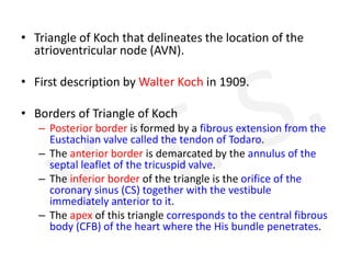
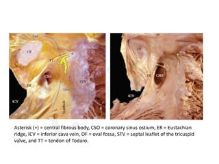
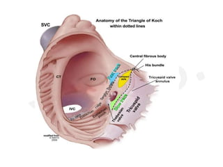
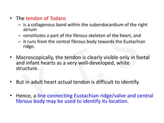
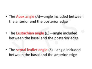
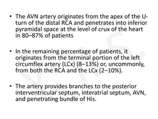
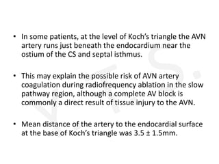
Koch’s Triangle
Dr. Yogesh Shilimkar
• Triangle of Koch that delineates the location of the
atrioventricular node (AVN).
• First description by Walter Koch in 1909.
• Borders of Triangle of Koch
– Posterior border is formed by a fibrous extension from the
Eustachian valve called the tendon of Todaro.
– The anterior border is demarcated by the annulus of the
septal leaflet of the tricuspid valve.
– The inferior border of the triangle is the orifice of the
coronary sinus (CS) together with the vestibule
immediately anterior to it.
– The apex of this triangle corresponds to the central fibrous
body (CFB) of the heart where the His bundle penetrates.
Asterisk (∗) = central fibrous body, CSO = coronary sinus ostium, ER = Eustachian
ridge, ICV = inferior cava vein, OF = oval fossa, STV = septal leaflet of the tricuspid
valve, and TT = tendon of Todaro.
• The tendon of Todaro
– is a collagenous band within the subendocardium of the right
atrium
– constitutes a part of the fibrous skeleton of the heart, and
– it runs from the central fibrous body towards the Eustachian
ridge.
• Macroscopically, the tendon is clearly visible only in foetal
and infant hearts as a very well-developed, white
structure.
• But in adult heart actual tendon is difficult to identify.
• Hence, a line connecting Eustachian ridge/valve and central
fibrous body may be used to identify its location.
• The Apex angle (A)—angle included between
the anterior and the posterior edge
• The Eustachian angle (E)—angle included
between the basal and the posterior edge
• The septal leaflet angle (S)—angle included
between the basal and the anterior edge
Fluroscopy
• In 45 degree RAO view the triangle is more or less in
same plane as the screen.
• Apex seen at fluroscopic RAO – at anterosuperior limit
of septal leaflet of tricuspid valve.
• However, anatomic apex is few mm posteroinferior to
that.
• It is here that bundle of His would penetrate into
central fibrous body.
Ablation
• The vestibular portion is the area often targeted for
ablation of the slow pathway in atrioventricular nodal
reentrant tachycardia (AVNRT).
• The so-called fast pathway corresponds to the area of
musculature close to the apex of the triangle of Koch.
• The dimensions of Koch’s triangle vary from one individual
to another.
• This variation in sizes is clinically relevant in catheter
ablation procedures of this area which are largely guided by
anatomic landmarks.
Structures in Koch’s triangle
• The body of the AVN is found near the apex of the triangle
and the His bundle penetrates the CFB at the apex.
• The penetrating His bundle can readily be distinguished
from the compact node at the point where the conduction
axis itself becomes completely surrounded by tissues of the
CFB and conducts electrical pulses to the ventricles.
• The AVN consists of a compact portion and an area of
transitional cells.
• The compact AV portion lies over the CFB.
• The transitional cells are surrounded by a greater
quantity of connective cells than that covering the
working cells, but they are not insulated from the
adjacent myocardium.
• Instead, they form a kind of bridge between the
working and nodal myocardium and collect electrical
information from the atrial walls, transmitting it to the
AV node.
• Toward the inferior portion of the triangle, there are
extensions of nodal tissues that extend inferiorly to the
right and left toward the tricuspid and mitral valves,
respectively.
• These nodal extensions have been implicated in
slow pathway conduction.
• Because this area also contains the zone of
transitional cells that feeds into the compact AV
node, this too may have a role in slow pathway
conduction.
• Low amplitude with fractionated electrograms in
the base of the triangle of Koch near the coronary
sinus and anterior aspects of the tricuspid
annulus is now the key guidance to identify the
slow pathway area.
• In some circumstances, the AV node components
can be displaced, increasing the risk of total AV
block when attempting slow pathway ablation.
• AV node displacement occurs in Ebstein
malformation and persistent left SVC with a
grossly enlarged CS ostium.
• In both abnormalities the size of triangle of Koch
is reduced, resulting in shorter distance between
the compact AV node and the CS ostium.
• The inferior pyramidal space is the extracardiac
fibroadipose tissue wedging between the atrial
and ventricular musculatures.
• Important structures located around or inside the
inferior pyramidal space
– the orifice of the coronary sinus,
– atrioventricular node,
– atrioventricular nodal artery,
– muscular atrioventricular sandwich, and
– aortic and atrioventricular valvar attachments.
• These relationships can be easily shown with CT
angiography of the heart.
• The AVN artery originates from the apex of the U-
turn of the distal RCA and penetrates into inferior
pyramidal space at the level of crux of the heart
in 80–87% of patients
• In the remaining percentage of patients, it
originates from the terminal portion of the left
circumflex artery (LCx) (8–13%) or, uncommonly,
from both the RCA and the LCx (2–10%).
• The artery provides branches to the posterior
interventricular septum, interatrial septum, AVN,
and penetrating bundle of His.
• In some patients, at the level of Koch’s triangle the AVN
artery runs just beneath the endocardium near the
ostium of the CS and septal isthmus.
• This may explain the possible risk of AVN artery
coagulation during radiofrequency ablation in the slow
pathway region, although a complete AV block is
commonly a direct result of tissue injury to the AVN.
• Mean distance of the artery to the endocardial surface
at the base of Koch’s triangle was 3.5 ± 1.5mm.

Triangle of koch

  • 1.
  • 2.
    • Triangle ofKoch that delineates the location of the atrioventricular node (AVN). • First description by Walter Koch in 1909. • Borders of Triangle of Koch – Posterior border is formed by a fibrous extension from the Eustachian valve called the tendon of Todaro. – The anterior border is demarcated by the annulus of the septal leaflet of the tricuspid valve. – The inferior border of the triangle is the orifice of the coronary sinus (CS) together with the vestibule immediately anterior to it. – The apex of this triangle corresponds to the central fibrous body (CFB) of the heart where the His bundle penetrates.
  • 3.
    Asterisk (∗) =central fibrous body, CSO = coronary sinus ostium, ER = Eustachian ridge, ICV = inferior cava vein, OF = oval fossa, STV = septal leaflet of the tricuspid valve, and TT = tendon of Todaro.
  • 6.
    • The tendonof Todaro – is a collagenous band within the subendocardium of the right atrium – constitutes a part of the fibrous skeleton of the heart, and – it runs from the central fibrous body towards the Eustachian ridge. • Macroscopically, the tendon is clearly visible only in foetal and infant hearts as a very well-developed, white structure. • But in adult heart actual tendon is difficult to identify. • Hence, a line connecting Eustachian ridge/valve and central fibrous body may be used to identify its location.
  • 7.
    • The Apexangle (A)—angle included between the anterior and the posterior edge • The Eustachian angle (E)—angle included between the basal and the posterior edge • The septal leaflet angle (S)—angle included between the basal and the anterior edge
  • 8.
    Fluroscopy • In 45degree RAO view the triangle is more or less in same plane as the screen. • Apex seen at fluroscopic RAO – at anterosuperior limit of septal leaflet of tricuspid valve. • However, anatomic apex is few mm posteroinferior to that. • It is here that bundle of His would penetrate into central fibrous body.
  • 9.
    Ablation • The vestibularportion is the area often targeted for ablation of the slow pathway in atrioventricular nodal reentrant tachycardia (AVNRT). • The so-called fast pathway corresponds to the area of musculature close to the apex of the triangle of Koch. • The dimensions of Koch’s triangle vary from one individual to another. • This variation in sizes is clinically relevant in catheter ablation procedures of this area which are largely guided by anatomic landmarks.
  • 10.
    Structures in Koch’striangle • The body of the AVN is found near the apex of the triangle and the His bundle penetrates the CFB at the apex. • The penetrating His bundle can readily be distinguished from the compact node at the point where the conduction axis itself becomes completely surrounded by tissues of the CFB and conducts electrical pulses to the ventricles. • The AVN consists of a compact portion and an area of transitional cells. • The compact AV portion lies over the CFB.
  • 11.
    • The transitionalcells are surrounded by a greater quantity of connective cells than that covering the working cells, but they are not insulated from the adjacent myocardium. • Instead, they form a kind of bridge between the working and nodal myocardium and collect electrical information from the atrial walls, transmitting it to the AV node. • Toward the inferior portion of the triangle, there are extensions of nodal tissues that extend inferiorly to the right and left toward the tricuspid and mitral valves, respectively.
  • 12.
    • These nodalextensions have been implicated in slow pathway conduction. • Because this area also contains the zone of transitional cells that feeds into the compact AV node, this too may have a role in slow pathway conduction. • Low amplitude with fractionated electrograms in the base of the triangle of Koch near the coronary sinus and anterior aspects of the tricuspid annulus is now the key guidance to identify the slow pathway area.
  • 13.
    • In somecircumstances, the AV node components can be displaced, increasing the risk of total AV block when attempting slow pathway ablation. • AV node displacement occurs in Ebstein malformation and persistent left SVC with a grossly enlarged CS ostium. • In both abnormalities the size of triangle of Koch is reduced, resulting in shorter distance between the compact AV node and the CS ostium.
  • 14.
    • The inferiorpyramidal space is the extracardiac fibroadipose tissue wedging between the atrial and ventricular musculatures. • Important structures located around or inside the inferior pyramidal space – the orifice of the coronary sinus, – atrioventricular node, – atrioventricular nodal artery, – muscular atrioventricular sandwich, and – aortic and atrioventricular valvar attachments. • These relationships can be easily shown with CT angiography of the heart.
  • 15.
    • The AVNartery originates from the apex of the U- turn of the distal RCA and penetrates into inferior pyramidal space at the level of crux of the heart in 80–87% of patients • In the remaining percentage of patients, it originates from the terminal portion of the left circumflex artery (LCx) (8–13%) or, uncommonly, from both the RCA and the LCx (2–10%). • The artery provides branches to the posterior interventricular septum, interatrial septum, AVN, and penetrating bundle of His.
  • 16.
    • In somepatients, at the level of Koch’s triangle the AVN artery runs just beneath the endocardium near the ostium of the CS and septal isthmus. • This may explain the possible risk of AVN artery coagulation during radiofrequency ablation in the slow pathway region, although a complete AV block is commonly a direct result of tissue injury to the AVN. • Mean distance of the artery to the endocardial surface at the base of Koch’s triangle was 3.5 ± 1.5mm.