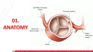
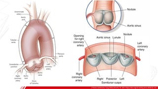
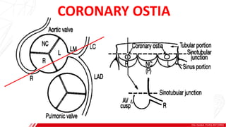
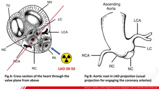
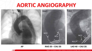
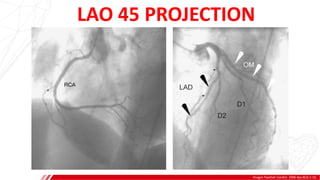
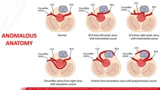
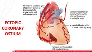
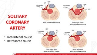
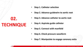
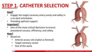
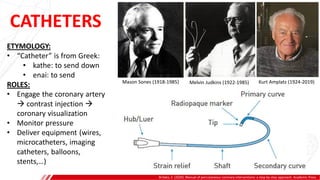
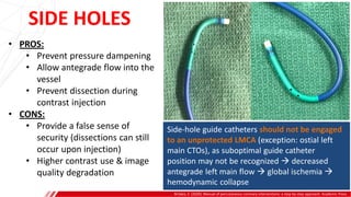
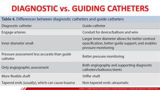
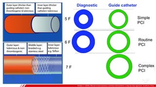
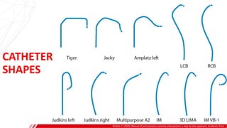
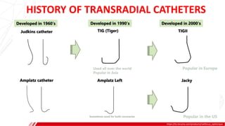
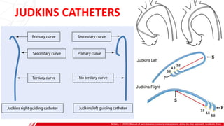
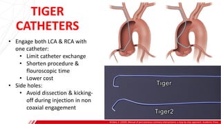
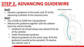
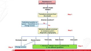
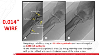
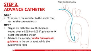
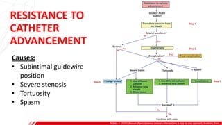
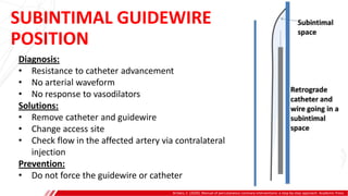
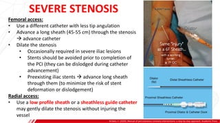
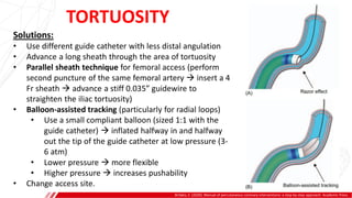
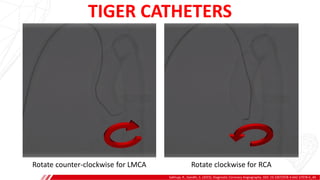
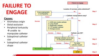
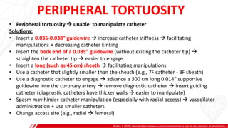
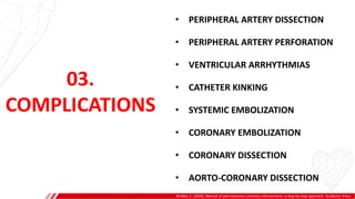
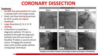
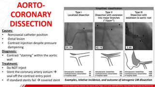
CORONARY ENGAGEMENT
Dr. Thiều Minh Sơn, MD
Interventional Cardiology Fellow at
Khánh Hòa Provincial General Hospital
Nha Trang, May 28th 2023
CONTENTS
1. Anatomy of coronary ostium
2. Coronary engaging technique
3. Complications
01.
ANATOMY
Loukas, M., Sharma, A., Blaak, C. et al. The Clinical Anatomy of the Coronary Arteries. J. of Cardiovasc. Trans. Res. 6, 197–207 (2013). DOI: https://doi.org/10.1007/s12265-013-9452-5
J. of Cardiovasc. Trans. Res. 6, 197–207 (2013). DOI: https://doi.org/10.1007/s12265-013-9452-5
AORTIC ROOT
• The initial part of the ascending aorta
• Composed of:
• 3 semilunar leaflets
• 3 interleaflet triangles
• 3 sinuses of Valsalva
• Sinutubular junction (thickened area of the aortic
wall) separates the root from the ascending aorta.
J Thorac Cardiovasc Surg 2018;156:e41-74. DOI: https://doi.org/10.1016/j.jtcvs.2018.02.115
Clin. Cardiol. 15,451-457 (1992)
CORONARY OSTIA
Sakhuja, R., Gandhi, S. (2015). Diagnostic Coronary Angiography. DOI: 10.1007/978-3-642-37078-6_40.
LAO 30-50
Fig B: Aortic root in LAO projection (usual
projection for engaging the coronary arteries)
Fig A: Cross section of the heart through the
valve plane from above
Topol, E. J., & Teirstein, P. S. (2015). Textbook of interventional cardiology E-Book. Elsevier Health Sciences.
AP RAO 20 – CAU 20 LAO 40 – CAU 20
AORTIC ANGIOGRAPHY
Images Paediatr Cardiol. 2006 Apr;8(2):1-16.
LAO 45 PROJECTION
ANOMALOUS
ANATOMY
Loukas, M., Sharma, A., Blaak, C. et al. The Clinical Anatomy of the Coronary Arteries. J. of Cardiovasc. Trans. Res. 6, 197–207 (2013). DOI: https://doi.org/10.1007/s12265-013-9452-5
ECTOPIC
CORONARY
OSTIUM
Loukas, M., Sharma, A., Blaak, C. et al. The Clinical Anatomy of the Coronary Arteries. J. of Cardiovasc. Trans. Res. 6, 197–207 (2013). DOI: https://doi.org/10.1007/s12265-013-9452-5
SOLITARY
CORONARY
ARTERY
• Interarterial course
• Retroaortic course
Loukas, M., Sharma, A., Blaak, C. et al. The Clinical Anatomy of the Coronary Arteries. J. of Cardiovasc. Trans. Res. 6, 197–207 (2013). DOI: https://doi.org/10.1007/s12265-013-9452-5
02.
TECHNIQUE
• Step 1. Catheter selection
• Step 2. Advance guidewire to aortic root
• Step 3. Advance catheter to aortic root
• Step 4. Aspirate guide catheter
• Step 5. Connect with manifold
• Step 6. Check pressure waveform
• Step 7. Manipulate to engage coronary ostia
Brilakis, E. (2020). Manual of percutaneous coronary interventions: a step-by-step approach. Academic Press.
STEP 1. CATHETER SELECTION
Brilakis, E. (2020). Manual of percutaneous coronary interventions: a step-by-step approach. Academic Press.
Goal?
• Engage the target coronary artery easily and safely in
a co-axial orientation
• Providing optimal support.
Important?
• One of the most critical decisions to ensure
procedural success, efficiency, and safety.
How?
• Based on:
• Arterial access site (radial vs femoral)
• Target coronary vessel
• Size of the aorta
CATHETERS
Brilakis, E. (2020). Manual of percutaneous coronary interventions: a step-by-step approach. Academic Press.
ETYMOLOGY:
• “Catheter” is from Greek:
• kathe: to send down
• enai: to send
ROLES:
• Engage the coronary artery
 contrast injection 
coronary visualization
• Monitor pressure
• Deliver equipment (wires,
microcatheters, imaging
catheters, balloons,
stents,…)
Mason Sones (1918-1985) Melvin Judkins (1922-1985) Kurt Amplatz (1924-2019)
CATHETER
LENGTH
Brilakis, E. (2020). Manual of percutaneous coronary interventions: a step-by-step approach. Academic Press.
• Most catheters are 100 cm long
• Shorter catheters (usually 90
cm):
• Retrograde CTO PCI
• Deliver equipment to very
distal lesions (e.g., through
bypass grafts)
• Longer catheters (125 cm):
• Very tall patients
• Very tortuous aorta
SIDE HOLES
Brilakis, E. (2020). Manual of percutaneous coronary interventions: a step-by-step approach. Academic Press.
• PROS:
• Prevent pressure dampening
• Allow antegrade flow into the
vessel
• Prevent dissection during
contrast injection
• CONS:
• Provide a false sense of
security (dissections can still
occur upon injection)
• Higher contrast use & image
quality degradation
Side-hole guide catheters should not be engaged
to an unprotected LMCA (exception: ostial left
main CTOs), as suboptimal guide catheter
position may not be recognized  decreased
antegrade left main flow  global ischemia 
hemodynamic collapse
DIAGNOSTIC vs. GUIDING CATHETERS
Brilakis, E. (2020). Manual of percutaneous coronary interventions: a step-by-step approach. Academic Press.
CATHETER
SHAPES
Brilakis, E. (2020). Manual of percutaneous coronary interventions: a step-by-step approach. Academic Press.
HISTORY OF TRANSRADIAL CATHETERS
https://tis.terumo.com/products/radifocus_optitorque
GUIDE CATHETER SHAPES
Intervent Cardiol Clin 4 (2015) 145–159. DOI: http://dx.doi.org/10.1016/j.iccl.2014.12.001
GUIDE CATHETER SHAPES
Intervent Cardiol Clin 4 (2015) 145–159. DOI: http://dx.doi.org/10.1016/j.iccl.2014.12.001
TYPES
&
SIZES
Sakhuja, R., Gandhi, S. (2015). Diagnostic Coronary Angiography. DOI: 10.1007/978-3-642-37078-6_40.
JUDKINS CATHETERS
Brilakis, E. (2020). Manual of percutaneous coronary interventions: a step-by-step approach. Academic Press.
TIGER
CATHETERS
Brilakis, E. (2020). Manual of percutaneous coronary interventions: a step-by-step approach. Academic Press.
• Engage both LCA & RCA with
one catheter:
• Limit catheter exchange
• Shorten procedure &
flouroscopic time
• Lower cost
• Side holes:
• Avoid dissection & kicking-
off during injection in non
coaxial engagement
STEP 2. ADVANCING GUIDEWIRE
Brilakis, E. (2020). Manual of percutaneous coronary interventions: a step-by-step approach. Academic Press.
Goal?
• Advance a guidewire to the aortic root  rail for
advancing a catheter to the coronary ostia
How?
• Use a 0.035 or 0.038 inch J-tip guidewire
• Advance the guidewire together with the catheter
(over the arterial sheath)
• Guidewire tip should always stay ahead of the tip
of the catheter
• Under fluoroscopic guidance
• Advance guidewire to the aortic cusps  fix the
guidewire (usually by the assistant)  advance
the catheter over it
Brilakis, E. (2020). Manual of percutaneous coronary interventions: a step-by-step approach. Academic Press.
0.035-0.038” GUIDEWIRE
Brilakis, E. (2020). Manual of percutaneous coronary interventions: a step-by-step approach. Academic Press.
For tight lesions, tortuous vessels, or when entering a side branch or crossing an aneurysm  repeat
wiring attempts can be performed using various 0.035 - 0.038 inch guidewires (polymer jacketed or with
very soft tip)
0.014”
WIRE
Moscucci, M. (2013). Grossman & Baim's cardiac catheterization, angiography, and intervention. Lippincott Williams & Wilkins.
Navigating a radial loop using an 0.014 inch guidewire and then exchange for
an 0.035 inch guidewire
 The loop usually straightens as the 0.035 inch guidewire passes through or
with gentle pullback and counterclockwise torque of the entire system
STEP 3.
ADVANCE CATHETER
Brilakis, E. (2020). Manual of percutaneous coronary interventions: a step-by-step approach. Academic Press.
Goal?
• To advance the catheter to the aortic root,
next to the coronary ostia
How?
• Diagnostic catheters are flushed and
loaded over a 0.035 or 0.038” guidewire 
insert through the sheath
• Advance the catheter under fluoroscopic
guidance to the aortic root, while the
guidewire is fixed
Brilakis, E. (2020). Manual of percutaneous coronary interventions: a step-by-step approach. Academic Press.
Causes:
• Subintimal guidewire
position
• Severe stenosis
• Tortuosity
• Spasm
RESISTANCE TO
CATHETER
ADVANCEMENT
Brilakis, E. (2020). Manual of percutaneous coronary interventions: a step-by-step approach. Academic Press.
Diagnosis:
• Resistance to catheter advancement
• No arterial waveform
• No response to vasodilators
Solutions:
• Remove catheter and guidewire
• Change access site
• Check flow in the affected artery via contralateral
injection
Prevention:
• Do not force the guidewire or catheter
SUBINTIMAL GUIDEWIRE
POSITION
Brilakis, E. (2020). Manual of percutaneous coronary interventions: a step-by-step approach. Academic Press.
Femoral access:
• Use a different catheter with less tip angulation
• Advance a long sheath (45-55 cm) through the stenosis
 advance catheter
• Dilate the stenosis
• Occasionally required in severe iliac lesions
• Stents should be avoided prior to completion of
the PCI (they can be dislodged during catheter
advancement)
• Preexisting iliac stents  advance long sheath
through them (to minimize the risk of stent
deformation or dislodgement)
Radial access:
• Use a low profile sheath or a sheathless guide catheter
may gently dilate the stenosis without injuring the
vessel
SEVERE STENOSIS
Brilakis, E. (2020). Manual of percutaneous coronary interventions: a step-by-step approach. Academic Press.
Solutions:
• Use different guide catheter with less distal angulation
• Advance a long sheath through the area of tortuosity
• Parallel sheath technique for femoral access (perform
second puncture of the same femoral artery  insert a 4
Fr sheath  advance a stiff 0.035” guidewire to
straighten the iliac tortuosity)
• Balloon-assisted tracking (particularly for radial loops)
• Use a small compliant balloon (sized 1:1 with the
guide catheter)  inflated halfway in and halfway
out the tip of the guide catheter at low pressure (3-
6 atm)
• Lower pressure  more flexible
• Higher pressure  increases pushability
• Change access site.
TORTUOSITY
EuroIntervention 2014;10:231-235. DOI: 10.4244/EIJV10I2A37
PARALLEL SHEATH TECHNIQUE
A) Right iliac artery kinking despite 8 Fr - 45 cm
sheath and a 0.035” extra-stiff guidewire. A
diagnostic 5 Fr catheter was unable to be
advanced beyond the abdominal aorta because of
excessive friction.
B) A second long 5 Fr - 45 cm sheath parallel to the 8
Fr sheath and a stiff 0.035” guidewire via the 5 Fr
sheath straightened the artery. The angiography and
intervention were finished successfully without major
friction.
Brilakis, E. (2020). Manual of percutaneous coronary interventions: a step-by-step approach. Academic Press.
• Specific to radial access
• More common in women and smokers
Causes:
• Small radial artery size
• High origin radial artery
• Radial artery tortuosity
• Multiple needle passes to obtain access
• Extensive catheter manipulations
• Multiple catheter exchanges
• Inadequate sedation
Prevention:
• Avoid using small radial arteries (ultrasound guidance
while obtaining radial access)
• Vasodilator administration after sheath insertion
• Avoid using large catheters
• Adequate sedation prior to obtaining access.
SPASM
Spasm in a high origin radial artery
Cardiovascular Revascularization Medicine 14 (2013) 321–324. DOI: https://doi.org/10.1016/j.carrev.2013.08.009
Diagnosis:
• Resistance to catheter advancement
• ± arterial waveform
• Angiography (only when there is no arterial pressure
dampening)
Solutions:
• Administer intra-arterial vasodilators
• Subcutaneous nitroglycerin injection
• More sedation
• Use smaller guide catheters (such as 5 French)
• Use Heartrail (Terumo) guide catheters (with dimpled
surface that decreases friction)
• Use a sheathless guide catheter
• Use of hydrophilic sheaths and catheters
• Use of low profile sheaths for radial access.
SPASM
Brilakis, E. (2020). Manual of percutaneous coronary interventions: a step-by-step approach. Academic Press.
Causes:
• Very tall patients
• Severe subclavian or iliac/aortic
tortuosity
• Distal radial access
Solutions:
• Use longer catheters (may need
balloons and stents with long shaft)
• Use guide catheter extensions
• Parallel sheath technique (to straighten
the artery)
• Change access site (femoral route is
usually shorter)
FAILURE TO REACH THE
AORTIC ROOT
STEP 4.
ASPIRATE GUIDE CATHETER
Brilakis, E. (2020). Manual of percutaneous coronary interventions: a step-by-step approach. Academic Press.
Goal?
• To clear the catheter of any air or other
material (thrombus or plaque) before
engaging the coronary artery
How?
• Aspirated 2-3 mL of blood
• Discard over a white gauze to determine if
thrombus (usually from the sheath) or
plaque scraped from the aortic wall (often
has iridescent appearance due to
cholesterol crystals), has been retrieved
A
B
A: Thrombus. B: Plaque (debris scraped from
the aortic wall by coronary guiding catheters)
INABILITY TO
ASPIRATE BLOOD
Brilakis, E. (2020). Manual of percutaneous coronary interventions: a step-by-step approach. Academic Press.
A
B
Causes:
• Catheter tip:
• Against the aortic/
coronary wall
• Next to a ostial lesion
• Too large for the ostium
• Catheter is kinked
• Catheter contains plaque,
thrombus, air, etc
Warning: do not insert a guidewire and do not flush
the catheter if you cannot aspirate back, as this may
cause embolization of thrombus/plaque in the aorta,
potentially causing stroke or acute coronary occlusion.
STEP 5.
CONNECT WITH MANIFOLD
Brilakis, E. (2020). Manual of percutaneous coronary interventions: a step-by-step approach. Academic Press.
Goal?
• To connect the catheter with the
manifold, enabling pressure monitoring
and contrast injection
How?
• Connect the catheter hub with the
manifold
• Aspirate blood into the manifold syringe
• If there is any air or visible debris 
discard
• Inject contrast until it exits from the tip
of the guide catheter
A
B
STEP 6.
PRESSURE WAVEFORM
JACC: Cardiovascular Interventions 12.20 (2019): 2093-2101. DOI: https://doi.org/10.1016/j.jcin.2019.06.036
Goal?
• To ensure that:
• Catheter tip is free (in the aortic
root)
• Catheter lumen is clear of any
foreign material (thrombus,
plaque, air)
How?
• Inspect the pressure waveform:
• Damping?
• Ventricularization?
A
B
DAMPED
WAVEFORMS
J INVASIVE CARDIOL 2017;29(11):387-389.
A
B
“Damping” = overdamping
Characteristics:
• Abrupt decline of mean pressure
• Narrow pulse pressure
• Delayed upstroke and
downstroke
• Loss of dicrotic notch
Meaning:
• Severe partial obstruction
Figure 1: Pressure-wave damping. Note the abrupt decline of
coronary pressure with narrow pulse pressre and a delayed
upstroke and downstroke
Figure 2: Immediate pullback of the catheter  pressure
waveform returns back to baseline
DAMPED
WAVEFORMS
Brilakis, E. (2020). Manual of percutaneous coronary interventions: a step-by-step approach. Academic Press.
Causes?
• Catheter is:
• Against the aortic/coronary wall
• Deeply or subselectively
engaged
• Positioned next to a ostial lesion
• Too large
• Catheter contains plaque, thrombus,
air,…
• Catheter is kinked
• Pressure transducer malfunction
A
B
WARNING !!!
Brilakis, E. (2020). Manual of percutaneous coronary interventions: a step-by-step approach. Academic Press.
• Failure to recognize pressure
dampening followed by contrast
injection can cause potentially
catastrophic complications:
• Systemic embolization
• Coronary embolization
• Coronary dissection
• Aorto-coronary dissection
A
B
VENTRICULARIZED
WAVEFORMS
American Heart Journal, 01 Dec 1989, 118(6):1160-1166. DOI: 10.1016/0002-8703(89)90004-5
A
B
The term “ventricularization” is actually a
misnomer!
“Ventricularized” coronary pressure has no
etiologic relationship to the LV pressure!
Characteristics:
• Steep decline of diastolic pressure
• Decline of systolic pressure (to a lesser
extent)
• Large pulse pressure
• Absence of the dicrotic notch
• Presystolic positive deflection
CORONARY BLOOD FLOW
Alex Yartsev (2020). Coronary blood flow. Deranged Physiology.
A
B
• 5% of cardiac output
• 75% LM flow and 50% RCA flow occurs in diastole
• In systole, LV contraction  high chamber pressure 
blood flow is reduced
• RV systolic pressure is lower  blood flow is less affected
• In diastole, ventricular relaxation  sucks the blood (in the
aorta and the catheter) into coronary artery
VENTRICULARIZED
WAVEFORMS
American Heart Journal, 01 Dec 1989, 118(6):1160-1166. DOI: 10.1016/0002-8703(89)90004-5
A
B
Causes:
• Catheter is:
• Positioned next to a ostial lesion
• Too large
Mechanism:
• Very small residual space between the catheter
and the coronary artery  restricts the diastolic
blood flow into the coronary artery 
ventricular suction  steep decline of the
diastolic pressure
• Motion of the ascending aorta during atria
systole  presystolic positive deflection (easier
to identify because in these patients the
intracoronary pressure is flat at the time of this
wave)
VENTRICULARIZATION
Am Heart J. 1989 Dec;118(6):1160-6. doi: 10.1016/0002-8703(89)90004-5.
A
B
• Ventricularized pressure waveform can be considered a hybrid
between:
• Coronary arterial pressure, and
• Coronary wedge pressure
• Important clue to the presence of left main coronary artery
disease
VENTRICULARIZED WAVEFORMS
J INVASIVE CARDIOL 2017;29(11):387-389.
A
B
Significance:
• Continuing to inject contrast despite
ventricularization may lead to:
• Ventricular fibrillation
• Dissection of the proximal
coronary artery
Management:
• Withdraw the catheter and
reposition it, or
• Gently inject contrast while
withdrawing the catheter (hit and
run technique – experts only!)
• Spasm  nitroglycerin may help
Figure 1: Aortic (Ao) pressure from the coronary angiographic
catheter tip showing damping with “ventricularization”, which
requires quick injection and removal of the catheter, a “hit-and-
run” maneuver. To reduce the chances of a coronary dissection,
one should realign the catheter in a more coaxial position, if
possible.
The Cardiac Catheterization Handbook. 6th ed. Philadelphia, PA:
Elsevier; 2016: 137.
STEP 7.
ENGAGING CORONARY OSTIA
Brilakis, E. (2020). Manual of percutaneous coronary interventions: a step-by-step approach. Academic Press.
Goal?
• Insert the tip of the catheter into the
target coronary artery
• Optimal catheter engagement:
• No pressure dampening
• Coaxial orientation
• 2-3 mm engagement depth
A
B
JUDKINS LEFT (JL)
http://cardiaccathpro.com/LeftCoronaryAngio.html
A
B
• JL4 is the most commonly used
catheter for Left Coronary Angiography
• Pre-shaped to minimize the need for
manipulation
• The 4 reflects the length of the
segment between the primary and
secondary curve
• Patients with a small or narrow aortic
arch will usually need a JL3.5 catheter
• Catheter is too short  tip pointed
upward
• Catheter is too long  tip pointed
downward
JUDKINS LEFT (JL)
http://cardiaccathpro.com/LeftCoronaryAngio.html
A
B
• Advance JL catheter into the ascending aorta over a guidewire in the LAO view (30-50 degree)
 catheter tip positioned just below the coronary ostium
• Gentle pull back  catheter fall into the LM ostium  noted on screen by a sudden “dive” of
the catheter tip
• Slight rotation may be helpful (counterclockwise, as the left main ostium usually lies
posteriorly), but avoid excessive rotation
JUDKINS LEFT (JL)
Brilakis, E. (2020). Manual of percutaneous coronary interventions: a step-by-step approach. Academic Press.
A
B
• Advance the catheter tip 2-3 cm above the
upward deflection of the J wire
• Withdrawn the J wire  catheter tip fall
into the left cusp  then into the left main
ostium
• Advance the catheter tip too deep into
the right coronary cusp  prevents it
from entering the left coronary cusp
JUDKINS LEFT (JL)
Sakhuja, R., Gandhi, S. (2015). Diagnostic Coronary Angiography. DOI: 10.1007/978-3-642-37078-6_40.
A
B
1. Often, simply advancing the JL catheter without manipulation will
intubate the LM ostium
2. If the catheter tip lies below the coronary ostium despite gentle retraction
and torque  downsize (e.g., JL4 to JL3.5).
3. If the catheter folds on itself  upsize (e.g., JL4 to JL5).
JUDKINS RIGHT (JR)
Sakhuja, R., Gandhi, S. (2015). Diagnostic Coronary Angiography. DOI: 10.1007/978-3-642-37078-6_40.
A
B
• Advance the catheter is over a guidewire in the LAO view to the valve plane (the bottom
of the right coronary cusp)
• Slowly withdrawn from the valve plane 2–4 cm while simultaneously rotating clockwise
(anterior) approximately 180 degree
TIGER CATHETERS
Sakhuja, R., Gandhi, S. (2015). Diagnostic Coronary Angiography. DOI: 10.1007/978-3-642-37078-6_40.
A
B
Rotate counter-clockwise for LMCA Rotate clockwise for RCA
FAILURE TO
ENGAGE
Brilakis, E. (2020). Manual of percutaneous coronary interventions: a step-by-step approach. Academic Press.
A
B
Causes:
• Anomalous origin
• Ostial occlusion
• Peripheral tortuosity
 unable to
manipulate catheter
• Suboptimal catheter
size
• Suboptimal catheter
shape
PERIPHERAL TORTUOSITY
Brilakis, E. (2020). Manual of percutaneous coronary interventions: a step-by-step approach. Academic Press.
A
B
• Peripheral tortuosity  unable to manipulate catheter
Solutions:
• Insert a 0.035-0.038” guidewire  increase catheter stiffness  facilitating
manipulations + decreasing catheter kinking
• Insert the back end of a 0.035” guidewire (without exiting the catheter tip) 
straighten the catheter tip  easier to engage
• Insert a long (such as 45 cm) sheath  facilitating manipulations
• Use a catheter that slightly smaller than the sheath (e.g., 7F catheter - 8F sheath)
• Use a diagnostic catheter to engage  advance a 300 cm long 0.014” supportive
guidewire into the coronary artery  remove diagnostic catheter  insert guiding
catheter (diagnostic catheters have thicker walls  easier to manipulate)
• Spasm may hinder catheter manipulation (especially with radial access)  vasodilator
administration + use smaller catheters
• Change access site (e.g., radial  femoral)
03.
COMPLICATIONS
• PERIPHERAL ARTERY DISSECTION
• PERIPHERAL ARTERY PERFORATION
• VENTRICULAR ARRHYTHMIAS
• CATHETER KINKING
• SYSTEMIC EMBOLIZATION
• CORONARY EMBOLIZATION
• CORONARY DISSECTION
• AORTO-CORONARY DISSECTION
Brilakis, E. (2020). Manual of percutaneous coronary interventions: a step-by-step approach. Academic Press.
PERIPHERAL ARTERY
DISSECTION
Brilakis, E. (2020). Manual of percutaneous coronary interventions: a step-by-step approach. Academic Press.
Causes:
• Subintimal guidewire advancement
• Forceful sheath/catheter advancement
• Advance catheter without leading
guidewire
Diagnosis:
• Challenging catheter advancement 
angiography (only when there is no
arterial pressure dampening)
PERIPHERAL ARTERY DISSECTION
Brilakis, E. (2020). Manual of percutaneous coronary interventions: a step-by-step approach. Academic Press.
Treatment:
• Most often no specific treatment is required
• Small retrograde dissections are usually sealed off
by antegrade blood flow
• Large dissections may require endovascular repair
(usually with stent)
• Switch to different access site
Prevention:
• NEVER use force when advancing guidewires and
catheters
• Use long sheaths in patients with severe iliac
tortuosity
• Perform femoral angiography with a guidewire in
place (to keep the sheath tip away from the
arterial wall)
PERIPHERAL ARTERY
PERFORATION
Brilakis, E. (2020). Manual of percutaneous coronary interventions: a step-by-step approach. Academic Press.
Causes:
• Forceful guidewire or catheter
advancement into a side branch
• Small caliber artery
• Sheath advancement past the tip of the
guidewire
Diagnosis:
• Perform angiography when the guidewire
or catheter follows an unexpected course
or when resistance is felt during
advancement
PERIPHERAL
ARTERY
PERFORATION
Circ Cardiovasc Interv. 2019;12:e007386. DOI: 10.1161/CIRCINTERVENTIONS.119.007386
Radial artery:
• Advance a catheter over
the perforated segment 
usually suffices to achieve
hemostasis
Femoral artery:
• Balloon occlusion 
covered stent
Small branch:
• Coil (or fat or thrombus)
embolization
VENTRICULAR
ARRHYTHMIAS
Brilakis, E. (2020). Manual of percutaneous coronary interventions: a step-by-step approach. Academic Press.
Causes:
• Entry of the guidewire into the left ventricle
Solution:
• Remove guidewire from the left ventricle
Prevention:
• Monitor and adjust the position of the tip
of the guidewire to prevent entry into the
left ventricle
CATHETER
KINKING
Brilakis, E. (2020). Manual of percutaneous coronary interventions: a step-by-step approach. Academic Press.
A
B
Causes:
• Severe iliac/aortic/subclavian
tortuosity
• Extensive catheter
manipulation
Diagnosis:
• Catheter tip not moving
when rotating
• Dampened/lost pressure
waveform
• Fluoroscopy  confirm
kinking location
CATHETER KINKING
Brilakis, E. (2020). Manual of percutaneous coronary interventions: a step-by-step approach. Academic Press.
A
B
STEP 1:
• Gently untwist the catheter (opposite direction)  advance a 0.035” guidewire
through kinked segment (may use the guidewire back end)
• Kinking below the brachial artery (radial access)  inflate a blood pressure cuff at the
brachial artery to “fix” the kinked catheter  untwist  remove
STEP 2:
• Obtain another arterial access  snare the tip of the kinked catheter  straightening
the kinked segment  untwist
STEP 3:
• Attach an indeflator to the catheter hub  inflate to 3-4 atm
STEP 4:
• Cut the catheter hub  advance a sheath (same size or 1-2F larger than the catheter
size)  encase the kinked segment  withdrawal
STEP 5:
• Arteriotomy (pull knot through skin)
SYSTEMIC EMBOLIZATION
Circulation: Cardiovascular Interventions. 2019;12:e007791. DOI: https://doi.org/10.1161/CIRCINTERVENTIONS.119.007791
• One of the most feared complications
• Embolization material can be air, plaque,
thrombus
Causes:
• Not aspirate the guide catheter before
coronary artery engagement
• Plaque dislodgement (in patients with
significant aortic atherosclerosis) during
catheter advancement
Diagnosis:
• Depends on the area affected
• Stroke  acute neurologic deficit
• Lower extremity  pulses may decrease or
disappear, lower extremity discomfort
Major Complications With Diagnostic Left Heart Catheterization
(Study on 43.768 LHC procedures)
SYSTEMIC EMBOLIZATION
J Am Coll Cardiol 2018;71:1910–20. DOI: https://doi.org/10.1016/j.jacc.2018.02.065
Treatment:
• Stroke: emergency head CT + neurology
evaluation  may need emergent
neurointerventional treatment
• Other sites: usually conservative management,
sometimes endovascular treatment may be
required
Prevention:
• Aspirate and flush sheath before catheter
insertion
• Aspirate catheter before contrast injection
• Do not inject if there is dampening of the
pressure waveform
• Use a sheathless guide (with an inner dilator) 
prevents the “razor effect” and does not scrape
plaques
CORONARY EMBOLIZATION
Brilakis, E. (2020). Manual of percutaneous coronary interventions: a step-by-step approach. Academic Press.
Causes:
• Embolization material can be air,
thrombus (from catheters), or plaque
(from aortic plague dislodgement)
Diagnosis:
• Chest pain
• ST-segment elevation
• May develop cardiogenic shock or
cardiac arrest
• Angiography: decrease/cessation of
coronary flow
CORONARY EMBOLIZATION
Brilakis, E. (2020). Manual of percutaneous coronary interventions: a step-by-step approach. Academic Press.
Treatment:
• Air embolism:
• Administer 100% oxygen  helps absorb the
embolized air
• If antegrade flow stops  aspiration through guide
or thrombectomy catheter
• Repetitive forceful saline injections  “unclog”
vessels
• Intracoronary epinephrine (0.05 mg)
• Plaque/thrombus embolism:
• Anticoagulation and intravenous antiplatelet agents
(GP IIb/IIIa inhibitors or cangrelor)
• Thrombectomy (if large visible thrombus)
• Intracoronary vasodilators (nicardipine, nitroprusside,
verapamil, adenosine)
CORONARY DISSECTION
A. Lindsay et al. (2016). Complications of Percutaneous Coronary Intervention,DOI 10.1007/978-1-4471-4959-0_17
Causes:
• Non-coaxial catheter position
• Contrast injection despite pressure
dampening
• Ostial lesion
Diagnosis:
• Large dissections  impede coronary flow
 STEMI  possibly lead to cardiogenic
shock or cardiac arrest (especially LM
dissections)
• Small dissections may not (at least
immediately) impede coronary flow and
may be harder to detect.
• Angiography  filling defects
• IVUS, OCT may help confirm the diagnosis
CORONARY DISSECTION
American Heart Journal, Volume 159, Issue 6, 2010, Pages 1147-1153, ISSN 0002-8703, https://doi.org/10.1016/j.ahj.2010.03.012.
Treatment:
• Do NOT lose wire position (if a wire
is already within the target vessel)
• Small, non-flow limiting dissections
(A, B) usually not require
treatment
• Large dissections (C, D, E, F) 
stenting
• If dissection is caused by a
diagnostic catheter  insert a
guidewire (through the diagnostic
catheter) into the true lumen (if
feasible)  obtain a second arterial
access  engage the coronary
artery with another guide catheter
(“ping pong” technique)
Examples and relative incidence (percentage) of types of iatrogenic LM dissection
AORTO-
CORONARY
DISSECTION
American Heart Journal, Volume 159, Issue 6, 2010, Pages 1147-1153, ISSN 0002-8703, https://doi.org/10.1016/j.ahj.2010.03.012.
Causes:
• Noncoaxial catheter position
• Ostial lesion
• Contrast injection despite pressure
dampening
Diagnosis:
• Contrast “staining” within the aortic
wall
Treatment:
• Do NOT inject
• Stent the coronary artery ostium 
seal off the contrast entry point
• If standard stents fail  covered stent Examples, relative incidence, and outcome of iatrogenic LM dissection
J Thorac Cardiovasc Surg 2006;131:230-10022-5223. DOi:10.1016/j.jtcvs.2005.08.051
Fig 1. Proximal aortic dissection, with contrast
clearly seen filling the false lumen on both sides of
the aortic wall (arrows)
Fig 2. Coronary dissection at the RCA ostium (white
arrow), with retrograde contrast seen in the aortic wall
(black arrow).
Baumann et al. BMC Medical Imaging (2017) 17:64. DOI:10.1186/s12880-017-0227-3
CONCLUSIONS
1. Engaging the coronary artery ostia is one of the most essential steps of
diagnostic angiography and PCI
2. Using multiple catheters (Judkins, Amplatz) or single catheter (Tiger, Jacky)
3. Requirements of an optimal catheter engagement: no pressure dampening,
coaxial orientation, 2-3 mm engagement depth
4. Pressure waveform monitor is of the utmost importance. Failure to recognize
pressure damping/ventricularization followed by contrast injection can cause
catastrophic complications.
FINAL MESSAGE
“Never take your eyes off the monitor and the pressure curve!”
“Serious complications in the cath lab often happen not out of
ignorance or lack of expertise, but because of ignoring some
basic principles and lack of cath lab discipline.”

CORONARY ENGAGEMENT.pdf

  • 1.
    CORONARY ENGAGEMENT Dr. ThiềuMinh Sơn, MD Interventional Cardiology Fellow at Khánh Hòa Provincial General Hospital Nha Trang, May 28th 2023
  • 2.
    CONTENTS 1. Anatomy ofcoronary ostium 2. Coronary engaging technique 3. Complications
  • 3.
    01. ANATOMY Loukas, M., Sharma,A., Blaak, C. et al. The Clinical Anatomy of the Coronary Arteries. J. of Cardiovasc. Trans. Res. 6, 197–207 (2013). DOI: https://doi.org/10.1007/s12265-013-9452-5
  • 4.
    J. of Cardiovasc.Trans. Res. 6, 197–207 (2013). DOI: https://doi.org/10.1007/s12265-013-9452-5 AORTIC ROOT • The initial part of the ascending aorta • Composed of: • 3 semilunar leaflets • 3 interleaflet triangles • 3 sinuses of Valsalva • Sinutubular junction (thickened area of the aortic wall) separates the root from the ascending aorta.
  • 5.
    J Thorac CardiovascSurg 2018;156:e41-74. DOI: https://doi.org/10.1016/j.jtcvs.2018.02.115
  • 6.
    Clin. Cardiol. 15,451-457(1992) CORONARY OSTIA
  • 7.
    Sakhuja, R., Gandhi,S. (2015). Diagnostic Coronary Angiography. DOI: 10.1007/978-3-642-37078-6_40. LAO 30-50 Fig B: Aortic root in LAO projection (usual projection for engaging the coronary arteries) Fig A: Cross section of the heart through the valve plane from above
  • 8.
    Topol, E. J.,& Teirstein, P. S. (2015). Textbook of interventional cardiology E-Book. Elsevier Health Sciences. AP RAO 20 – CAU 20 LAO 40 – CAU 20 AORTIC ANGIOGRAPHY
  • 9.
    Images Paediatr Cardiol.2006 Apr;8(2):1-16. LAO 45 PROJECTION
  • 10.
    ANOMALOUS ANATOMY Loukas, M., Sharma,A., Blaak, C. et al. The Clinical Anatomy of the Coronary Arteries. J. of Cardiovasc. Trans. Res. 6, 197–207 (2013). DOI: https://doi.org/10.1007/s12265-013-9452-5
  • 11.
    ECTOPIC CORONARY OSTIUM Loukas, M., Sharma,A., Blaak, C. et al. The Clinical Anatomy of the Coronary Arteries. J. of Cardiovasc. Trans. Res. 6, 197–207 (2013). DOI: https://doi.org/10.1007/s12265-013-9452-5
  • 12.
    SOLITARY CORONARY ARTERY • Interarterial course •Retroaortic course Loukas, M., Sharma, A., Blaak, C. et al. The Clinical Anatomy of the Coronary Arteries. J. of Cardiovasc. Trans. Res. 6, 197–207 (2013). DOI: https://doi.org/10.1007/s12265-013-9452-5
  • 13.
    02. TECHNIQUE • Step 1.Catheter selection • Step 2. Advance guidewire to aortic root • Step 3. Advance catheter to aortic root • Step 4. Aspirate guide catheter • Step 5. Connect with manifold • Step 6. Check pressure waveform • Step 7. Manipulate to engage coronary ostia Brilakis, E. (2020). Manual of percutaneous coronary interventions: a step-by-step approach. Academic Press.
  • 14.
    STEP 1. CATHETERSELECTION Brilakis, E. (2020). Manual of percutaneous coronary interventions: a step-by-step approach. Academic Press. Goal? • Engage the target coronary artery easily and safely in a co-axial orientation • Providing optimal support. Important? • One of the most critical decisions to ensure procedural success, efficiency, and safety. How? • Based on: • Arterial access site (radial vs femoral) • Target coronary vessel • Size of the aorta
  • 15.
    CATHETERS Brilakis, E. (2020).Manual of percutaneous coronary interventions: a step-by-step approach. Academic Press. ETYMOLOGY: • “Catheter” is from Greek: • kathe: to send down • enai: to send ROLES: • Engage the coronary artery  contrast injection  coronary visualization • Monitor pressure • Deliver equipment (wires, microcatheters, imaging catheters, balloons, stents,…) Mason Sones (1918-1985) Melvin Judkins (1922-1985) Kurt Amplatz (1924-2019)
  • 16.
    CATHETER LENGTH Brilakis, E. (2020).Manual of percutaneous coronary interventions: a step-by-step approach. Academic Press. • Most catheters are 100 cm long • Shorter catheters (usually 90 cm): • Retrograde CTO PCI • Deliver equipment to very distal lesions (e.g., through bypass grafts) • Longer catheters (125 cm): • Very tall patients • Very tortuous aorta
  • 17.
    SIDE HOLES Brilakis, E.(2020). Manual of percutaneous coronary interventions: a step-by-step approach. Academic Press. • PROS: • Prevent pressure dampening • Allow antegrade flow into the vessel • Prevent dissection during contrast injection • CONS: • Provide a false sense of security (dissections can still occur upon injection) • Higher contrast use & image quality degradation Side-hole guide catheters should not be engaged to an unprotected LMCA (exception: ostial left main CTOs), as suboptimal guide catheter position may not be recognized  decreased antegrade left main flow  global ischemia  hemodynamic collapse
  • 18.
    DIAGNOSTIC vs. GUIDINGCATHETERS Brilakis, E. (2020). Manual of percutaneous coronary interventions: a step-by-step approach. Academic Press.
  • 19.
    Brilakis, E. (2020).Manual of percutaneous coronary interventions: a step-by-step approach. Academic Press.
  • 20.
    CATHETER SHAPES Brilakis, E. (2020).Manual of percutaneous coronary interventions: a step-by-step approach. Academic Press.
  • 21.
    HISTORY OF TRANSRADIALCATHETERS https://tis.terumo.com/products/radifocus_optitorque
  • 22.
    GUIDE CATHETER SHAPES InterventCardiol Clin 4 (2015) 145–159. DOI: http://dx.doi.org/10.1016/j.iccl.2014.12.001
  • 23.
    GUIDE CATHETER SHAPES InterventCardiol Clin 4 (2015) 145–159. DOI: http://dx.doi.org/10.1016/j.iccl.2014.12.001
  • 24.
    TYPES & SIZES Sakhuja, R., Gandhi,S. (2015). Diagnostic Coronary Angiography. DOI: 10.1007/978-3-642-37078-6_40.
  • 25.
    JUDKINS CATHETERS Brilakis, E.(2020). Manual of percutaneous coronary interventions: a step-by-step approach. Academic Press.
  • 26.
    TIGER CATHETERS Brilakis, E. (2020).Manual of percutaneous coronary interventions: a step-by-step approach. Academic Press. • Engage both LCA & RCA with one catheter: • Limit catheter exchange • Shorten procedure & flouroscopic time • Lower cost • Side holes: • Avoid dissection & kicking- off during injection in non coaxial engagement
  • 27.
    STEP 2. ADVANCINGGUIDEWIRE Brilakis, E. (2020). Manual of percutaneous coronary interventions: a step-by-step approach. Academic Press. Goal? • Advance a guidewire to the aortic root  rail for advancing a catheter to the coronary ostia How? • Use a 0.035 or 0.038 inch J-tip guidewire • Advance the guidewire together with the catheter (over the arterial sheath) • Guidewire tip should always stay ahead of the tip of the catheter • Under fluoroscopic guidance • Advance guidewire to the aortic cusps  fix the guidewire (usually by the assistant)  advance the catheter over it
  • 28.
    Brilakis, E. (2020).Manual of percutaneous coronary interventions: a step-by-step approach. Academic Press.
  • 29.
    0.035-0.038” GUIDEWIRE Brilakis, E.(2020). Manual of percutaneous coronary interventions: a step-by-step approach. Academic Press. For tight lesions, tortuous vessels, or when entering a side branch or crossing an aneurysm  repeat wiring attempts can be performed using various 0.035 - 0.038 inch guidewires (polymer jacketed or with very soft tip)
  • 30.
    0.014” WIRE Moscucci, M. (2013).Grossman & Baim's cardiac catheterization, angiography, and intervention. Lippincott Williams & Wilkins. Navigating a radial loop using an 0.014 inch guidewire and then exchange for an 0.035 inch guidewire  The loop usually straightens as the 0.035 inch guidewire passes through or with gentle pullback and counterclockwise torque of the entire system
  • 31.
    STEP 3. ADVANCE CATHETER Brilakis,E. (2020). Manual of percutaneous coronary interventions: a step-by-step approach. Academic Press. Goal? • To advance the catheter to the aortic root, next to the coronary ostia How? • Diagnostic catheters are flushed and loaded over a 0.035 or 0.038” guidewire  insert through the sheath • Advance the catheter under fluoroscopic guidance to the aortic root, while the guidewire is fixed
  • 32.
    Brilakis, E. (2020).Manual of percutaneous coronary interventions: a step-by-step approach. Academic Press. Causes: • Subintimal guidewire position • Severe stenosis • Tortuosity • Spasm RESISTANCE TO CATHETER ADVANCEMENT
  • 33.
    Brilakis, E. (2020).Manual of percutaneous coronary interventions: a step-by-step approach. Academic Press. Diagnosis: • Resistance to catheter advancement • No arterial waveform • No response to vasodilators Solutions: • Remove catheter and guidewire • Change access site • Check flow in the affected artery via contralateral injection Prevention: • Do not force the guidewire or catheter SUBINTIMAL GUIDEWIRE POSITION
  • 34.
    Brilakis, E. (2020).Manual of percutaneous coronary interventions: a step-by-step approach. Academic Press. Femoral access: • Use a different catheter with less tip angulation • Advance a long sheath (45-55 cm) through the stenosis  advance catheter • Dilate the stenosis • Occasionally required in severe iliac lesions • Stents should be avoided prior to completion of the PCI (they can be dislodged during catheter advancement) • Preexisting iliac stents  advance long sheath through them (to minimize the risk of stent deformation or dislodgement) Radial access: • Use a low profile sheath or a sheathless guide catheter may gently dilate the stenosis without injuring the vessel SEVERE STENOSIS
  • 35.
    Brilakis, E. (2020).Manual of percutaneous coronary interventions: a step-by-step approach. Academic Press. Solutions: • Use different guide catheter with less distal angulation • Advance a long sheath through the area of tortuosity • Parallel sheath technique for femoral access (perform second puncture of the same femoral artery  insert a 4 Fr sheath  advance a stiff 0.035” guidewire to straighten the iliac tortuosity) • Balloon-assisted tracking (particularly for radial loops) • Use a small compliant balloon (sized 1:1 with the guide catheter)  inflated halfway in and halfway out the tip of the guide catheter at low pressure (3- 6 atm) • Lower pressure  more flexible • Higher pressure  increases pushability • Change access site. TORTUOSITY
  • 36.
    EuroIntervention 2014;10:231-235. DOI:10.4244/EIJV10I2A37 PARALLEL SHEATH TECHNIQUE A) Right iliac artery kinking despite 8 Fr - 45 cm sheath and a 0.035” extra-stiff guidewire. A diagnostic 5 Fr catheter was unable to be advanced beyond the abdominal aorta because of excessive friction. B) A second long 5 Fr - 45 cm sheath parallel to the 8 Fr sheath and a stiff 0.035” guidewire via the 5 Fr sheath straightened the artery. The angiography and intervention were finished successfully without major friction.
  • 37.
    Brilakis, E. (2020).Manual of percutaneous coronary interventions: a step-by-step approach. Academic Press. • Specific to radial access • More common in women and smokers Causes: • Small radial artery size • High origin radial artery • Radial artery tortuosity • Multiple needle passes to obtain access • Extensive catheter manipulations • Multiple catheter exchanges • Inadequate sedation Prevention: • Avoid using small radial arteries (ultrasound guidance while obtaining radial access) • Vasodilator administration after sheath insertion • Avoid using large catheters • Adequate sedation prior to obtaining access. SPASM Spasm in a high origin radial artery
  • 38.
    Cardiovascular Revascularization Medicine14 (2013) 321–324. DOI: https://doi.org/10.1016/j.carrev.2013.08.009 Diagnosis: • Resistance to catheter advancement • ± arterial waveform • Angiography (only when there is no arterial pressure dampening) Solutions: • Administer intra-arterial vasodilators • Subcutaneous nitroglycerin injection • More sedation • Use smaller guide catheters (such as 5 French) • Use Heartrail (Terumo) guide catheters (with dimpled surface that decreases friction) • Use a sheathless guide catheter • Use of hydrophilic sheaths and catheters • Use of low profile sheaths for radial access. SPASM
  • 39.
    Brilakis, E. (2020).Manual of percutaneous coronary interventions: a step-by-step approach. Academic Press. Causes: • Very tall patients • Severe subclavian or iliac/aortic tortuosity • Distal radial access Solutions: • Use longer catheters (may need balloons and stents with long shaft) • Use guide catheter extensions • Parallel sheath technique (to straighten the artery) • Change access site (femoral route is usually shorter) FAILURE TO REACH THE AORTIC ROOT
  • 40.
    STEP 4. ASPIRATE GUIDECATHETER Brilakis, E. (2020). Manual of percutaneous coronary interventions: a step-by-step approach. Academic Press. Goal? • To clear the catheter of any air or other material (thrombus or plaque) before engaging the coronary artery How? • Aspirated 2-3 mL of blood • Discard over a white gauze to determine if thrombus (usually from the sheath) or plaque scraped from the aortic wall (often has iridescent appearance due to cholesterol crystals), has been retrieved A B A: Thrombus. B: Plaque (debris scraped from the aortic wall by coronary guiding catheters)
  • 41.
    INABILITY TO ASPIRATE BLOOD Brilakis,E. (2020). Manual of percutaneous coronary interventions: a step-by-step approach. Academic Press. A B Causes: • Catheter tip: • Against the aortic/ coronary wall • Next to a ostial lesion • Too large for the ostium • Catheter is kinked • Catheter contains plaque, thrombus, air, etc Warning: do not insert a guidewire and do not flush the catheter if you cannot aspirate back, as this may cause embolization of thrombus/plaque in the aorta, potentially causing stroke or acute coronary occlusion.
  • 42.
    STEP 5. CONNECT WITHMANIFOLD Brilakis, E. (2020). Manual of percutaneous coronary interventions: a step-by-step approach. Academic Press. Goal? • To connect the catheter with the manifold, enabling pressure monitoring and contrast injection How? • Connect the catheter hub with the manifold • Aspirate blood into the manifold syringe • If there is any air or visible debris  discard • Inject contrast until it exits from the tip of the guide catheter A B
  • 43.
    STEP 6. PRESSURE WAVEFORM JACC:Cardiovascular Interventions 12.20 (2019): 2093-2101. DOI: https://doi.org/10.1016/j.jcin.2019.06.036 Goal? • To ensure that: • Catheter tip is free (in the aortic root) • Catheter lumen is clear of any foreign material (thrombus, plaque, air) How? • Inspect the pressure waveform: • Damping? • Ventricularization? A B
  • 44.
    DAMPED WAVEFORMS J INVASIVE CARDIOL2017;29(11):387-389. A B “Damping” = overdamping Characteristics: • Abrupt decline of mean pressure • Narrow pulse pressure • Delayed upstroke and downstroke • Loss of dicrotic notch Meaning: • Severe partial obstruction Figure 1: Pressure-wave damping. Note the abrupt decline of coronary pressure with narrow pulse pressre and a delayed upstroke and downstroke Figure 2: Immediate pullback of the catheter  pressure waveform returns back to baseline
  • 45.
    DAMPED WAVEFORMS Brilakis, E. (2020).Manual of percutaneous coronary interventions: a step-by-step approach. Academic Press. Causes? • Catheter is: • Against the aortic/coronary wall • Deeply or subselectively engaged • Positioned next to a ostial lesion • Too large • Catheter contains plaque, thrombus, air,… • Catheter is kinked • Pressure transducer malfunction A B
  • 46.
    WARNING !!! Brilakis, E.(2020). Manual of percutaneous coronary interventions: a step-by-step approach. Academic Press. • Failure to recognize pressure dampening followed by contrast injection can cause potentially catastrophic complications: • Systemic embolization • Coronary embolization • Coronary dissection • Aorto-coronary dissection A B
  • 47.
    VENTRICULARIZED WAVEFORMS American Heart Journal,01 Dec 1989, 118(6):1160-1166. DOI: 10.1016/0002-8703(89)90004-5 A B The term “ventricularization” is actually a misnomer! “Ventricularized” coronary pressure has no etiologic relationship to the LV pressure! Characteristics: • Steep decline of diastolic pressure • Decline of systolic pressure (to a lesser extent) • Large pulse pressure • Absence of the dicrotic notch • Presystolic positive deflection
  • 48.
    CORONARY BLOOD FLOW AlexYartsev (2020). Coronary blood flow. Deranged Physiology. A B • 5% of cardiac output • 75% LM flow and 50% RCA flow occurs in diastole • In systole, LV contraction  high chamber pressure  blood flow is reduced • RV systolic pressure is lower  blood flow is less affected • In diastole, ventricular relaxation  sucks the blood (in the aorta and the catheter) into coronary artery
  • 49.
    VENTRICULARIZED WAVEFORMS American Heart Journal,01 Dec 1989, 118(6):1160-1166. DOI: 10.1016/0002-8703(89)90004-5 A B Causes: • Catheter is: • Positioned next to a ostial lesion • Too large Mechanism: • Very small residual space between the catheter and the coronary artery  restricts the diastolic blood flow into the coronary artery  ventricular suction  steep decline of the diastolic pressure • Motion of the ascending aorta during atria systole  presystolic positive deflection (easier to identify because in these patients the intracoronary pressure is flat at the time of this wave)
  • 50.
    VENTRICULARIZATION Am Heart J.1989 Dec;118(6):1160-6. doi: 10.1016/0002-8703(89)90004-5. A B • Ventricularized pressure waveform can be considered a hybrid between: • Coronary arterial pressure, and • Coronary wedge pressure • Important clue to the presence of left main coronary artery disease
  • 51.
    VENTRICULARIZED WAVEFORMS J INVASIVECARDIOL 2017;29(11):387-389. A B Significance: • Continuing to inject contrast despite ventricularization may lead to: • Ventricular fibrillation • Dissection of the proximal coronary artery Management: • Withdraw the catheter and reposition it, or • Gently inject contrast while withdrawing the catheter (hit and run technique – experts only!) • Spasm  nitroglycerin may help Figure 1: Aortic (Ao) pressure from the coronary angiographic catheter tip showing damping with “ventricularization”, which requires quick injection and removal of the catheter, a “hit-and- run” maneuver. To reduce the chances of a coronary dissection, one should realign the catheter in a more coaxial position, if possible. The Cardiac Catheterization Handbook. 6th ed. Philadelphia, PA: Elsevier; 2016: 137.
  • 52.
    STEP 7. ENGAGING CORONARYOSTIA Brilakis, E. (2020). Manual of percutaneous coronary interventions: a step-by-step approach. Academic Press. Goal? • Insert the tip of the catheter into the target coronary artery • Optimal catheter engagement: • No pressure dampening • Coaxial orientation • 2-3 mm engagement depth A B
  • 53.
    JUDKINS LEFT (JL) http://cardiaccathpro.com/LeftCoronaryAngio.html A B •JL4 is the most commonly used catheter for Left Coronary Angiography • Pre-shaped to minimize the need for manipulation • The 4 reflects the length of the segment between the primary and secondary curve • Patients with a small or narrow aortic arch will usually need a JL3.5 catheter • Catheter is too short  tip pointed upward • Catheter is too long  tip pointed downward
  • 54.
    JUDKINS LEFT (JL) http://cardiaccathpro.com/LeftCoronaryAngio.html A B •Advance JL catheter into the ascending aorta over a guidewire in the LAO view (30-50 degree)  catheter tip positioned just below the coronary ostium • Gentle pull back  catheter fall into the LM ostium  noted on screen by a sudden “dive” of the catheter tip • Slight rotation may be helpful (counterclockwise, as the left main ostium usually lies posteriorly), but avoid excessive rotation
  • 55.
    JUDKINS LEFT (JL) Brilakis,E. (2020). Manual of percutaneous coronary interventions: a step-by-step approach. Academic Press. A B • Advance the catheter tip 2-3 cm above the upward deflection of the J wire • Withdrawn the J wire  catheter tip fall into the left cusp  then into the left main ostium • Advance the catheter tip too deep into the right coronary cusp  prevents it from entering the left coronary cusp
  • 56.
    JUDKINS LEFT (JL) Sakhuja,R., Gandhi, S. (2015). Diagnostic Coronary Angiography. DOI: 10.1007/978-3-642-37078-6_40. A B 1. Often, simply advancing the JL catheter without manipulation will intubate the LM ostium 2. If the catheter tip lies below the coronary ostium despite gentle retraction and torque  downsize (e.g., JL4 to JL3.5). 3. If the catheter folds on itself  upsize (e.g., JL4 to JL5).
  • 57.
    JUDKINS RIGHT (JR) Sakhuja,R., Gandhi, S. (2015). Diagnostic Coronary Angiography. DOI: 10.1007/978-3-642-37078-6_40. A B • Advance the catheter is over a guidewire in the LAO view to the valve plane (the bottom of the right coronary cusp) • Slowly withdrawn from the valve plane 2–4 cm while simultaneously rotating clockwise (anterior) approximately 180 degree
  • 58.
    TIGER CATHETERS Sakhuja, R.,Gandhi, S. (2015). Diagnostic Coronary Angiography. DOI: 10.1007/978-3-642-37078-6_40. A B Rotate counter-clockwise for LMCA Rotate clockwise for RCA
  • 59.
    FAILURE TO ENGAGE Brilakis, E.(2020). Manual of percutaneous coronary interventions: a step-by-step approach. Academic Press. A B Causes: • Anomalous origin • Ostial occlusion • Peripheral tortuosity  unable to manipulate catheter • Suboptimal catheter size • Suboptimal catheter shape
  • 60.
    PERIPHERAL TORTUOSITY Brilakis, E.(2020). Manual of percutaneous coronary interventions: a step-by-step approach. Academic Press. A B • Peripheral tortuosity  unable to manipulate catheter Solutions: • Insert a 0.035-0.038” guidewire  increase catheter stiffness  facilitating manipulations + decreasing catheter kinking • Insert the back end of a 0.035” guidewire (without exiting the catheter tip)  straighten the catheter tip  easier to engage • Insert a long (such as 45 cm) sheath  facilitating manipulations • Use a catheter that slightly smaller than the sheath (e.g., 7F catheter - 8F sheath) • Use a diagnostic catheter to engage  advance a 300 cm long 0.014” supportive guidewire into the coronary artery  remove diagnostic catheter  insert guiding catheter (diagnostic catheters have thicker walls  easier to manipulate) • Spasm may hinder catheter manipulation (especially with radial access)  vasodilator administration + use smaller catheters • Change access site (e.g., radial  femoral)
  • 61.
    03. COMPLICATIONS • PERIPHERAL ARTERYDISSECTION • PERIPHERAL ARTERY PERFORATION • VENTRICULAR ARRHYTHMIAS • CATHETER KINKING • SYSTEMIC EMBOLIZATION • CORONARY EMBOLIZATION • CORONARY DISSECTION • AORTO-CORONARY DISSECTION Brilakis, E. (2020). Manual of percutaneous coronary interventions: a step-by-step approach. Academic Press.
  • 62.
    PERIPHERAL ARTERY DISSECTION Brilakis, E.(2020). Manual of percutaneous coronary interventions: a step-by-step approach. Academic Press. Causes: • Subintimal guidewire advancement • Forceful sheath/catheter advancement • Advance catheter without leading guidewire Diagnosis: • Challenging catheter advancement  angiography (only when there is no arterial pressure dampening)
  • 63.
    PERIPHERAL ARTERY DISSECTION Brilakis,E. (2020). Manual of percutaneous coronary interventions: a step-by-step approach. Academic Press. Treatment: • Most often no specific treatment is required • Small retrograde dissections are usually sealed off by antegrade blood flow • Large dissections may require endovascular repair (usually with stent) • Switch to different access site Prevention: • NEVER use force when advancing guidewires and catheters • Use long sheaths in patients with severe iliac tortuosity • Perform femoral angiography with a guidewire in place (to keep the sheath tip away from the arterial wall)
  • 64.
    PERIPHERAL ARTERY PERFORATION Brilakis, E.(2020). Manual of percutaneous coronary interventions: a step-by-step approach. Academic Press. Causes: • Forceful guidewire or catheter advancement into a side branch • Small caliber artery • Sheath advancement past the tip of the guidewire Diagnosis: • Perform angiography when the guidewire or catheter follows an unexpected course or when resistance is felt during advancement
  • 65.
    PERIPHERAL ARTERY PERFORATION Circ Cardiovasc Interv.2019;12:e007386. DOI: 10.1161/CIRCINTERVENTIONS.119.007386 Radial artery: • Advance a catheter over the perforated segment  usually suffices to achieve hemostasis Femoral artery: • Balloon occlusion  covered stent Small branch: • Coil (or fat or thrombus) embolization
  • 66.
    VENTRICULAR ARRHYTHMIAS Brilakis, E. (2020).Manual of percutaneous coronary interventions: a step-by-step approach. Academic Press. Causes: • Entry of the guidewire into the left ventricle Solution: • Remove guidewire from the left ventricle Prevention: • Monitor and adjust the position of the tip of the guidewire to prevent entry into the left ventricle
  • 67.
    CATHETER KINKING Brilakis, E. (2020).Manual of percutaneous coronary interventions: a step-by-step approach. Academic Press. A B Causes: • Severe iliac/aortic/subclavian tortuosity • Extensive catheter manipulation Diagnosis: • Catheter tip not moving when rotating • Dampened/lost pressure waveform • Fluoroscopy  confirm kinking location
  • 68.
    CATHETER KINKING Brilakis, E.(2020). Manual of percutaneous coronary interventions: a step-by-step approach. Academic Press. A B STEP 1: • Gently untwist the catheter (opposite direction)  advance a 0.035” guidewire through kinked segment (may use the guidewire back end) • Kinking below the brachial artery (radial access)  inflate a blood pressure cuff at the brachial artery to “fix” the kinked catheter  untwist  remove STEP 2: • Obtain another arterial access  snare the tip of the kinked catheter  straightening the kinked segment  untwist STEP 3: • Attach an indeflator to the catheter hub  inflate to 3-4 atm STEP 4: • Cut the catheter hub  advance a sheath (same size or 1-2F larger than the catheter size)  encase the kinked segment  withdrawal STEP 5: • Arteriotomy (pull knot through skin)
  • 69.
    SYSTEMIC EMBOLIZATION Circulation: CardiovascularInterventions. 2019;12:e007791. DOI: https://doi.org/10.1161/CIRCINTERVENTIONS.119.007791 • One of the most feared complications • Embolization material can be air, plaque, thrombus Causes: • Not aspirate the guide catheter before coronary artery engagement • Plaque dislodgement (in patients with significant aortic atherosclerosis) during catheter advancement Diagnosis: • Depends on the area affected • Stroke  acute neurologic deficit • Lower extremity  pulses may decrease or disappear, lower extremity discomfort Major Complications With Diagnostic Left Heart Catheterization (Study on 43.768 LHC procedures)
  • 70.
    SYSTEMIC EMBOLIZATION J AmColl Cardiol 2018;71:1910–20. DOI: https://doi.org/10.1016/j.jacc.2018.02.065 Treatment: • Stroke: emergency head CT + neurology evaluation  may need emergent neurointerventional treatment • Other sites: usually conservative management, sometimes endovascular treatment may be required Prevention: • Aspirate and flush sheath before catheter insertion • Aspirate catheter before contrast injection • Do not inject if there is dampening of the pressure waveform • Use a sheathless guide (with an inner dilator)  prevents the “razor effect” and does not scrape plaques
  • 71.
    CORONARY EMBOLIZATION Brilakis, E.(2020). Manual of percutaneous coronary interventions: a step-by-step approach. Academic Press. Causes: • Embolization material can be air, thrombus (from catheters), or plaque (from aortic plague dislodgement) Diagnosis: • Chest pain • ST-segment elevation • May develop cardiogenic shock or cardiac arrest • Angiography: decrease/cessation of coronary flow
  • 72.
    CORONARY EMBOLIZATION Brilakis, E.(2020). Manual of percutaneous coronary interventions: a step-by-step approach. Academic Press. Treatment: • Air embolism: • Administer 100% oxygen  helps absorb the embolized air • If antegrade flow stops  aspiration through guide or thrombectomy catheter • Repetitive forceful saline injections  “unclog” vessels • Intracoronary epinephrine (0.05 mg) • Plaque/thrombus embolism: • Anticoagulation and intravenous antiplatelet agents (GP IIb/IIIa inhibitors or cangrelor) • Thrombectomy (if large visible thrombus) • Intracoronary vasodilators (nicardipine, nitroprusside, verapamil, adenosine)
  • 73.
    CORONARY DISSECTION A. Lindsayet al. (2016). Complications of Percutaneous Coronary Intervention,DOI 10.1007/978-1-4471-4959-0_17 Causes: • Non-coaxial catheter position • Contrast injection despite pressure dampening • Ostial lesion Diagnosis: • Large dissections  impede coronary flow  STEMI  possibly lead to cardiogenic shock or cardiac arrest (especially LM dissections) • Small dissections may not (at least immediately) impede coronary flow and may be harder to detect. • Angiography  filling defects • IVUS, OCT may help confirm the diagnosis
  • 74.
    CORONARY DISSECTION American HeartJournal, Volume 159, Issue 6, 2010, Pages 1147-1153, ISSN 0002-8703, https://doi.org/10.1016/j.ahj.2010.03.012. Treatment: • Do NOT lose wire position (if a wire is already within the target vessel) • Small, non-flow limiting dissections (A, B) usually not require treatment • Large dissections (C, D, E, F)  stenting • If dissection is caused by a diagnostic catheter  insert a guidewire (through the diagnostic catheter) into the true lumen (if feasible)  obtain a second arterial access  engage the coronary artery with another guide catheter (“ping pong” technique) Examples and relative incidence (percentage) of types of iatrogenic LM dissection
  • 75.
    AORTO- CORONARY DISSECTION American Heart Journal,Volume 159, Issue 6, 2010, Pages 1147-1153, ISSN 0002-8703, https://doi.org/10.1016/j.ahj.2010.03.012. Causes: • Noncoaxial catheter position • Ostial lesion • Contrast injection despite pressure dampening Diagnosis: • Contrast “staining” within the aortic wall Treatment: • Do NOT inject • Stent the coronary artery ostium  seal off the contrast entry point • If standard stents fail  covered stent Examples, relative incidence, and outcome of iatrogenic LM dissection
  • 76.
    J Thorac CardiovascSurg 2006;131:230-10022-5223. DOi:10.1016/j.jtcvs.2005.08.051 Fig 1. Proximal aortic dissection, with contrast clearly seen filling the false lumen on both sides of the aortic wall (arrows) Fig 2. Coronary dissection at the RCA ostium (white arrow), with retrograde contrast seen in the aortic wall (black arrow).
  • 77.
    Baumann et al.BMC Medical Imaging (2017) 17:64. DOI:10.1186/s12880-017-0227-3
  • 78.
    CONCLUSIONS 1. Engaging thecoronary artery ostia is one of the most essential steps of diagnostic angiography and PCI 2. Using multiple catheters (Judkins, Amplatz) or single catheter (Tiger, Jacky) 3. Requirements of an optimal catheter engagement: no pressure dampening, coaxial orientation, 2-3 mm engagement depth 4. Pressure waveform monitor is of the utmost importance. Failure to recognize pressure damping/ventricularization followed by contrast injection can cause catastrophic complications.
  • 79.
    FINAL MESSAGE “Never takeyour eyes off the monitor and the pressure curve!” “Serious complications in the cath lab often happen not out of ignorance or lack of expertise, but because of ignoring some basic principles and lack of cath lab discipline.”