EMG
ELECTROMYOGRAPHY
Prepared by Atheena Pandian
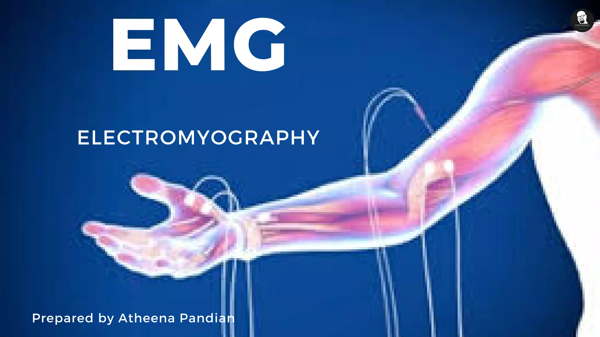
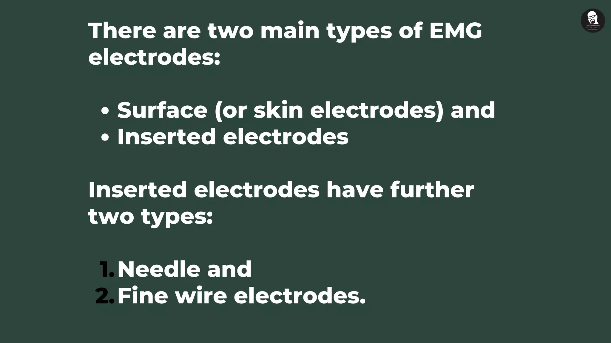
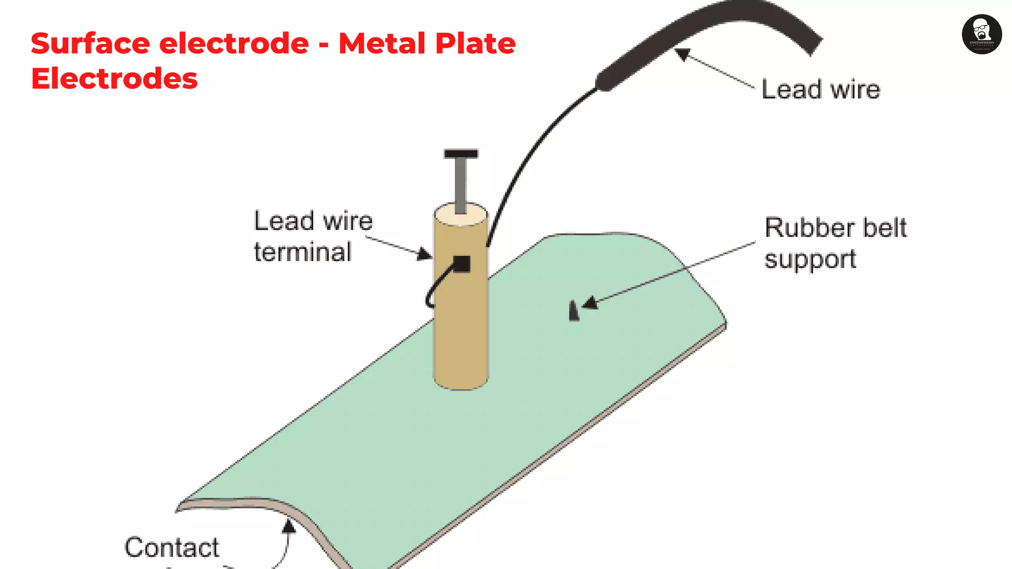
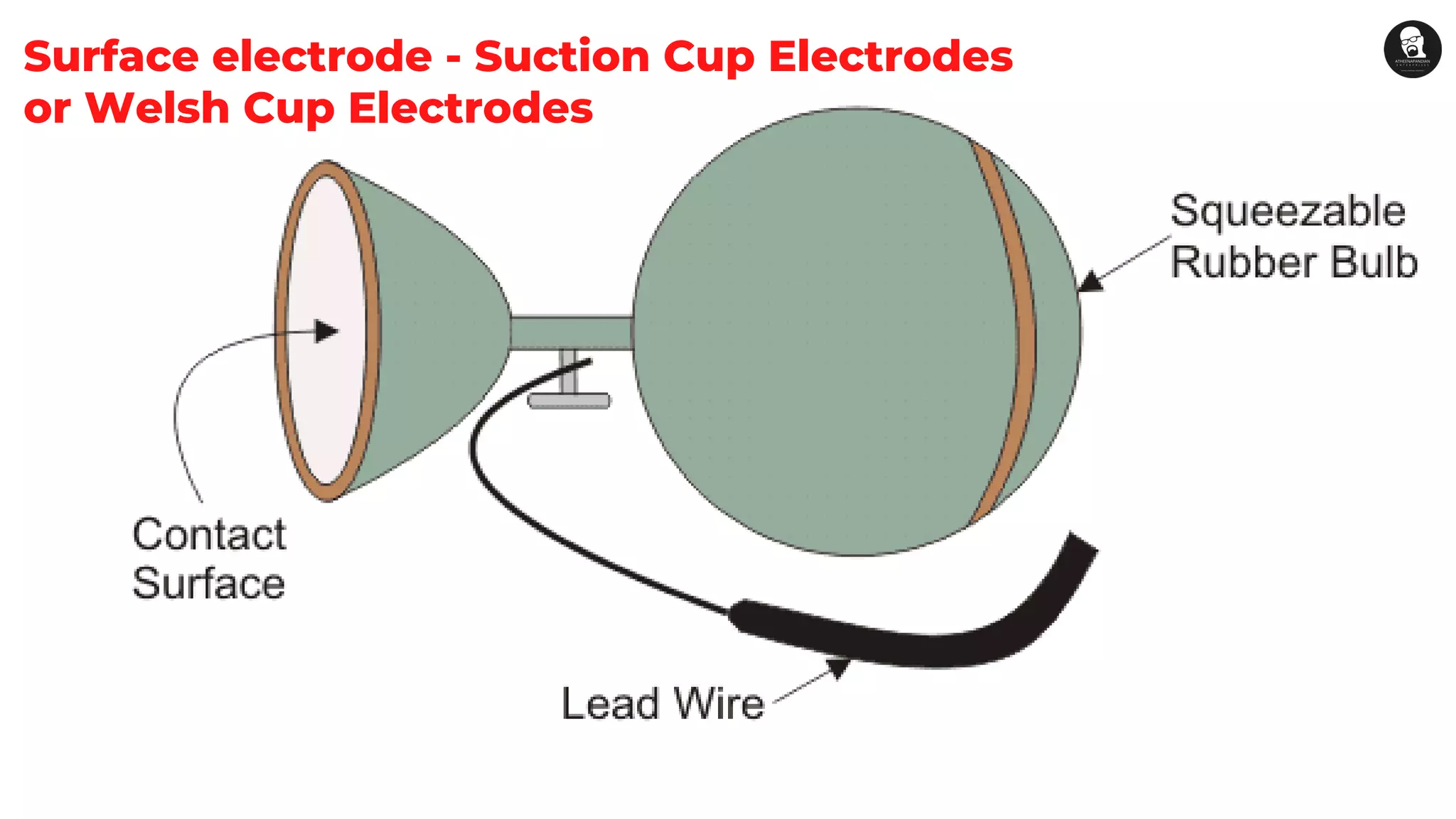
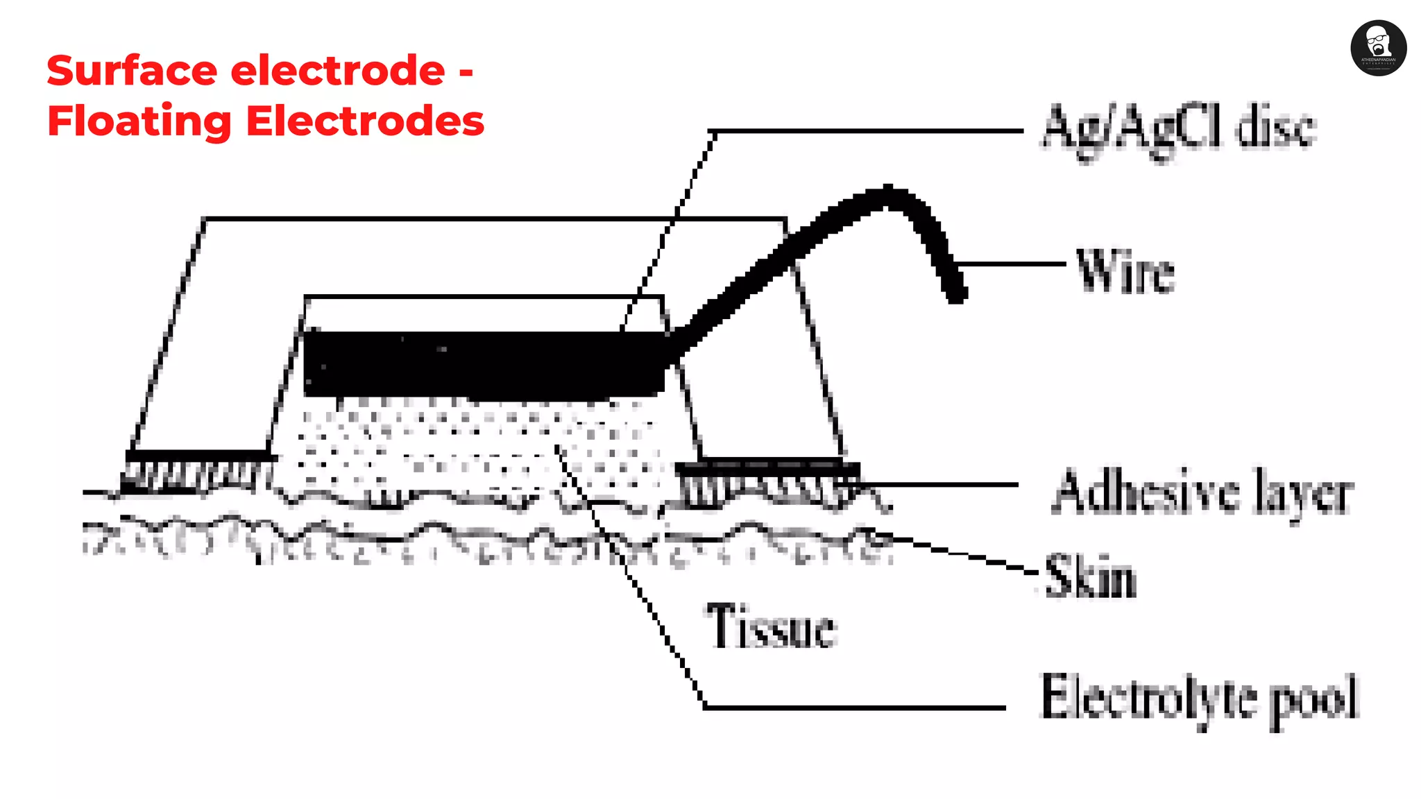
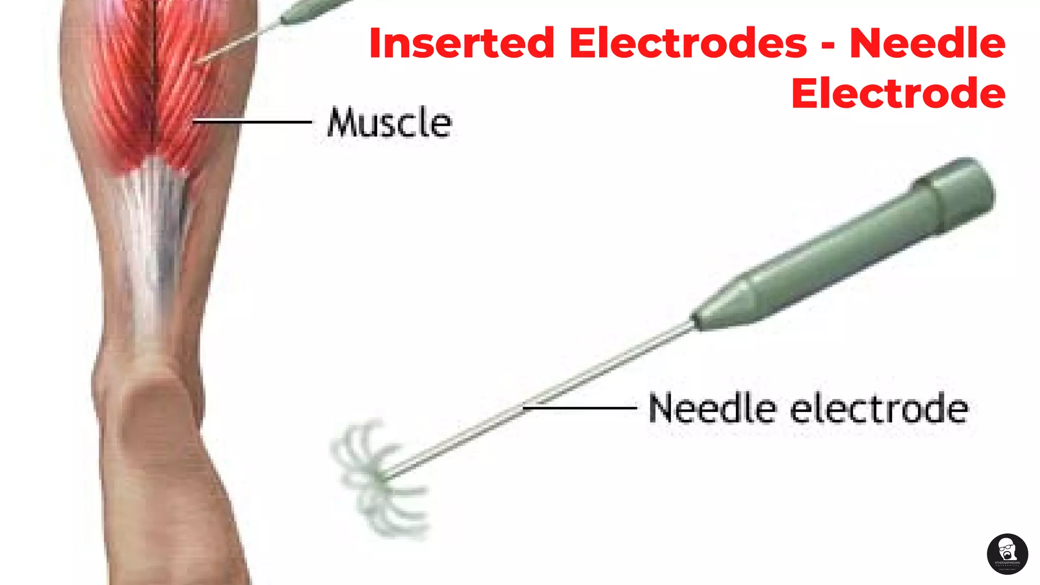
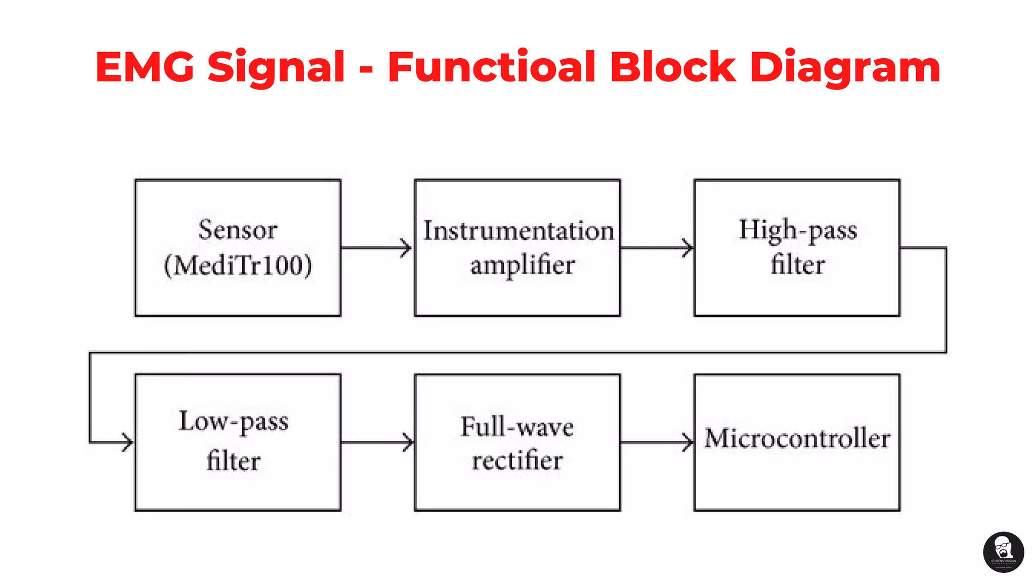
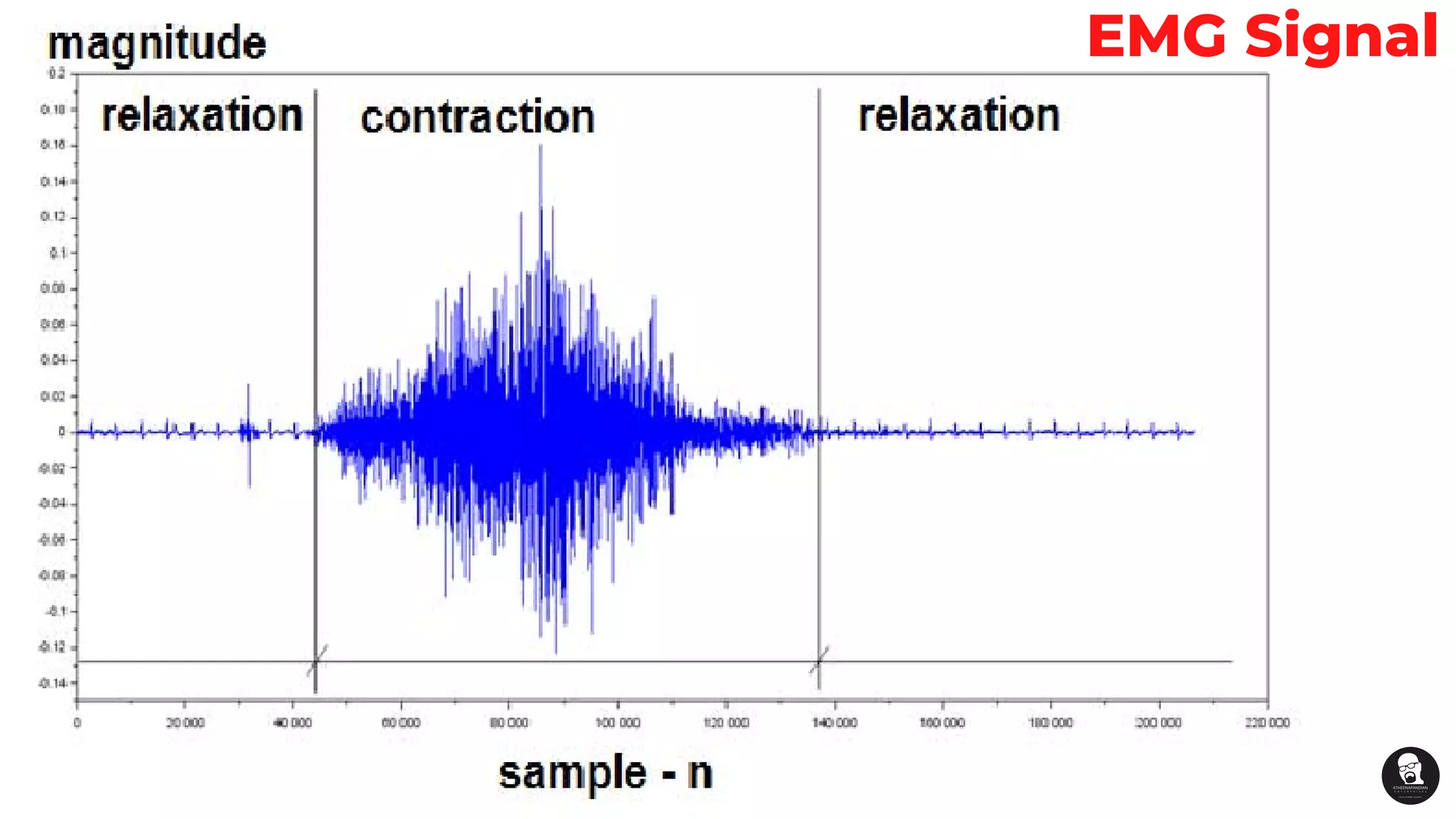
Electromyography (EMG) is an electrodiagnostic
medicine technique for evaluating and recording
the electrical activity produced by skeletal muscles
EMG is performed using an instrument
called an electromyograph to produce
a record called an electromyogram
An electromyograph
detects the electric
potential generated by
muscle cells
The signals can be
analyzed to detect
medical abnormalities,
activation level, or
recruitment order, or to
analyze
the biomechanics of
human or animal
movement
EMG
electrodes
Surface (or skin electrodes) and
Inserted electrodes
Needle and
Fine wire electrodes.
There are two main types of EMG
electrodes:
Inserted electrodes have further
two types:
1.
2.
Surface electrode measures the
potential available from the
surface of the skin.
Metal Plate Electrodes
Suction Cup Electrodes or Welsh Cup Electrodes
Adhesive Type Electrodes
Multipoint Type Electrodes
Floating Electrodes
Ear Clips and Scalp Electrodes
1.
2.
3.
4.
5.
6.
Surface electrode - Metal Plate
Electrodes
Surface electrode - Suction Cup Electrodes
or Welsh Cup Electrodes
Surface electrode -
Adhesive Type Electrodes
Surface electrode -Multipoint Type Electrodes
Surface electrode -
Floating Electrodes
Surface electrode -
Ear Clips and Scalp
Electrodes
Needle Electrode
Fine Wire Electrode
Inserted Electrodes
1.
2.
Inserted Electrodes - Needle
Electrode
Inserted Electrodes - Fine wire
Electrode
EMG Signal - Functioal Block Diagram
EMG Signal
Good Luck

Electromyography (emg) basics