Downloaded 284 times
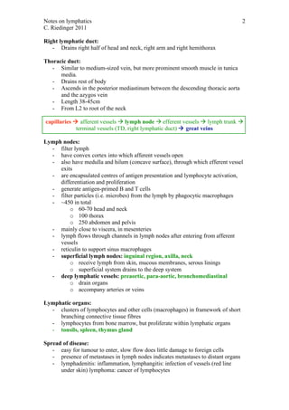

The lymphatic system consists of lymph, lymphatic vessels, lymph nodes, and lymphatic organs. It functions to maintain fluid balance, absorb fats, and aid the immune system. Lymph is clear fluid that forms from tissue fluid and is transported through lymphatic vessels and lymph nodes before draining into the bloodstream. The right lymphatic duct drains the right side of the body while the larger thoracic duct drains the rest of the body before both vessels drain into large veins in the neck. Lymph nodes filter lymph and contain immune cells that fight pathogens. Disease can spread through the lymphatic system as lymph nodes indicate metastasis and infections can occur in vessels.


