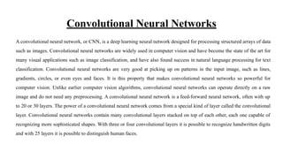
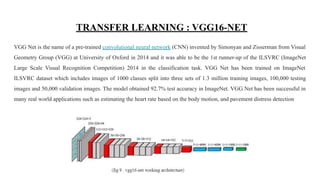
Lung cancer is one of the deadliest cancers worldwide. However, the early detection of lung cancer significantly improves survival rate. Cancerous (malignant) and noncancerous (benign) pulmonary nodules are the small growths of cells inside the lung. Detection of malignant lung nodules at an early stage is necessary for the crucial prognosis.
Since its launch in mid-January, the Data Science Bowl Lung Cancer Detection Competition has attracted more than 1,000 submissions. To be successful in this competition, data scientists need to be able to get started quickly and make rapid iterative changes. In this talk, we show how to compute features of the scanned images in the competition with a pre-trained Convolutional Neural Network (CNN) with Cognitive Toolkit (previously named CNTK), and use these features to classify the scans into cancerous or not cancerous, using a boosted tree with Light-GBM library, all in one hour.
Blog post: https://blogs.technet.microsoft.com/machinelearning/2017/02/17/quick-start-guide-to-the-data-science-bowl-lung-cancer-detection-challenge-using-deep-learning-microsoft-cognitive-toolkit-and-azure-gpu-vms/
Adaptive K-Means Clustering Algorithm for MR Breast Image Segmentation
3D Brain Tumor Segmentation Scheme using K-mean Clustering and Connected Component Labeling Algorithms
Volume Identification and Estimation of MRI Brain Tumor
MRI Breast cancer diagnosis hybrid approach using adaptive Ant-based segmentation and Multilayer Perceptron NN classifier
Brain tumor detection with the mri image and 54900 image Performance analysis of automated brain tumor detection from MR imaging and CT scan using basic image processing techniques based on various hard and soft .This repo is of segmentation and morphological operations which are the basic concepts of image processing. Detection and extraction of tumor from MRI scan images of the brain is done using python.
Performance analysis of automated brain tumor detection from MR imaging and CT scan using basic image processing techniques based on various hard and soft computing has been performed in our work. Moreover, we applied six traditional classifiers to detect brain tumor in the images. Then we applied CNN for brain tumor detection to include deep learning method in our work. We compared the result of the traditional one having the best accuracy (SVM) with the result of CNN. Furthermore, our work presents a generic method of tumor detection and extraction of its various features.
Talk @ ACM SF Bayarea Chapter on Deep Learning for medical imaging space.
The talk covers use cases, special challenges and solutions for Deep Learning for Medical Image Analysis using Tensorflow+Keras. You will learn about:
- Use cases for Deep Learning in Medical Image Analysis
- Different DNN architectures used for Medical Image Analysis
- Special purpose compute / accelerators for Deep Learning (in the Cloud / On-prem)
- How to parallelize your models for faster training of models and serving for inferenceing.
- Optimization techniques to get the best performance from your cluster (like Kubernetes/ Apache Mesos / Spark)
- How to build an efficient Data Pipeline for Medical Image Analysis using Deep Learning
- Resources to jump start your journey - like public data sets, common models used in Medical Image Analysis
image classification is a common problem in Artificial Intelligence , we used CIFR10 data set and tried a lot of methods to reach a high test accuracy like neural networks and Transfer learning techniques .
you can view the source code and the papers we read on github : https://github.com/Asma-Hawari/Machine-Learning-Project-
Brain Tumor Segmentation using Enhanced U-Net Model with Empirical AnalysisMD Abdullah Al Nasim
Cancer of the brain is deadly and requires careful surgical segmentation. The brain tumors were segmented using U-Net using a Convolutional Neural Network (CNN). When looking for overlaps of necrotic, edematous, growing, and healthy tissue, it might be hard to get relevant information from the images. The 2D U-Net network was improved and trained with the BraTS datasets to find these four areas. U-Net can set up many encoder and decoder routes that can be used to get information from images that can be used in different ways. To reduce computational time, we use image segmentation to exclude insignificant background details. Experiments on the BraTS datasets show that our proposed model for segmenting brain tumors from MRI (MRI) works well. In this study, we demonstrate that the BraTS datasets for 2017, 2018, 2019, and 2020 do not significantly differ from the BraTS 2019 dataset's attained dice scores of 0.8717 (necrotic), 0.9506 (edema), and 0.9427 (enhancing).
BRAIN TUMOR MRI IMAGE SEGMENTATION AND DETECTION IN IMAGE PROCESSINGDharshika Shreeganesh
Image processing is an active research area in which medical image processing is a highly challenging field. Medical imaging
techniques are used to image the inner portions of the human body for medical diagnosis. Brain tumor is a serious life altering
disease condition. Image segmentation plays a significant role in image processing as it helps in the extraction of suspicious regions
from the medical images. In this paper we have proposed segmentation of brain MRI image using K-means clustering algorithm
followed by morphological filtering which avoids the misclustered regions that can inevitably be formed after segmentation of the brain MRI image for detection of tumor location.
Intro to Deep Learning for Medical Image Analysis, with Dan Lee from Dentuit AISeth Grimes
Dan Lee from Dentuit AI presented an Intro to Deep Learning for Medical Image Analysis at the Maryland AI meetup (https://www.meetup.com/Maryland-AI), May 27, 2020. Visit https://www.youtube.com/watch?v=xl8i7CGDQi0 for video.
details about brain tumor
literature survey on many reference papers related to brain tumor detection using various techniques
our proposed novel methodology for brain tumor detection
Brain tumor classification using artificial neural network on mri imageseSAT Journals
Abstract
In this paper, an attempt has been made to summarize the multi-resolution transformation and the different classifiers useful to
analyze the brain tumor using MRI. X-ray, MRI, Ultrasound etc. are different techniques used to scan brain tumor images.
Radiologist prefers MRI to get detail information about tumor to help him diagnoses. In this paper we have used MRI of brain
tumor for analysis. We have used Digital image processing tool for detection of the tumor. The identification, detection and
classification of brain tumor have been done by extracting features from MRI with the help of wavelet transformation. The MRI of
brain tumor is classified into two categories normal and abnormal brain. In this work Digital image processing has been used as
a tool for getting clear and exact details about tumor in earlier stages. This helps the physicians and practitioners for diagnoses.
Key word – Brain tumor, Wavelet transform, segmentation.
Brain Tumor Detection Using Image ProcessingSinbad Konick
The process of brain tumor detection using various filters and finding out the best possible approach. Processing the image and using other filters and find out the result.
Lung Cancer Detection using Machine Learningijtsrd
Modern three dimensional 3 D medical imaging offers the potential and promise for major advances in science and medicine as higher fidelity images are produced. Due to advances in computer aided diagnosis and continuous progress in the field of computerized medical image visualization, there is need to develop one of the most important fields within scientific imaging. From the early basis report on cancer patients it has been seen that a greater number of people die of lung cancer than from other cancers such as colon, breast and prostate cancers combined. Lung cancer are related to smoking or secondhand smoke , or less often to exposure to radon or other environmental factors that’s why this can be prevented. But still it is not yet clear if these cancers can be prevented or not. In this research work, approach of segmentation, feature extraction and Convolution Neural Network CNN will be applied for locating, characterizing cancer portion. Harpreet Singh | Er. Ravneet Kaur | "Lung Cancer Detection using Machine Learning" Published in International Journal of Trend in Scientific Research and Development (ijtsrd), ISSN: 2456-6470, Volume-4 | Issue-6 , October 2020, URL: https://www.ijtsrd.com/papers/ijtsrd33659.pdf Paper Url: https://www.ijtsrd.com/computer-science/computer-architecture/33659/lung-cancer-detection-using-machine-learning/harpreet-singh
Development of Computational Tool for Lung Cancer Prediction Using Data MiningEditor IJCATR
The requirement for computerization of detection of lung cancer disease arises ever since recent-techniques which involve
manual-examination of the blood smear as the first step toward diagnosis. This is quite time-consuming, and their accurateness depends
upon the ability of operator's. So, prevention of lung cancer is very essential. This paper has surveyed various techniques used by previous
authors like ANN (Artificial Neural Network), image processing, LDA (Linear Dependent Analysis), SOM (Self Organizing Map) etc.
Image segmentation and classification tasks in computer vision have proven to be highly effective using neural networks, specifically Convolutional Neural Networks (CNNs). These tasks have numerous
practical applications, such as in medical imaging, autonomous driving, and surveillance. CNNs are capable
of learning complex features directly from images and achieving outstanding performance across several
datasets. In this work, we have utilized three different datasets to investigate the efficacy of various preprocessing and classification techniques in accurssedately segmenting and classifying different structures
within the MRI and natural images. We have utilized both sample gradient and Canny Edge Detection
methods for pre-processing, and K-means clustering have been applied to segment the images. Image
augmentation improves the size and diversity of datasets for training the models for image classification
Image segmentation and classification tasks in computer vision have proven to be highly effective using neural networks, specifically Convolutional Neural Networks (CNNs). These tasks have numerous
practical applications, such as in medical imaging, autonomous driving, and surveillance. CNNs are capable
of learning complex features directly from images and achieving outstanding performance across several
datasets. In this work, we have utilized three different datasets to investigate the efficacy of various preprocessing and classification techniques in accurssedately segmenting and classifying different structures
within the MRI and natural images. We have utilized both sample gradient and Canny Edge Detection
methods for pre-processing, and K-means clustering have been applied to segment the images. Image
augmentation improves the size and diversity of datasets for training the models for image classification.
This work highlights transfer learning’s effectiveness in image classification using CNNs and VGG 16 that
provides insights into the selection of pre-trained models and hyper parameters for optimal performance.
We have proposed a comprehensive approach for image segmentation and classification, incorporating preprocessing techniques, the K-means algorithm for segmentation, and employing deep learning models such
as CNN and VGG 16 for classification.
3D Segmentation of Brain Tumor ImagingIJAEMSJORNAL
A brain tumor is a collection of anomalous cells that grow in or around the brain. Brain tumors affect the humans badly, it can disrupt proper brain function and be life-threatening. In this project, we have proposed a system to detect, segment, and classify the tumors present in the brain. Once the brain tumor is identified at the very beginning, proper treatments can be done and it may be cured.
Glioblastomas brain tumour segmentation based on convolutional neural network...IJECEIAES
Brain tumour segmentation can improve diagnostics efficiency, rise the prediction rate and treatment planning. This will help the doctors and experts in their work. Where many types of brain tumour may be classified easily, the gliomas tumour is challenging to be segmented because of the diffusion between the tumour and the surrounding edema. Another important challenge with this type of brain tumour is that the tumour may grow anywhere in the brain with different shape and size. Brain cancer presents one of the most famous diseases over the world, which encourage the researchers to find a high-throughput system for tumour detection and classification. Several approaches have been proposed to design automatic detection and classification systems. This paper presents an integrated framework to segment the gliomas brain tumour automatically using pixel clustering for the MRI images foreground and background and classify its type based on deep learning mechanism, which is the convolutional neural network. In this work, a novel segmentation and classification system is proposed to detect the tumour cells and classify the brain image if it is healthy or not. After collecting data for healthy and non-healthy brain images, satisfactory results are found and registered using computer vision approaches. This approach can be used as a part of a bigger diagnosis system for breast tumour detection and manipulation.
Brain tumor classification in magnetic resonance imaging images using convol...IJECEIAES
Deep learning (DL) is a subfield of artificial intelligence (AI) used in several sectors, such as cybersecurity, finance, marketing, automated vehicles, and medicine. Due to the advancement of computer performance, DL has become very successful. In recent years, it has processed large amounts of data, and achieved good results, especially in image analysis such as segmentation and classification. Manual evaluation of tumors, based on medical images, requires expensive human labor and can easily lead to misdiagnosis of tumors. Researchers are interested in using DL algorithms for automatic tumor diagnosis. convolutional neural network (CNN) is one such algorithm. It is suitable for medical image classification tasks. In this paper, we will focus on the development of four sequential CNN models to classify brain tumors in magnetic resonance imaging (MRI) images. We followed two steps, the first being data preprocessing and the second being automatic classification of preprocessed images using CNN. The experiments were conducted on a dataset of 3,000 MRI images, divided into two classes: tumor and normal. We obtained a good accuracy of 98,27%, which outperforms other existing models.
Exciting IoT projects for your final year.pdfjagan477830
The final year presents an opportunity for students to engage in exciting Internet of Things (IoT) projects. These projects offer a platform for students to apply their knowledge and skills in developing innovative solutions that address real-world challenges. The IoT projects provide a valuable learning experience that prepares students for the demands of the modern workplace.
Innovative IoT-Based Projects to Revolutionize Everyday Life.pdfjagan477830
Welcome to the presentation on Transforming Daily Living: Unleashing the Potential of Innovative IoT-Based Projects. Today, we will explore the exciting advancements and possibilities that arise from integrating Internet of Things (IoT) technologies into our everyday live
Welcome and brief overview of IoT (Internet of Things) .Highlight the significance of IoT in connecting devices and enabling automation. Introduce the focus of the presentation: IoT-based mini projects
Mini Projects for Computer Science Engineering.pdfjagan477830
This PowerPoint presentation showcases a variety of mini projects suitable for Computer Science Engineering (CSE) students. It covers projects in web development, mobile app development, data analysis, machine learning, IoT, network security, game development, natural language processing, cloud computing, and robotics. The presentation aims to inspire CSE students to explore different project ideas and technologies within their field.
Mini Projects for Electronics and Communication Engineering.pdfjagan477830
these mini projects are meant to enhance your practical skills, apply theoretical knowledge, and gain hands-on experience in the field of ECE. Feel free to choose a project that aligns with your interests and level of expertise.
Mini Projects for Computer Science Engineering Students.pdfjagan477830
"Welcome to the presentation on Mini Projects for Computer Science and Engineering (CSE)
This presentation highlights a selection of engaging and practical mini projects for CSE students."
Overview of Embedded Systems Projects Examples.pdfjagan477830
Definition of Embedded Systems: Embedded systems are computer systems that are designed to perform specific tasks, often with real-time computing constraints. They are found in a wide range of applications, from consumer electronics to industrial automation.
Importance of Embedded Systems: Embedded systems are critical to modern technology, enabling everything from smart homes to medical devices. They are often highly optimized for their specific task, making them more efficient and cost-effective than general-purpose computing systems.
The Future of CSE Projects_ Emerging Technologies to Watch Out For.pdfjagan477830
The future of CSE projects is looking brighter than ever with the emergence of new technologies. Artificial Intelligence (AI), Machine Learning, Big Data and Internet of Things (IoT) are some examples that have been gaining traction in recent years and will continue to be important for CSE projects. AI can help automate processes, while machine learning can help analyze data more accurately. Big data allows businesses to gain insights into customer behaviour which helps them make better decisions. Lastly, IoT enables devices to communicate with each other without manual intervention making it easier for businesses to manage their operations more efficiently. All these emerging technologies offer great potentials when used correctly in CSE projects so they should definitely be watched out for!
A Comprehensive Guide of Python Final Year Projects with Source Code.pdfjagan477830
Final-year projects are an integral part of a student's academic journey. It provides an opportunity for students to apply their knowledge and skills to real-world problems. Python, being a versatile programming language, is widely used in final-year projects across various fields. This presentation will explore some popular Python final-year projects with source code.
Top AI project ideas for engineering students.pdfjagan477830
Welcome to the presentation on Top AI Project Ideas for Engineering Students. Artificial intelligence (AI) is a rapidly growing field that has the potential to transform the way we live and work. From image recognition to natural language processing, and autonomous vehicles to predictive maintenance, AI is being applied in diverse fields with great success.
How to Choose the Perfect Mtech Project Topic for Your Interests and Career G...jagan477830
The introduction should provide an overview of the presentation and why it's important to choose the right Mtech project. It should emphasize the benefits of choosing a project based on your interests and career goals, such as increased motivation, better career prospects, and personal fulfillment.
Beginner-Friendly IoT Arduino Projects to Try.pdfjagan477830
The Arduino community provides a wealth of tutorials, examples, and libraries that you can use to learn how to use Arduino and build your own projects
Some basic concepts to understand when working with Arduino include digital and analog signals, input and output pins, and pulse width modulation (PWM) for controlling the brightness of LEDs or the speed of motors
Sentiment Analysis on social networking sites.pptx.pdfjagan477830
Sentiment Analysis is the Process of computationally identifying and categorizing opinions from piece of text, and determine whether the writer’s attitude towards a particular topic/product/event is positive or negative or neutral.
Sentiment analysis is often referred to with different names such as Opinion Mining, Sentient classification, Sentiment analysis, and Sentiment extraction.
Machine Learning statistical model using Transportation datajagan477830
As the world is growing rapidly the people and the vehicles we use to move from one place to another, so the transportation is playing a vital role in making human lives easiest to travel from one place to another, everyday more and more vehicles are being produced and being bought by the people around the world, be it Electric, Hydrogen, petrol, diesel or solar powered.
Diabetes Prediction Using Machine Learningjagan477830
Our proposed system aims at Predicting the number of Diabetes patients and eliminating the risk of False Negatives Drastically.
In proposed System, we use Random forest, Decision tree, Logistic Regression and Gradient Boosting Classifier to classify the Patients who are affected with Diabetes or not.
Random Forest and Decision Tree are the algorithms which can be used for both classification and regression.
The dataset is classified into trained and test dataset where the data can be trained individually, these algorithms are very easy to implement as well as very efficient in producing better results and can able to process large amount of data.
Even for large dataset these algorithms are extremely fast and can able to give accuracy of about over 90%.
Identifying and classifying unknown Network Disruptionjagan477830
Since the evolution of modern technology and with the drastic increase in the scale of network communication more and more network disruptions in traffic and private protocols have been taking place. Identifying and classifying the unknown network disruptions can provide support and even help to maintain the backup systems.
Detection of Retinal pigmentosa in paediatric agejagan477830
In order to register the user who wants to use the programme, the project Detection of Retinal Pigmentosa in Paediatric Age Patients combines deep learning with MySQL.
"The proposed system overcomes the above mentioned issue in an efficient way. It aims at analyzing the number of fraud transactions that are present in the dataset.
"
"Project Support for CSE, IT, ECE, and EEE students. Real-time projects under Industrial experts. Mini & Major project for Diploma, BTech, MTech, MS. Project guidance, Documentation support, and paper publications.
"
Mini Projects for ECE Students with Low Cost in Hyderabadjagan477830
Out of all of these factors, cost is the one that has the biggest impact on the little projects that ece students complete. You can spend a lot of your time working on construction projects as an engineering student and utilise the college's facilities for smaller projects, but for obvious reasons, you cannot spend more money on your project.
Any engineering student may effectively display their skill sets through a short project. You want to make an impression on the interviewer or the examiner with your projects, which genuinely showcase your technical experience.
Operation “Blue Star” is the only event in the history of Independent India where the state went into war with its own people. Even after about 40 years it is not clear if it was culmination of states anger over people of the region, a political game of power or start of dictatorial chapter in the democratic setup.
The people of Punjab felt alienated from main stream due to denial of their just demands during a long democratic struggle since independence. As it happen all over the word, it led to militant struggle with great loss of lives of military, police and civilian personnel. Killing of Indira Gandhi and massacre of innocent Sikhs in Delhi and other India cities was also associated with this movement.
We all have good and bad thoughts from time to time and situation to situation. We are bombarded daily with spiraling thoughts(both negative and positive) creating all-consuming feel , making us difficult to manage with associated suffering. Good thoughts are like our Mob Signal (Positive thought) amidst noise(negative thought) in the atmosphere. Negative thoughts like noise outweigh positive thoughts. These thoughts often create unwanted confusion, trouble, stress and frustration in our mind as well as chaos in our physical world. Negative thoughts are also known as “distorted thinking”.
Synthetic Fiber Construction in lab .pptxPavel ( NSTU)
Synthetic fiber production is a fascinating and complex field that blends chemistry, engineering, and environmental science. By understanding these aspects, students can gain a comprehensive view of synthetic fiber production, its impact on society and the environment, and the potential for future innovations. Synthetic fibers play a crucial role in modern society, impacting various aspects of daily life, industry, and the environment. ynthetic fibers are integral to modern life, offering a range of benefits from cost-effectiveness and versatility to innovative applications and performance characteristics. While they pose environmental challenges, ongoing research and development aim to create more sustainable and eco-friendly alternatives. Understanding the importance of synthetic fibers helps in appreciating their role in the economy, industry, and daily life, while also emphasizing the need for sustainable practices and innovation.
How to Make a Field invisible in Odoo 17Celine George
It is possible to hide or invisible some fields in odoo. Commonly using “invisible” attribute in the field definition to invisible the fields. This slide will show how to make a field invisible in odoo 17.
How to Split Bills in the Odoo 17 POS ModuleCeline George
Bills have a main role in point of sale procedure. It will help to track sales, handling payments and giving receipts to customers. Bill splitting also has an important role in POS. For example, If some friends come together for dinner and if they want to divide the bill then it is possible by POS bill splitting. This slide will show how to split bills in odoo 17 POS.
This is a presentation by Dada Robert in a Your Skill Boost masterclass organised by the Excellence Foundation for South Sudan (EFSS) on Saturday, the 25th and Sunday, the 26th of May 2024.
He discussed the concept of quality improvement, emphasizing its applicability to various aspects of life, including personal, project, and program improvements. He defined quality as doing the right thing at the right time in the right way to achieve the best possible results and discussed the concept of the "gap" between what we know and what we do, and how this gap represents the areas we need to improve. He explained the scientific approach to quality improvement, which involves systematic performance analysis, testing and learning, and implementing change ideas. He also highlighted the importance of client focus and a team approach to quality improvement.
2. Table of Contents
1. Introduction
2. Visualization of Dataset
3. Proposed Model
4. Convolutional neural network
5. Transfer Learning : VGG16-Net
6. Future work
7. Reference
3. INTRODUCTION
Lung cancer is one of the deadliest cancers worldwide. However, the early detection of lung cancer significantly improves
survival rate. Cancerous (malignant) and noncancerous (benign) pulmonary nodules are the small growths of cells inside the
lung. Detection of malignant lung nodules at an early stage is necessary for the crucial prognosis.
Early-stage cancerous lung nodules are very much similar to non-cancerous nodules and need a differential diagnosis on the
basis of slight morphological changes, locations, and clinical biomarkers. The challenging task is to measure the probability
of malignancy for the early cancerous lung nodules. Various diagnostic procedures are used by physicians, in connection, for
the early diagnosis of malignant lung nodules, such as clinical settings, computed tomography (CT) scan analysis
(morphological assessment), positron emission tomography (PET) (metabolic assessments), and needle prick biopsy analysis
For the input layer, lung nodule CT images are used and are collected for various steps of the project. The source of the
dataset is the LUNA16 dataset .
The LUNA16 dataset is a subset of LIDC-IDRI dataset, in which the heterogeneous scans are filtered by different criteria.
Since pulmonary nodules can be very small, a thin slice should be chosen. Therefore scans with a slice thickness greater than
2.5 mm were discarded.
4. VISUALIZATION OF DATASET
Visualization of dataset is an important part of training , it gives better understanding of dataset. But CT scan images are hard
to visualize for a normal pc or any window browser. Therefore we use the pydicom library to solve this problem. The
Pydicom library gives an image array and metadata information stored in CT images like patient’s name,patient’s id, patient’s
birth date,image position , image number , doctor’s name , doctor’s birth date etc.
6. PROPOSED MODELS
The proposed model is a convolutional neural network approach based on lung segmentation on CT scan images. At first we
preprocess the dataset of luna16. We tried three different models of Convolutional Neural Networks, which are based on the
comparative study of performance of each type model in different dataset and for different classification problems.
Convolutional Neural Networks
A convolutional neural network, or CNN, is a deep learning neural network designed for processing structured arrays of data
such as images. Convolutional neural networks are widely used in computer vision and have become the state of the art for
many visual applications such as image classification, and have also found success in natural language processing for text
classification. Convolutional neural networks are very good at picking up on patterns in the input image, such as lines,
gradients, circles, or even eyes and faces. It is this property that makes convolutional neural networks so powerful for
computer vision. Unlike earlier computer vision algorithms, convolutional neural networks can operate directly on a raw
image and do not need any preprocessing. A convolutional neural network is a feed-forward neural network, often with up to
20 or 30 layers. The power of a convolutional neural network comes from a special kind of layer called the convolutional
layer.
7. Convolutional Neural Networks
A convolutional neural network, or CNN, is a deep learning neural network designed for processing structured arrays of data
such as images. Convolutional neural networks are widely used in computer vision and have become the state of the art for
many visual applications such as image classification, and have also found success in natural language processing for text
classification. Convolutional neural networks are very good at picking up on patterns in the input image, such as lines,
gradients, circles, or even eyes and faces. It is this property that makes convolutional neural networks so powerful for
computer vision. Unlike earlier computer vision algorithms, convolutional neural networks can operate directly on a raw
image and do not need any preprocessing. A convolutional neural network is a feed-forward neural network, often with up
to 20 or 30 layers. The power of a convolutional neural network comes from a special kind of layer called the convolutional
layer. Convolutional neural networks contain many convolutional layers stacked on top of each other, each one capable of
recognizing more sophisticated shapes. With three or four convolutional layers it is possible to recognize handwritten digits
and with 25 layers it is possible to distinguish human faces.
8. TRANSFER LEARNING : VGG16-NET
VGG Net is the name of a pre-trained convolutional neural network (CNN) invented by Simonyan and Zisserman from Visual
Geometry Group (VGG) at University of Oxford in 2014 and it was able to be the 1st runner-up of the ILSVRC (ImageNet
Large Scale Visual Recognition Competition) 2014 in the classification task. VGG Net has been trained on ImageNet
ILSVRC dataset which includes images of 1000 classes split into three sets of 1.3 million training images, 100,000 testing
images and 50,000 validation images. The model obtained 92.7% test accuracy in ImageNet. VGG Net has been successful in
many real world applications such as estimating the heart rate based on the body motion, and pavement distress detection
9. VGG Net has learned to extract the features (feature extractor) that can distinguish the objects and is used to classify unseen
objects. VGG was invented with the purpose of enhancing classification accuracy by increasing the depth of the CNNs. VGG
16 and VGG 19, having 16 and 19 weight layers, respectively, have been used for object recognition. VGG Net takes input of
224×224 RGB images and passes them through a stack of convolutional layers with the fixed filter size of 3×3 and the stride
of 1. There are five max pooling filters embedded between convolutional layers in order to down-sample the input
representation (image, hidden-layer output matrix, etc.). The stack of convolutional layers are followed by 3 fully connected
layers, having 4096, 4096 and 1000 channels, respectively. The last layer is a soft-max layer . Below figure shows VGG
network structure.
But in our approach we have images with the shape of (512,512) . so we build our own model using vgg16-net architecture.
And compile the model with a powerful adam optimizer , learning rate is 0.0001 , entropy is binary_crossentropy and
accuracy metrics. The below figure shows model summary , convolution layers, max-pooling layers and params.
10. FUTURE WORK
So, in order to increase the accuracy of the model we will try to do more efficient data-preprocessing techniques are to be
implemented now after and before the image segmentation process which will mainly focus on efficient division of data into
cancerous and non-cancerous classes and making the dataset compatible to be processed with computer vision library of
python otherwise implementing the algorithms on the dataset from self defined functions.
Also a new data processing, training and classification pipeline is to be proposed which will help the models to predict the
data more accurately.
Current Suggestions includes the use of some other transfer learning models from imagenet in keras including the one
proposed above and implementation of Feature Extraction Algorithms like BRISK and SIFT from Computer Vision Library
and also integrating the ML training methods.
11. REFERENCES
1. Bjerager M., Palshof T., Dahl R., Vedsted P., Olesen F. Delay in diagnosis of lung cancer in general practice. Br. J. Gen. Pract.
2006;56:863–868. [PMC free article] [PubMed] [Google Scholar]
2. Nair M., Sandhu S.S., Sharma A.K. Cancer molecular markers: A guide to cancer detection and management. Semin. Cancer Biol.
2018;52:39–55. doi: 10.1016/j.semcancer.2018.02.002. [PubMed] [Google Scholar]
3. Silvestri G.A., Tanner N.T., Kearney P., Vachani A., Massion P.P., Porter A., Springmeyer S.C., Fang K.C., Midthun D., Mazzone P.J.
Assessment of plasma proteomics biomarker’s ability to distinguish benign from malignant lung nodules: Results of the PANOPTIC
(Pulmonary Nodule Plasma Proteomic Classifier) trial. Chest. 2018;154:491–500. doi: 10.1016/j.chest.2018.02.012. [PMC free article]
[PubMed] [Google Scholar]
4. Shi Z., Zhao J., Han X., Pei B., Ji G., Qiang Y. A new method of detecting pulmonary nodules with PET/CT based on an improved
watershed algorithm. PLoS ONE. 2015;10:e0123694. [PMC free article] [PubMed] [Google Scholar]
5. Lee K.S., Mayo J.R., Mehta A.C., Powell C.A., Rubin G.D., Prokop C.M.S., Travis W.D. Incidental Pulmonary Nodules Detected on
CT Images: Fleischner 2017. Radiology. 2017;284:228–243. [PubMed] [Google Scholar]
12. ABOUT TechieYan Technologies
TechieYan Technologies offers a special platform where you can study all the most cutting-edge technologies directly from
industry professionals and get certifications. TechieYan collaborates closely with engineering schools, engineering students,
academic institutions, the Indian Army, and businesses.
Address: 16-11-16/V/24, Sri Ram Sadan, Moosarambagh, Hyderabad 500036
Phone : +91 7075575787
Website: https://techieyantechnologies.com/