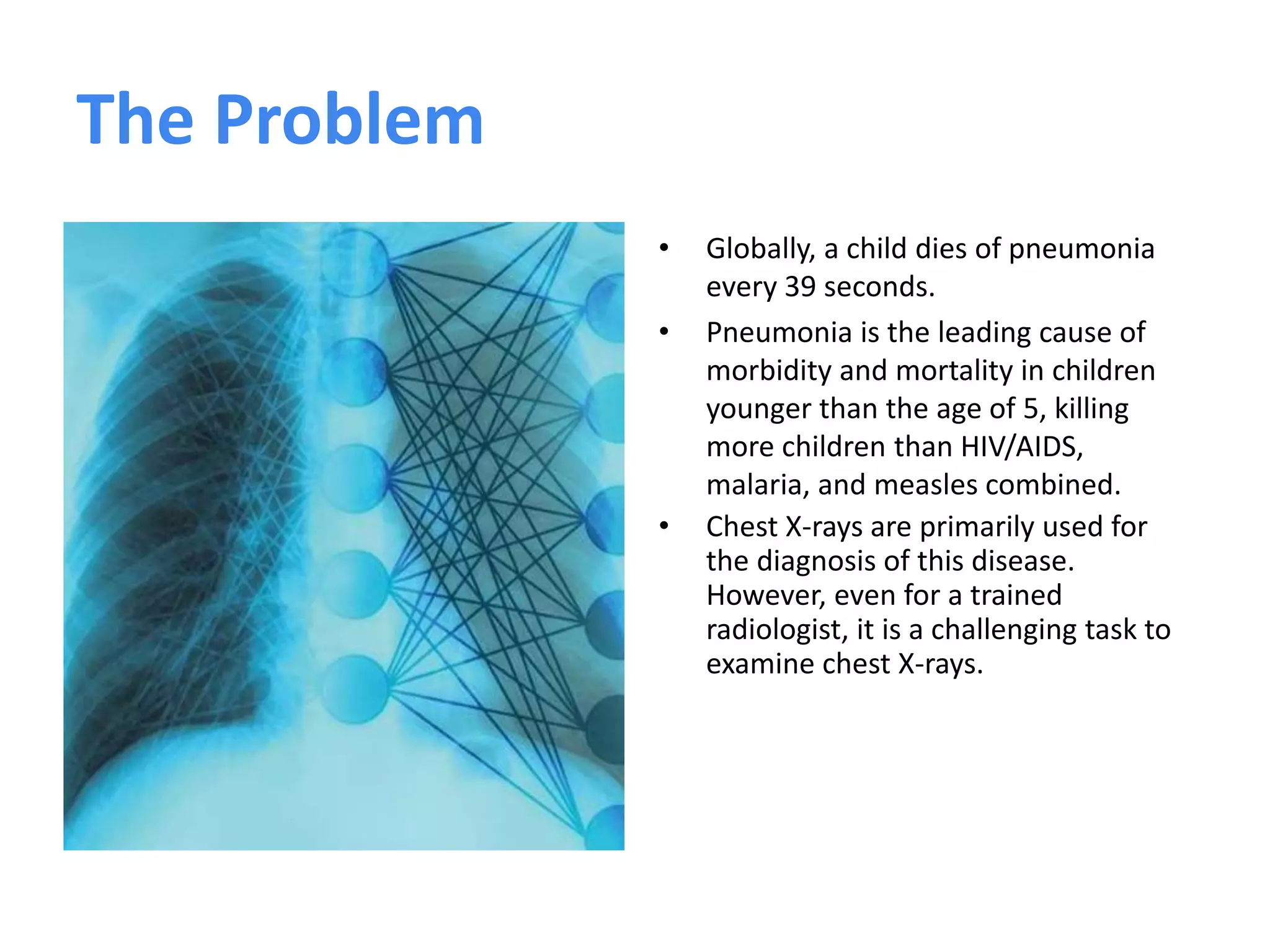
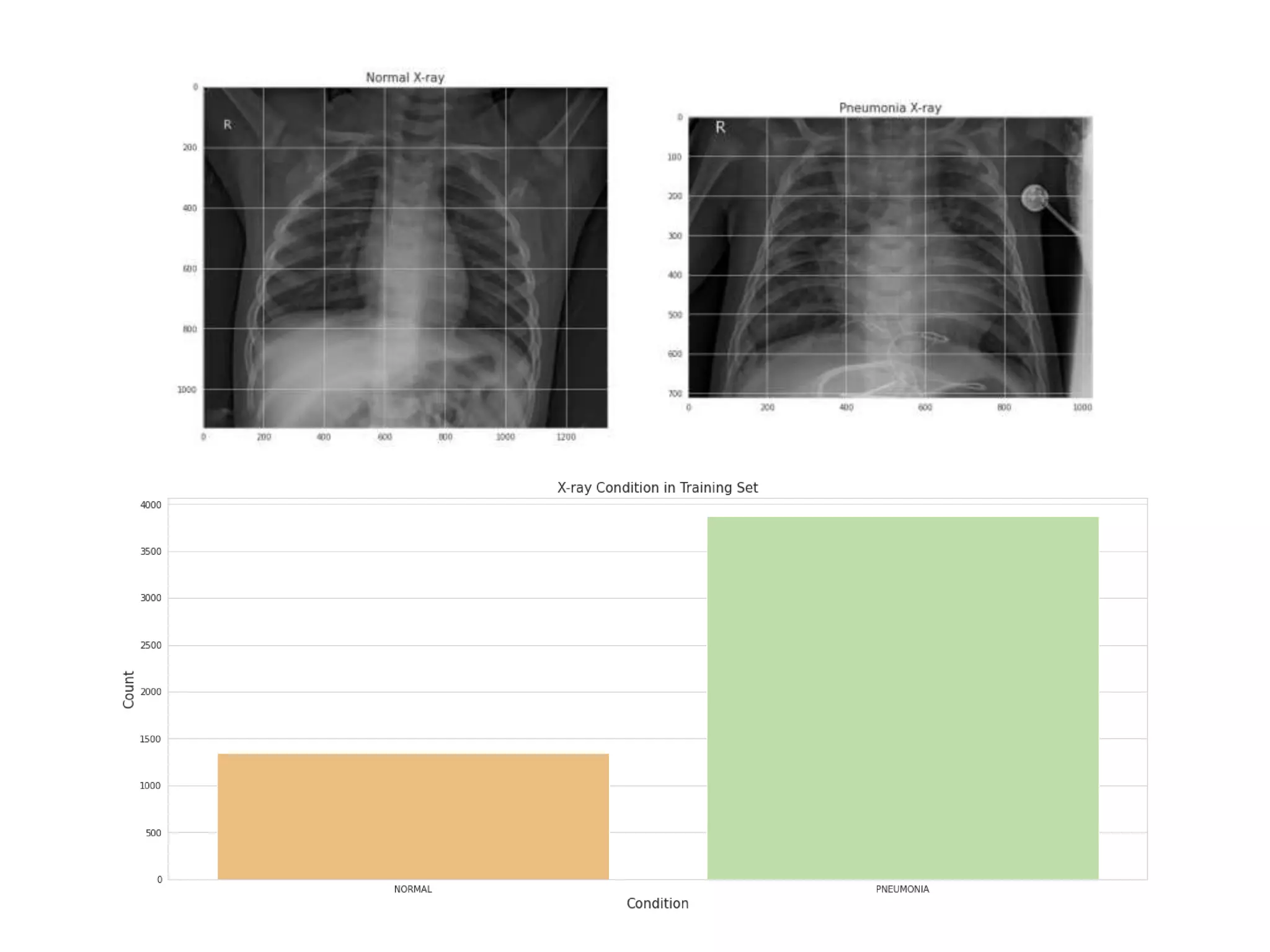
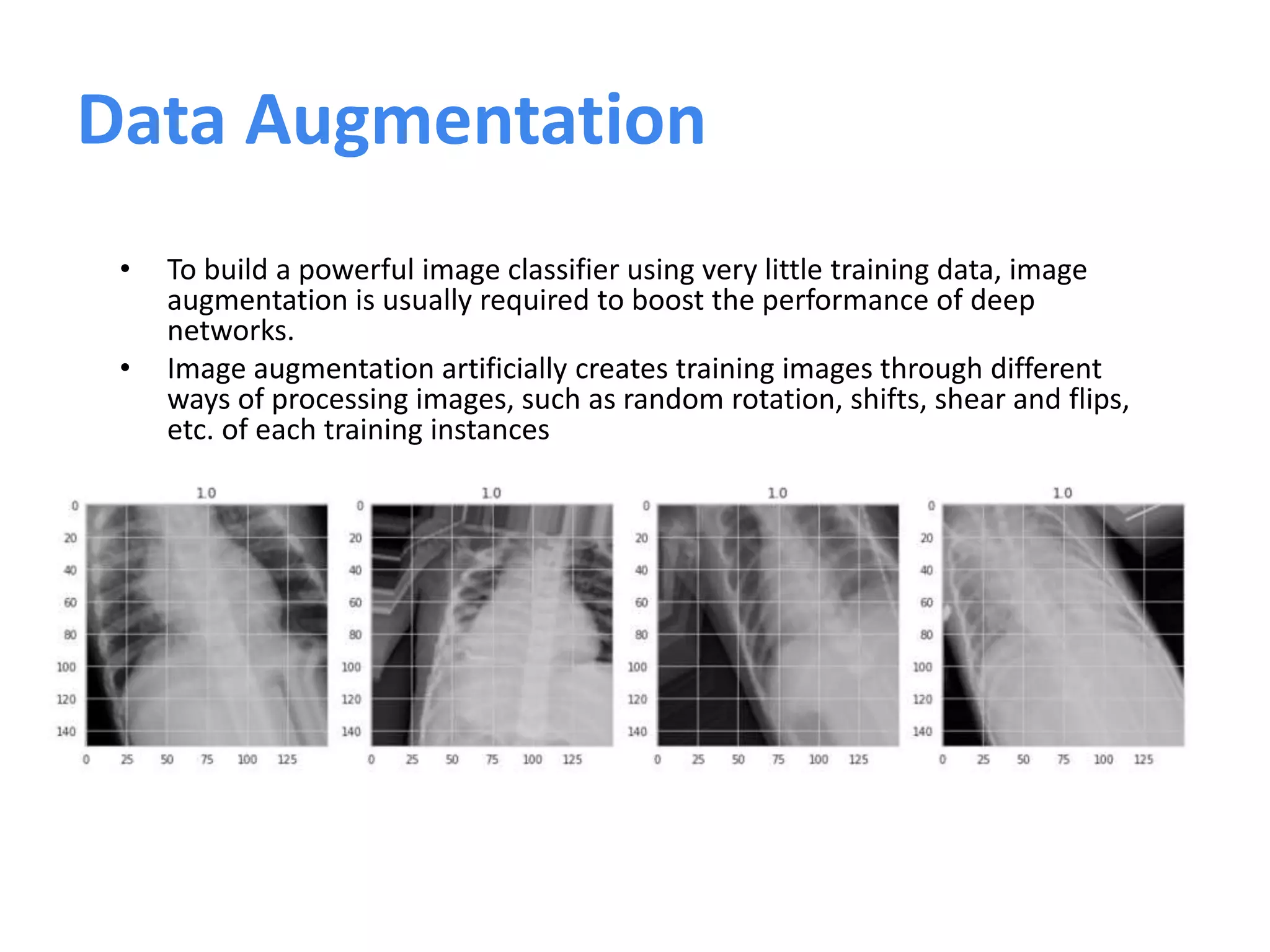
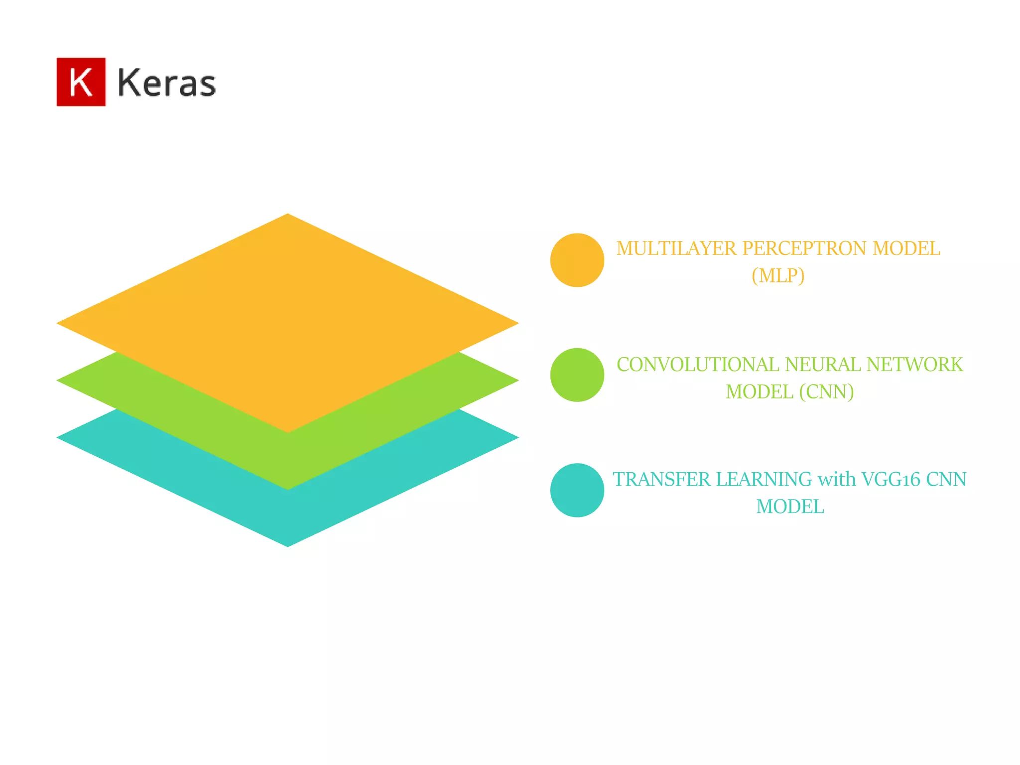
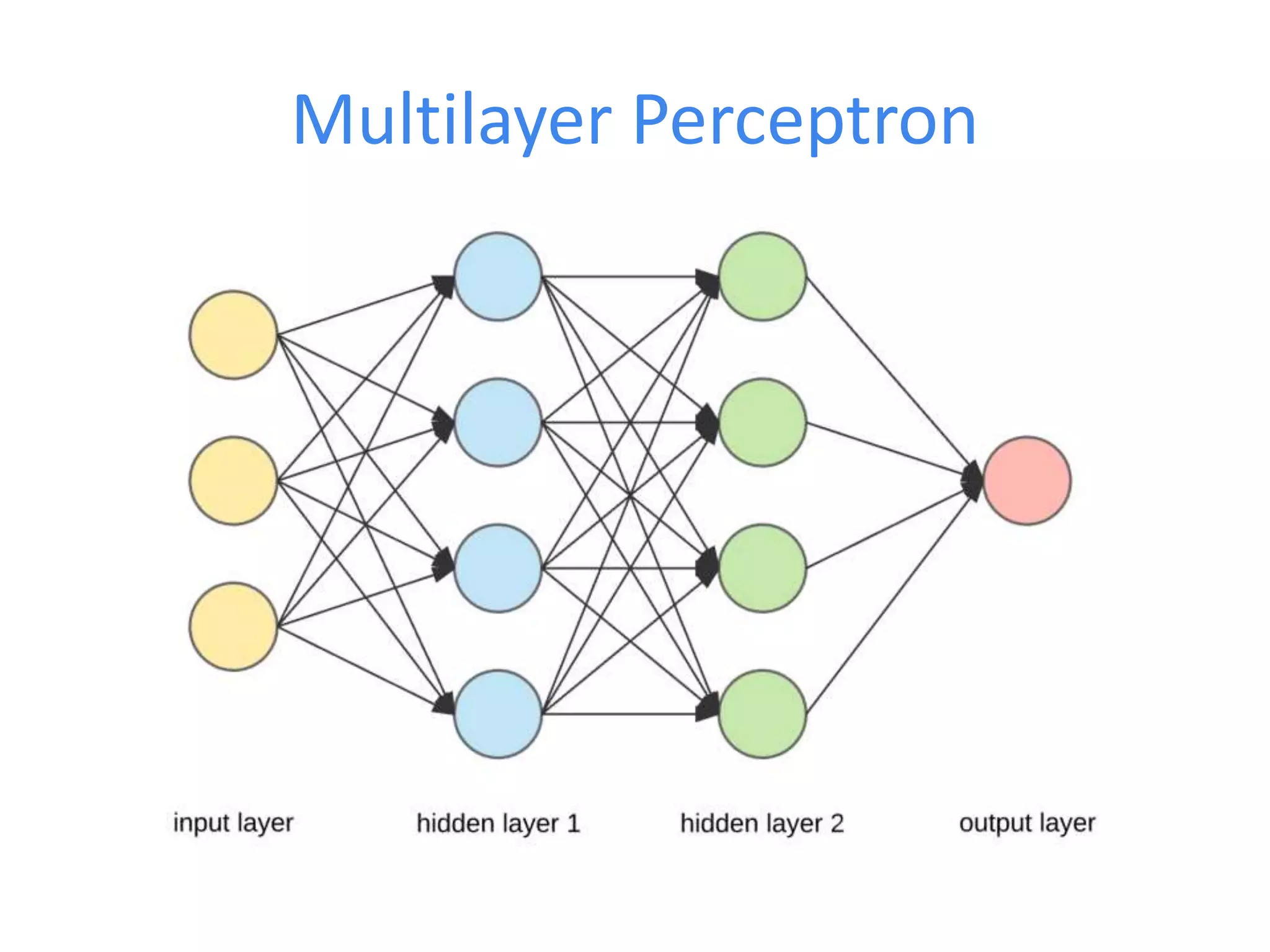
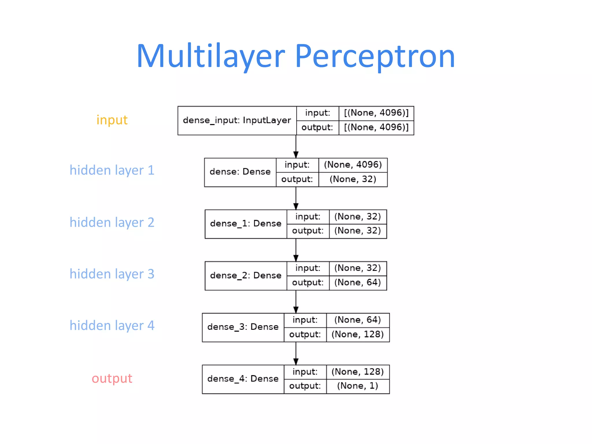
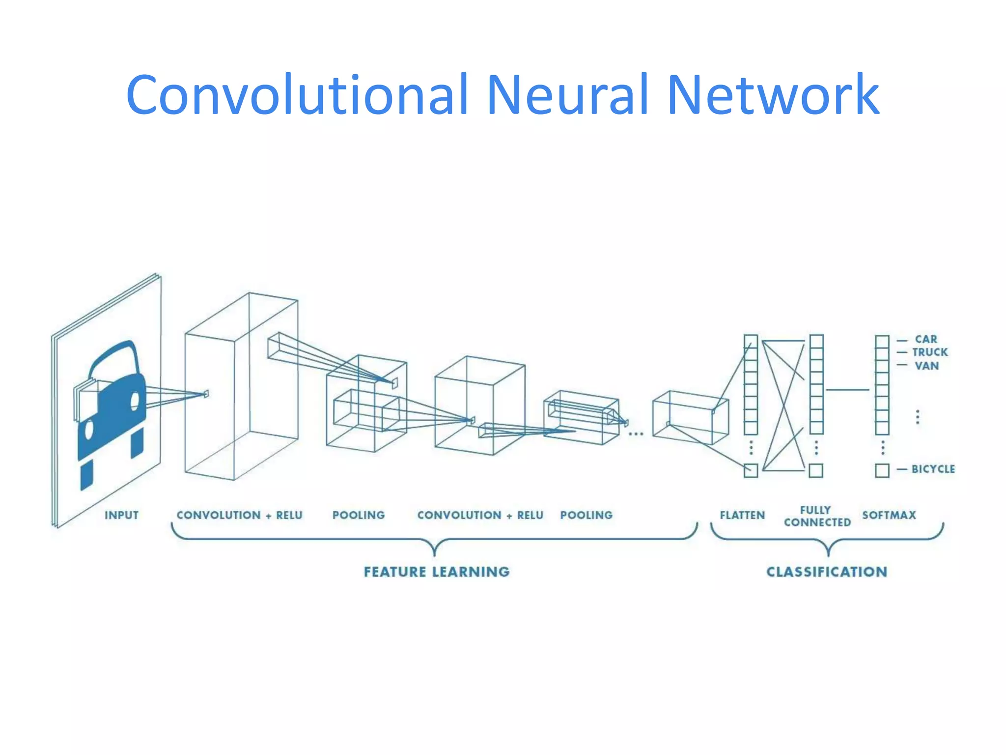
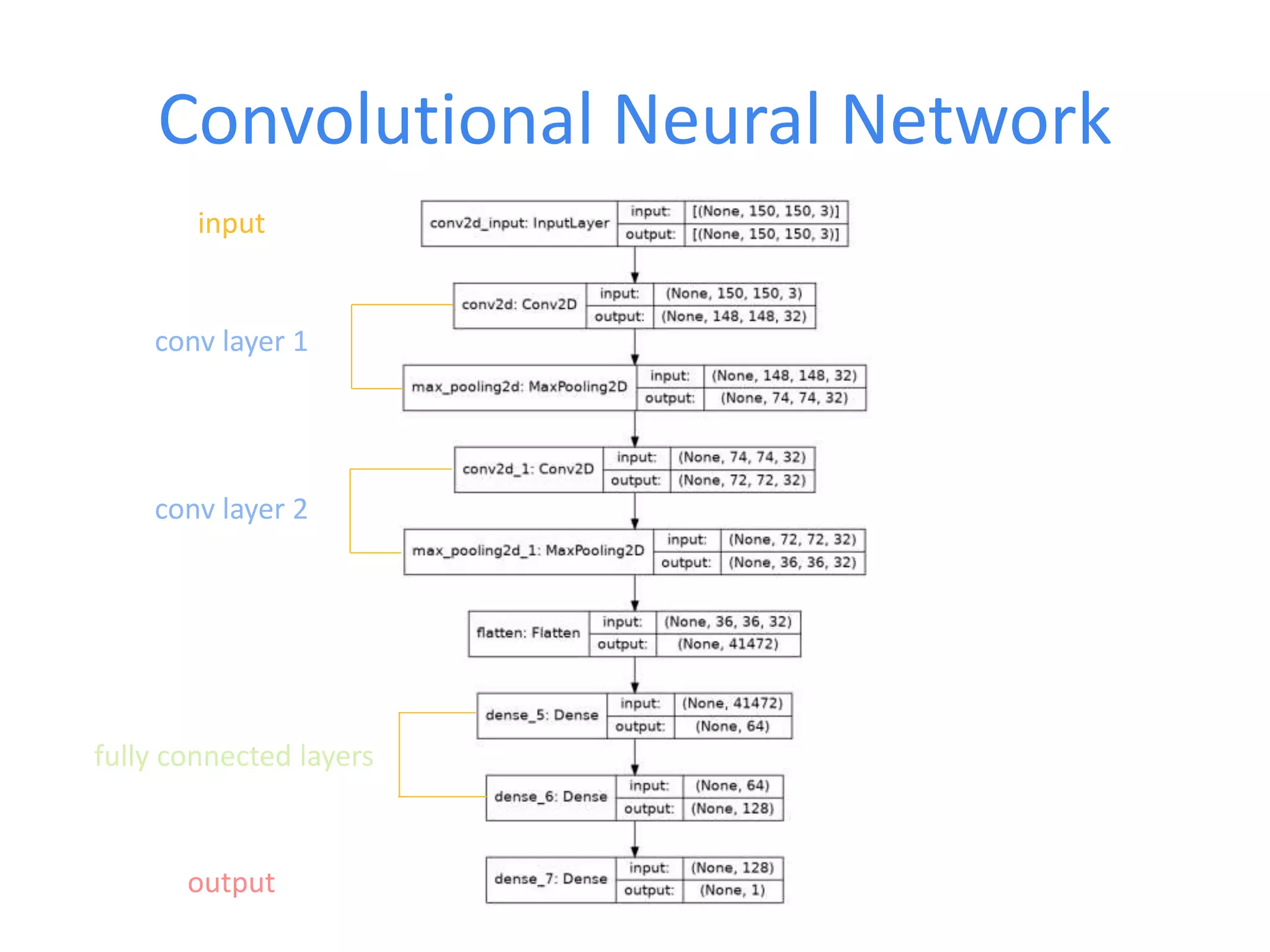
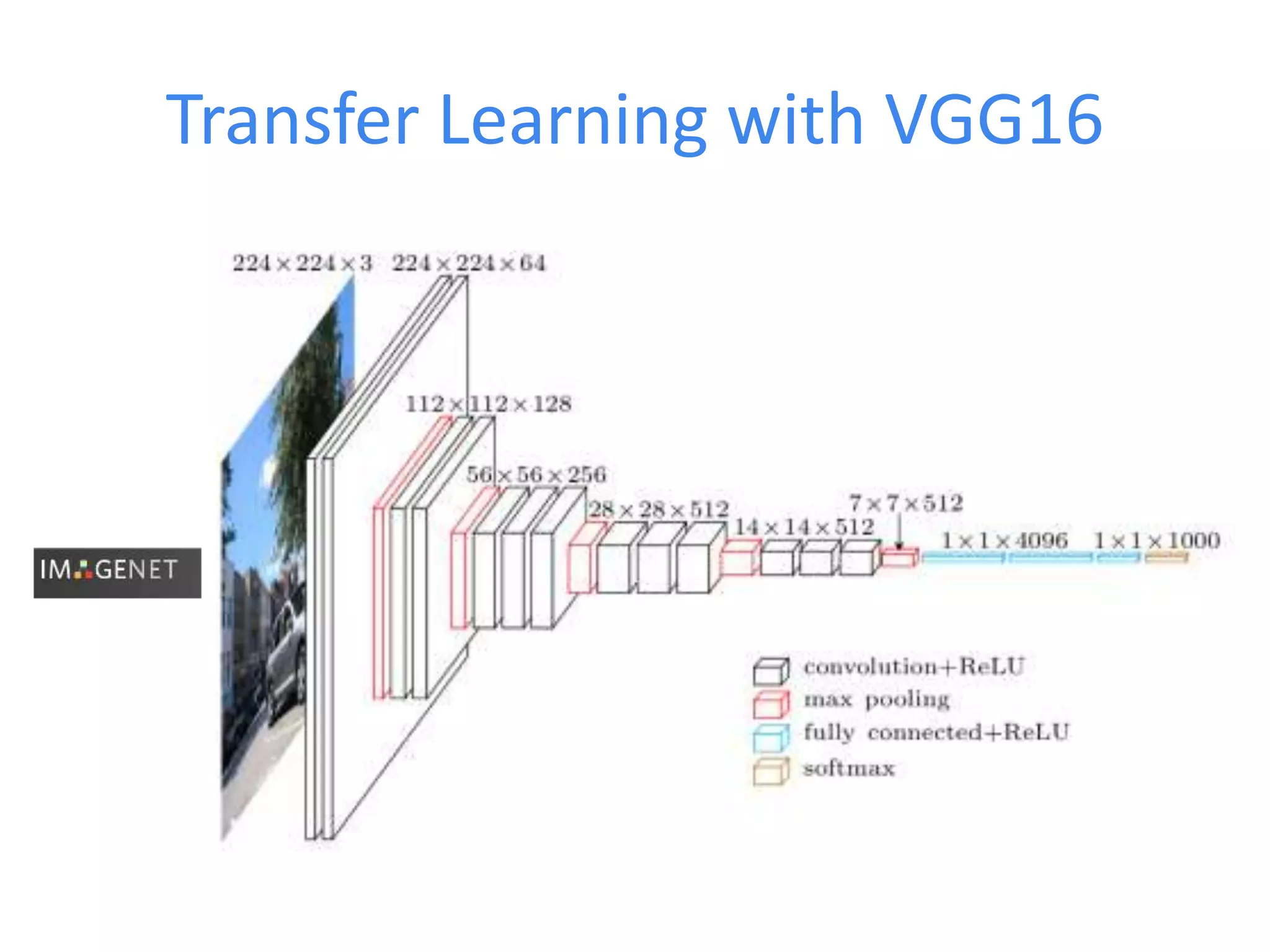
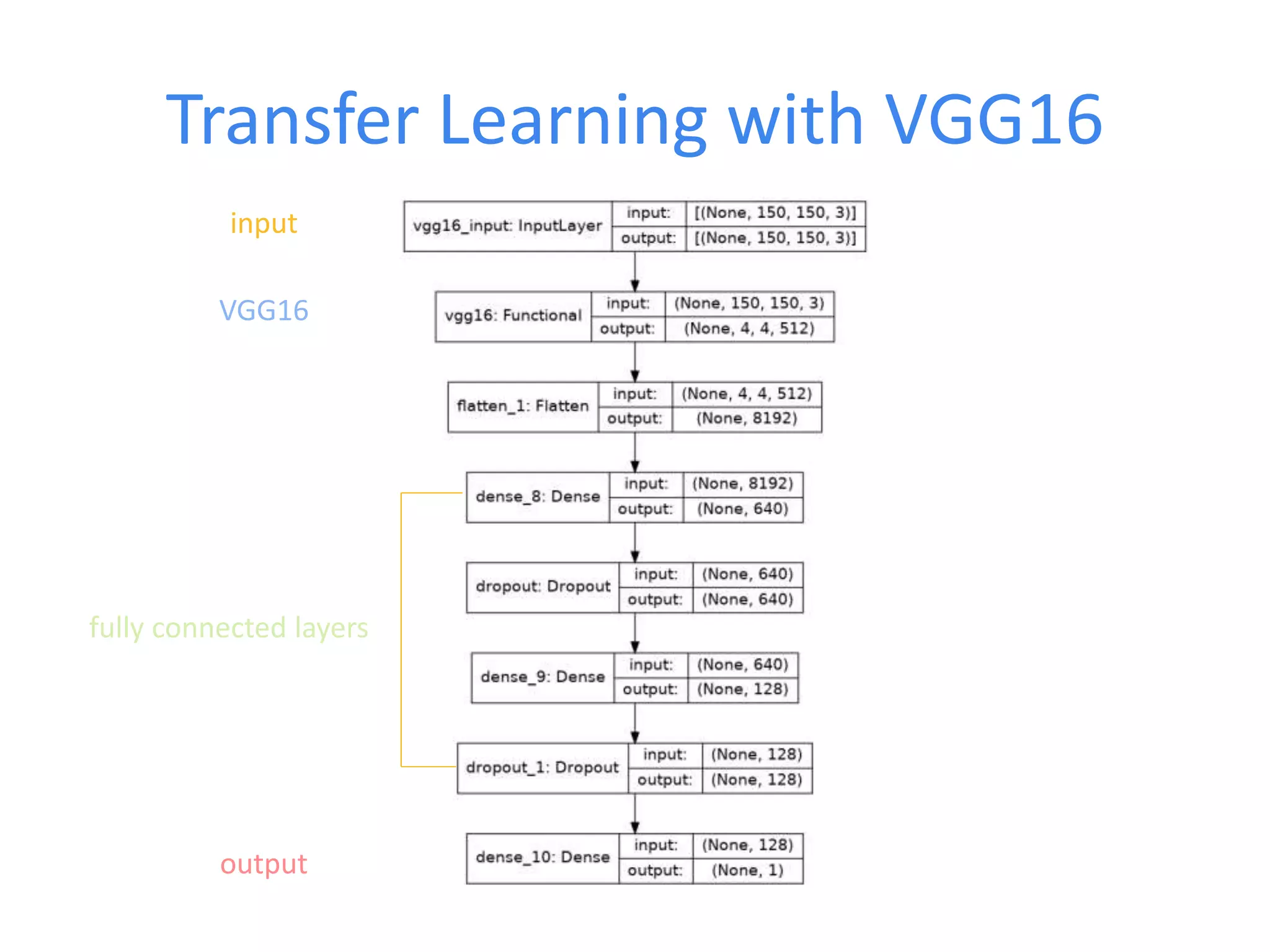
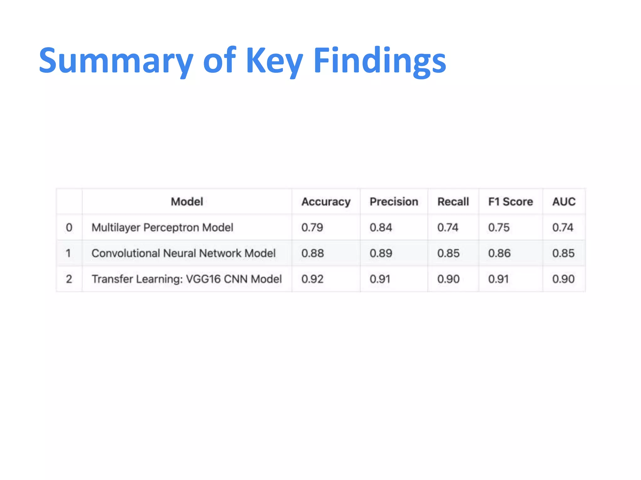
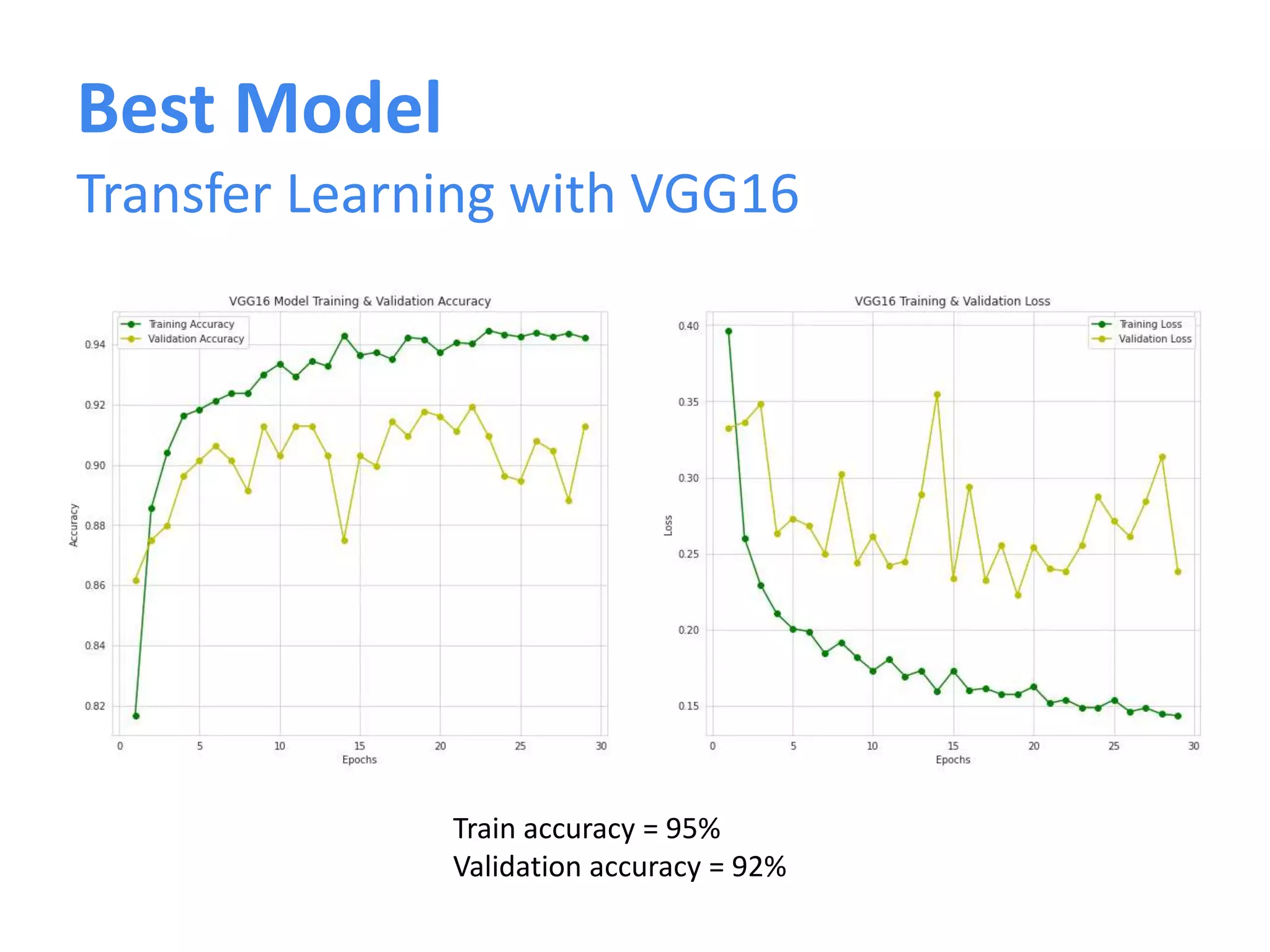
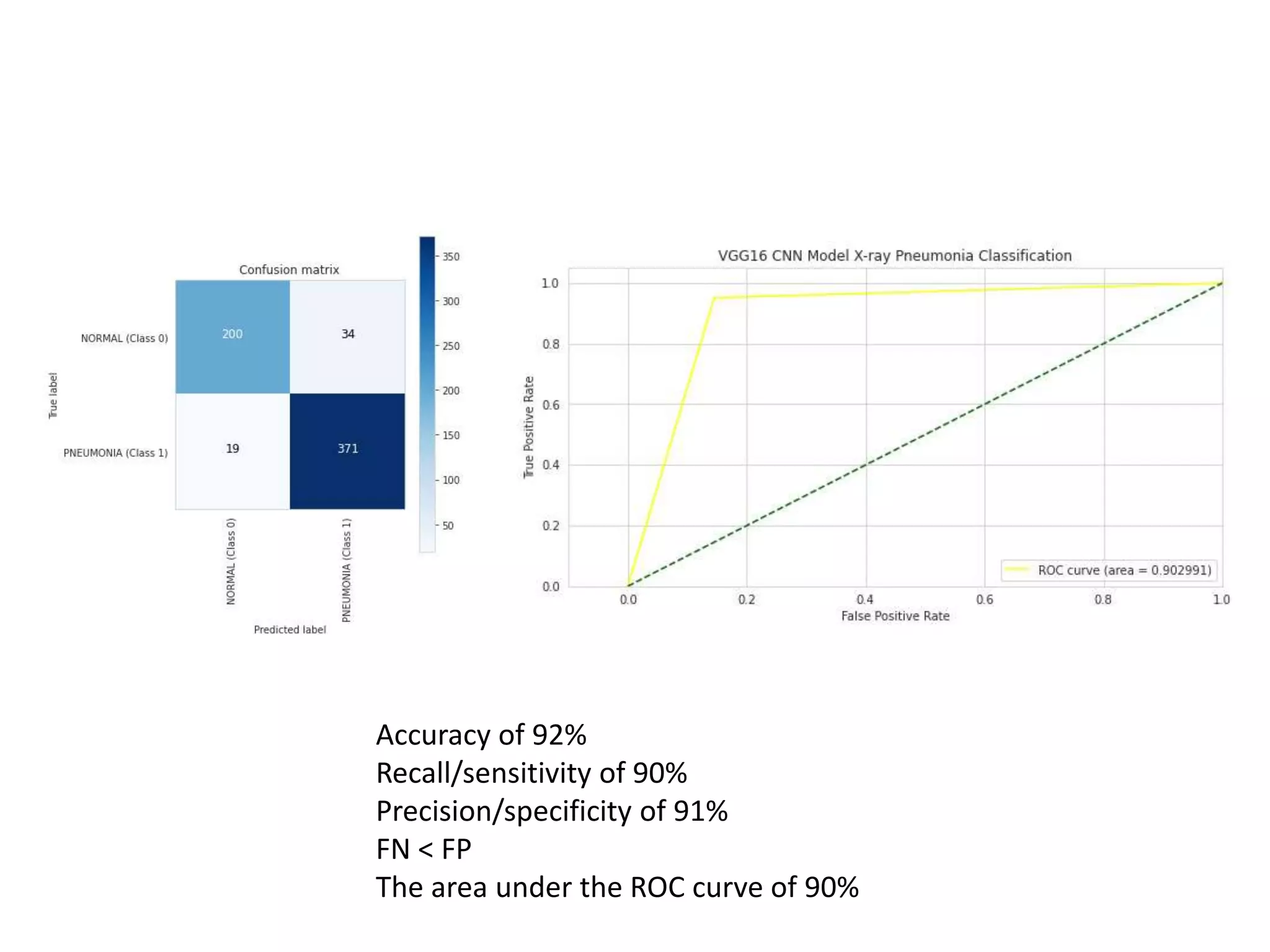
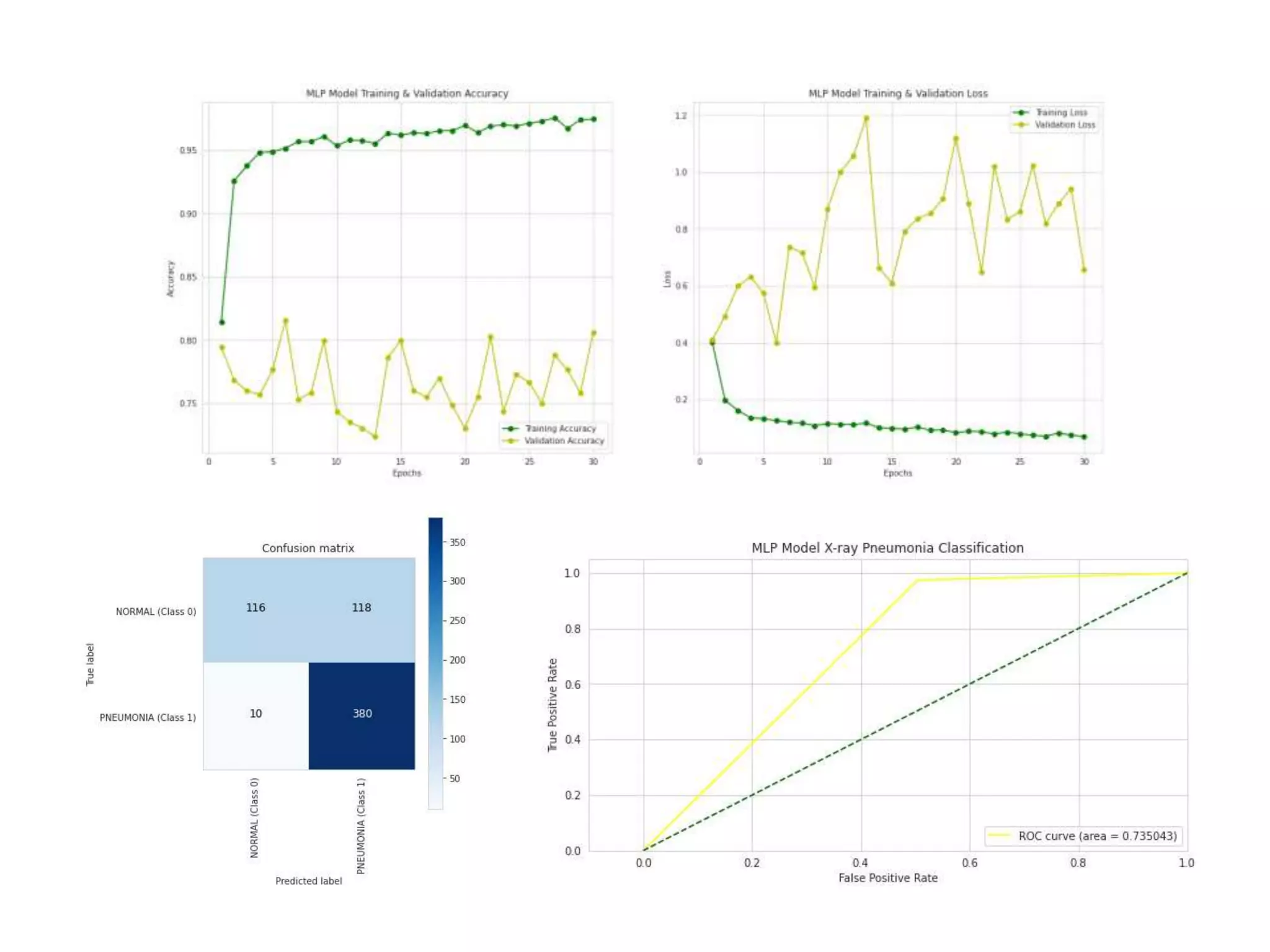
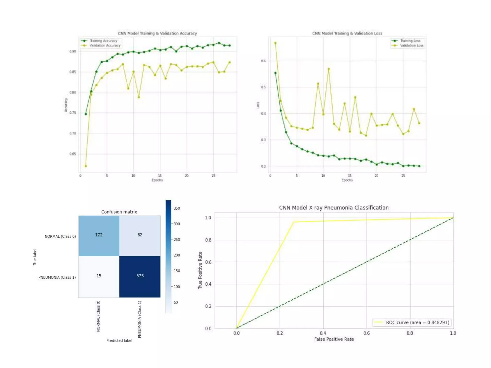
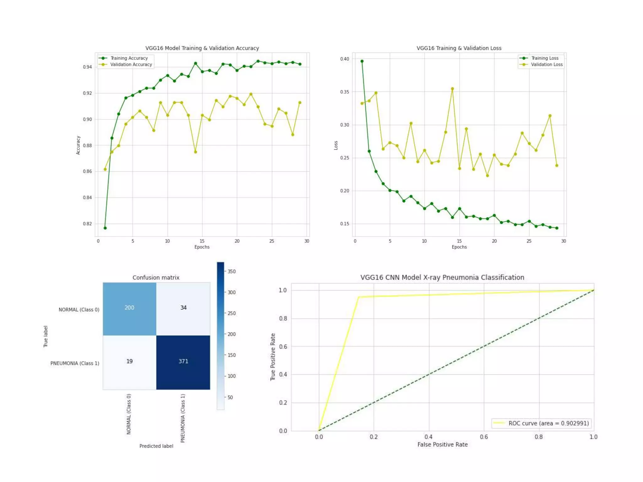
CHEST X-RAY PNEUMONIA
IMAGE CLASSIFICATION
WITH
DEEP LEARNING
Bao Tram Duong
The Problem
• Globally, a child dies of pneumonia
every 39 seconds.
• Pneumonia is the leading cause of
morbidity and mortality in children
younger than the age of 5, killing
more children than HIV/AIDS,
malaria, and measles combined.
• Chest X-rays are primarily used for
the diagnosis of this disease.
However, even for a trained
radiologist, it is a challenging task to
examine chest X-rays.
The Solution
To solve this, deep learning (DL), a branch of machine learning (ML),
is developed to detect hidden features in images which are not
apparent or cannot be detected even by medical experts.
With AI system aiding medical experts in expediting the diagnosis,
earlier treatment can be prescribed, resulting in improved clinical
outcomes.
OBTAIN
1
• Chest X-ray images (anterior-posterior) were
selected from pediatric patients of one to five years
old from Guangzhou Women and Children’s Medical
Center.
• There are 5,863 X-Ray images (JPEG)
• 2 categories: Normal & Pneumonia
EXPLORE
OBTAIN
1 2
• To build a powerful image classifier using very little training data, image
augmentation is usually required to boost the performance of deep
networks.
• Image augmentation artificially creates training images through different
ways of processing images, such as random rotation, shifts, shear and flips,
etc. of each training instances
Data Augmentation
EXPLORE
OBTAIN
MODEL
1 3
2
MULTILAYER PERCEPTRON MODEL
(MLP)
CONVOLUTIONAL NEURAL NETWORK
MODEL (CNN)
TRANSFER LEARNING with VGG16 CNN
MODEL
Multilayer Perceptron
output
input
hidden layer 1
hidden layer 2
hidden layer 3
hidden layer 4
Convolutional Neural Network
input
output
conv layer 1
conv layer 2
fully connected layers
Transfer Learning with VGG16
input
output
VGG16
fully connected layers
Summary of Key Findings
INTERPRETATION
EXPLORE
OBTAIN
MODEL
1 3 4
2
Best Model
Transfer Learning with VGG16
Train accuracy = 95%
Validation accuracy = 92%
Accuracy of 92%
Recall/sensitivity of 90%
Precision/specificity of 91%
FN < FP
The area under the ROC curve of 90%
1. Build a multi-class classification model to distinguish between Normal,
Viral Pneumonia, and Bacterial Pneumonia
2. Combine CNN models with other classifiers such as Support Vector
Machine (SVM)
3. Tune parameters such as learning rate, batch size, optimizer, number of
layers, types of layer, number of neurons per layer, and the type of
activation functions for each layer. GridSearchCV or
RandomizedSearchSV can be used to achieve this.
Future Work
Pneumonia are largely preventable in this age group and failure in long-
term management or preventing it will result in increases of the risk of
developing chronic pulmonary disorders in later adult life. Chang (2013)
suggests:
1. Focus on solving problems that are associated with increased risk of
pneumonia such as overcrowding, access to clean water, malnutrition,
anemia, young maternal age, low birth weight, and exposure to
tobacco smoke and other environmental pollutants
2. Invest in resources to collect data systematically, especially in poor
countries
3. Develop a universally agreed diagnostic gold standard for childhood
pneumonia, especially one that can also differentiate between
bacterial and non-bacterial pneumonia, which is currently a major
limitation in clinical research in this area
Recommendations
THANK YOU
APPENDIX
Chang, A. B., Ooi, M. H., Perera, D., & Grimwood, K. (2013). Improving the
Diagnosis, Management, and Outcomes of Children with Pneumonia: Where
are the Gaps?. Frontiers in pediatrics, 1,
29. https://doi.org/10.3389/fped.2013.00029
Geron, A. (2019). Hands-on machine learning with Scikit-Learn, Keras and
TensorFlow: concepts, tools, and techniques to build intelligent systems (2nd
ed.). O’Reilly.
Kermany, D. S., Goldbaum, M., Cai, W., Valentim, C., Liang, H., Baxter, S. L.,
McKeown, A., Yang, G., Wu, X., Yan, F., Dong, J., Prasadha, M. K., Pei, J., Ting,
M., Zhu, J., Li, C., Hewett, S., Dong, J., Ziyar, I., Shi, A., … Zhang, K. (2018).
Identifying Medical Diagnoses and Treatable Diseases by Image-Based Deep
Learning. Cell, 172(5), 1122–
1131.e9. https://doi.org/10.1016/j.cell.2018.02.010
Unicef. (2021, April 07). Pneumonia in children statistics. Retrieved April 13,
2021, from https://data.unicef.org/topic/child-
health/pneumonia/#:~:text=A%20child%20dies%20of%20pneumonia,of%20the
se%20deaths%20are%20preventable.
Reference
Chest X-ray Pneumonia Classification with Deep Learning

Chest X-ray Pneumonia Classification with Deep Learning

  • 1.
    CHEST X-RAY PNEUMONIA IMAGECLASSIFICATION WITH DEEP LEARNING Bao Tram Duong
  • 2.
    The Problem • Globally,a child dies of pneumonia every 39 seconds. • Pneumonia is the leading cause of morbidity and mortality in children younger than the age of 5, killing more children than HIV/AIDS, malaria, and measles combined. • Chest X-rays are primarily used for the diagnosis of this disease. However, even for a trained radiologist, it is a challenging task to examine chest X-rays.
  • 3.
    The Solution To solvethis, deep learning (DL), a branch of machine learning (ML), is developed to detect hidden features in images which are not apparent or cannot be detected even by medical experts. With AI system aiding medical experts in expediting the diagnosis, earlier treatment can be prescribed, resulting in improved clinical outcomes.
  • 4.
    OBTAIN 1 • Chest X-rayimages (anterior-posterior) were selected from pediatric patients of one to five years old from Guangzhou Women and Children’s Medical Center. • There are 5,863 X-Ray images (JPEG) • 2 categories: Normal & Pneumonia
  • 5.
  • 7.
    • To builda powerful image classifier using very little training data, image augmentation is usually required to boost the performance of deep networks. • Image augmentation artificially creates training images through different ways of processing images, such as random rotation, shifts, shear and flips, etc. of each training instances Data Augmentation
  • 8.
  • 9.
    MULTILAYER PERCEPTRON MODEL (MLP) CONVOLUTIONALNEURAL NETWORK MODEL (CNN) TRANSFER LEARNING with VGG16 CNN MODEL
  • 10.
  • 11.
    Multilayer Perceptron output input hidden layer1 hidden layer 2 hidden layer 3 hidden layer 4
  • 12.
  • 13.
    Convolutional Neural Network input output convlayer 1 conv layer 2 fully connected layers
  • 14.
  • 15.
    Transfer Learning withVGG16 input output VGG16 fully connected layers
  • 16.
  • 17.
  • 18.
    Best Model Transfer Learningwith VGG16 Train accuracy = 95% Validation accuracy = 92%
  • 19.
    Accuracy of 92% Recall/sensitivityof 90% Precision/specificity of 91% FN < FP The area under the ROC curve of 90%
  • 20.
    1. Build amulti-class classification model to distinguish between Normal, Viral Pneumonia, and Bacterial Pneumonia 2. Combine CNN models with other classifiers such as Support Vector Machine (SVM) 3. Tune parameters such as learning rate, batch size, optimizer, number of layers, types of layer, number of neurons per layer, and the type of activation functions for each layer. GridSearchCV or RandomizedSearchSV can be used to achieve this. Future Work
  • 21.
    Pneumonia are largelypreventable in this age group and failure in long- term management or preventing it will result in increases of the risk of developing chronic pulmonary disorders in later adult life. Chang (2013) suggests: 1. Focus on solving problems that are associated with increased risk of pneumonia such as overcrowding, access to clean water, malnutrition, anemia, young maternal age, low birth weight, and exposure to tobacco smoke and other environmental pollutants 2. Invest in resources to collect data systematically, especially in poor countries 3. Develop a universally agreed diagnostic gold standard for childhood pneumonia, especially one that can also differentiate between bacterial and non-bacterial pneumonia, which is currently a major limitation in clinical research in this area Recommendations
  • 22.
  • 23.
  • 24.
    Chang, A. B.,Ooi, M. H., Perera, D., & Grimwood, K. (2013). Improving the Diagnosis, Management, and Outcomes of Children with Pneumonia: Where are the Gaps?. Frontiers in pediatrics, 1, 29. https://doi.org/10.3389/fped.2013.00029 Geron, A. (2019). Hands-on machine learning with Scikit-Learn, Keras and TensorFlow: concepts, tools, and techniques to build intelligent systems (2nd ed.). O’Reilly. Kermany, D. S., Goldbaum, M., Cai, W., Valentim, C., Liang, H., Baxter, S. L., McKeown, A., Yang, G., Wu, X., Yan, F., Dong, J., Prasadha, M. K., Pei, J., Ting, M., Zhu, J., Li, C., Hewett, S., Dong, J., Ziyar, I., Shi, A., … Zhang, K. (2018). Identifying Medical Diagnoses and Treatable Diseases by Image-Based Deep Learning. Cell, 172(5), 1122– 1131.e9. https://doi.org/10.1016/j.cell.2018.02.010 Unicef. (2021, April 07). Pneumonia in children statistics. Retrieved April 13, 2021, from https://data.unicef.org/topic/child- health/pneumonia/#:~:text=A%20child%20dies%20of%20pneumonia,of%20the se%20deaths%20are%20preventable. Reference