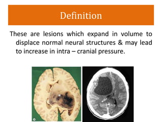
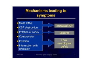
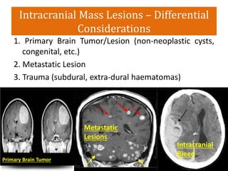
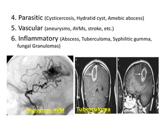
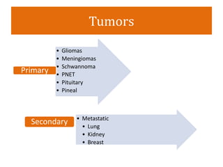
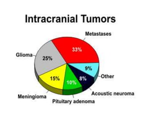
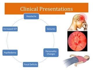
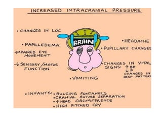
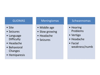
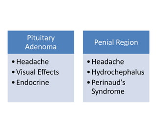
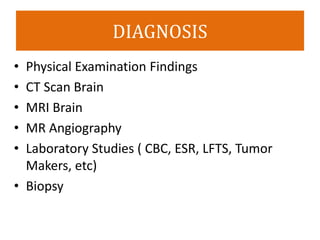
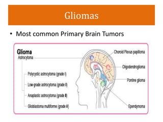
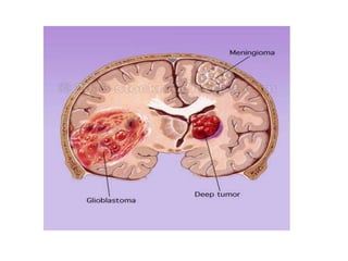
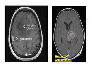
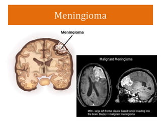
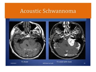
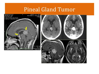
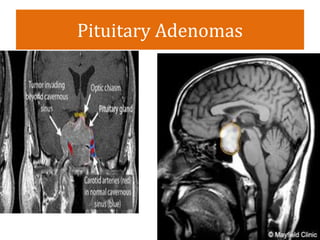
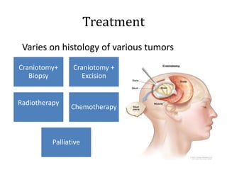
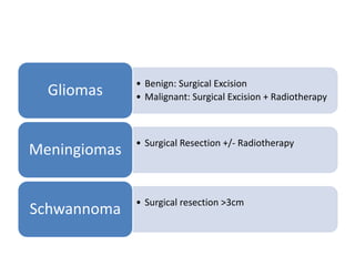
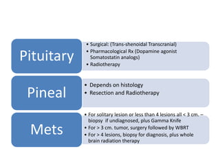
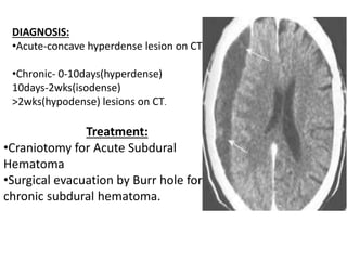
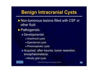
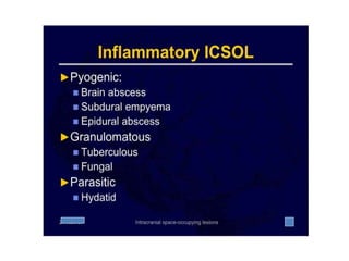
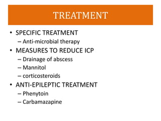
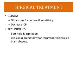
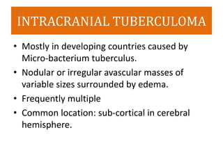
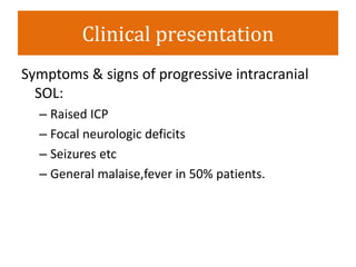
Intracranial Space Occupying
Lesions
Prof. Salman Sharif, FRCS
Chief of Neurosurgery
Liaquat National Hospital and
Medical College
Objectives
• Definition
• Types
• Clinical Presentations
• Diagnosis
• Treatment
Definition
These are lesions which expand in volume to
displace normal neural structures & may lead
to increase in intra – cranial pressure.
Intracranial Mass Lesions – Differential
Considerations
1. Primary Brain Tumor/Lesion (non-neoplastic cysts,
congenital, etc.)
2. Metastatic Lesion
3. Trauma (subdural, extra-dural haematomas)
• )
Primary Brain Tumor
Metastatic
Lesions
Intracranial
Bleed
4. Parasitic (Cysticercosis, Hydratid cyst, Amebic abscess)
5. Vascular (aneurysms, AVMs, stroke, etc.)
6. Inflammatory (Abscess, Tuberculoma, Syphilitic gumma,
fungal Granulomas)
Angiogram: AVM Tuberculoma
Tumors
• Gliomas
• Meningiomas
• Schwannoma
• PNET
• Pituitary
• Pineal
Primary
• Metastatic
• Lung
• Kidney
• Breast
Secondary
Clinical Presentations
Headache
Seizures
Personality
Changes
Focal Deficits
Papilledema
Increased ICP
GLIOMAS
• Site
• Seizures
• Language
Difficulty
• Headache
• Behavioral
Changes
• Hemiparesis
Meningiomas
• Middle age
• Slow growing
• Headache
• Seizures
Schwannomas
• Hearing
Problems
• Vertigo
• Headache
• Facial
weakness/numb
Pituitary
Adenoma
•Headache
•Visual Effects
•Endocrine
Penial Region
•Headache
•Hydrochephalus
•Perinaud’s
Syndrome
DIAGNOSIS
DIAGNOSIS
• Physical Examination Findings
• CT Scan Brain
• MRI Brain
• MR Angiography
• Laboratory Studies ( CBC, ESR, LFTS, Tumor
Makers, etc)
• Biopsy
Gliomas
• Most common Primary Brain Tumors
Grade III
Astrocytoma
Meningioma
Acoustic Schwannoma
Pineal Gland Tumor
Pituitary Adenomas
Treatment
Treatment
Varies on histology of various tumors
Craniotomy+
Biopsy
Craniotomy +
Excision
Radiotherapy
Chemotherapy
Palliative
• Benign: Surgical Excision
• Malignant: Surgical Excision + RadiotherapyGliomas
• Surgical Resection +/- Radiotherapy
Meningiomas
• Surgical resection >3cm
Schwannoma
• Surgical: (Trans-shenoidal Transcranial)
• Pharmacological Rx (Dopamine agonist
Somatostatin analogs)
• Radiotherapy
Pituitary
• Depends on histology
• Resection and RadiotherapyPineal
• For solitary lesion or less than 4 lesions all < 3 cm. –
biopsy if undiagnosed, plus Gamma Knife
• For > 3 cm. tumor, surgery followed by WBRT
• For > 4 lesions, biopsy for diagnosis, plus whole
brain radiation therapy
Mets
TRAUMA
• Intracranial haematomas
I. Extra dural haematomas :-
– between the dura & the skull
– middle meningeal artery
– Common site is temporal fossa.
TRAUMA
• Progressive deterioration of
level of consciousness
• Lucid Interval
• Pupillary changes :- called
Hutchinson’s pupillary
reaction.
Clinical
Features
EDH
INVESTIGATIONS:
CT (Biconvex hyperdense
lesion)
MRI
CEREBRAL ANGIOGRAPHY
Treatment:
Surgical evacuation followed by Craniotomy
• II. Subdural haematomas :-
–between the dura and the arachnoid.
–Common causes are bleeding from
superficial veins or venous sinuses.
–Anticoagulant treatment predispose to
intracranial bleeding and subdural
haematoma.
• Clinical features:
– Acute : Clinical features are similar to extra dural
hematoma.
– Chronic : Dementia, altered behaviour, psychiatric
manifestations or focal neurological deficits may
develop.
– In middle aged headache, contralateral
hemiplegia, papilledema
– children: vomiting, restlessness. Irritability, refusal
to feed, anaemia, seizures and failure to thrive.
Treatment:
•Craniotomy for Acute Subdural
Hematoma
•Surgical evacuation by Burr hole for
chronic subdural hematoma.
DIAGNOSIS:
•Acute-concave hyperdense lesion on CT
•Chronic- 0-10days(hyperdense)
10days-2wks(isodense)
>2wks(hypodense) lesions on CT.
BRAIN ABSCESS
• Mostly single may be multiple
• Majority Supratentorial, 10% infratentorial
• Metastatic:
– hematogenesis,direct spread from adjacent
structures or penetrating brain injury.
Clinical presentation
• Neurologic:
– Raised ICP(nausea,vomiting)
– Focal neurologic deficits(hemi-pariasis)
– Epileptic seizures
• Systemic toxicity(Fever,malaise)
• Symptoms of primary focus
infection(Otitis,sinusitis etc)
DIAGNOSIS
• Method of Choice- CT scan of Brain
– Ring enhancing Lesion
• Peripheral Blood smear
– Leukocytosis
– Raised ESR
TREATMENT
• SPECIFIC TREATMENT
– Anti-microbial therapy
• MEASURES TO REDUCE ICP
– Drainage of abscess
– Mannitol
– corticosteroids
• ANTI-EPILEPTIC TREATMENT
– Phenytoin
– Carbamazapine
SURGICAL TREATMENT
• GOALS:
– Obtain pus for culture & sensitivity
– Decrease ICP
• TECHNIQUES:
– Burr hole & aspiration
– Excision & craniotomy for recurrent, thickwalled
brain abscess.
INTRACRANIAL TUBERCULOMA
• Mostly in developing countries caused by
Micro-bacterium tuberculus.
• Nodular or irregular avascular masses of
variable sizes surrounded by edema.
• Frequently multiple
• Common location: sub-cortical in cerebral
hemisphere.
Clinical presentation
Symptoms & signs of progressive intracranial
SOL:
– Raised ICP
– Focal neurologic deficits
– Seizures etc
– General malaise,fever in 50% patients.
INVESTIGATIONS
• Lab work-up
– Leukocytosis
– ESR- raised or normal
– Mantox test- often+ve
• Chest X-ray
• Plain skull X-ray
• CT & MRI- Investigation of choice
Hyper-dense masses with ring and surroundind
edema, often”Target sign”
TREATMENT
• Anti-tubercular therapy
• Measures to reduce ICP
• Control seizure
INDICATIONS:
Intracranial lesions could not be specified
Progressive neurological detoriation
ALTERNATIVES:
Excision:
CSF-shunting: mandatory in complicating
obstructive hydrocephalus
SURGICAL TREATMENT
THANK YOU

Intracranial space occupying lesions