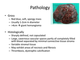
LIVER HEMANGIOMA
Dr. Uttam Laudari
JR III
Mentor- Dr. DRS
1.Definition/ classification
• Hepatic hemangiomas are benign tumors of the
liver consisting of clusters of blood-filled cavities,
lined by endothelial cells, fed by the hepatic
artery
• Small <3cm
• Medium 3-10cm
• Giant 10 and above
Location-wise they are most often found in the right
liver lobe, often in segment IV, often marginal
Epidemiology/ etiology
• Most common benign tumor of liver
• Autospy- 7% of population
• Asssociated with hemangioma of other organs
• Men=women
• Sex hormone no role
• MSKK – 115 patients, 35% were male
• No association between patients reproductiv
history and use of OCP
3. Pathology/pathogenesis
• The cause of HH is not known, it may be congenitally
determined
• reported cases of HH running in families, suggesting a
possible genetic connection,
• Infantile HH can be diagnosed prenatally or in
childhood. Reported frequency at 1 year is of 5-10%,
normally they regress in size over time, but not always.
Pathology
• Gross
– Red blue, soft, spongy mass
– Usually 1-2cm in diameter
– >4cm  giant hemangioma
• Histologically
– Sharply defined, not capsulated
– Large, cavernous vascular spaces partly of completely filled
with blood separated by minimal connective tissue stroma
– Variable stromal tissue
– May exhibit areas of necrosis and fibrosis
– Thrombosis, dystrophic calcification
Pathology
• Tumor parenchymal interface (Zimmermann)
1. Fibrous interface characteried by relatively avascular
capsule like fibrous lamellae
2. Interdigitating interface characterized by a misture
of parenchymal and hamangioma components
without capsule like interface
3. Compression interface characterized by direct
apposition between hemangioma and parenchyma
4. Irregular or spongy interface characcterized by
highly irregular border with numerous parenchymal
foci interspersed between dilated blood channels
Pathology
• Fibrous interface is the most common
interface in large hemangiomas and the
presence of this capsule like avascular fibrous
interface facilitated operative enucleation of
these lesion
4.Presentation and diagnosis
• Asymptomatic- most of the times
• Sypmtomatic
– Most of time nonspecific, digestive symptoms
– Pain RUQ
– decreased appetite, premature satiation
sensation, nausea, vomiting, abdominal
discomfort: sense of fullness, postprandial
bloating, early or late
• complications
4.Presentation and diagnosis
• Complications
• Inflammatory
– Acute- fever and chronic
• Mechanical
– rupture, spontaneous or traumatic: intraabdominal mass
disruption trauma, or marginal trauma when located in the
proximity of the costal margin, hence more exposed to
trauma
– compression of adjacent structures: stomach, resulting in
gastric obstruction (early feeling of fullness),
– Compression of bile ducts leading to jaundice,
Presentation and diagnosis
• haemobilia, volvulus / torsion / infarction for pedunculated
hepatic hemangiomas
• bleeding:
– intratumoral or intraperitoneal, with or without consumptive
coagulopathy
– Kassalbach-Merritt syndrome (HH giant, thrombocytopenia,
intravascular coagulation)
– Osler-Rendu-Weber disease (hereditary telangiectasia: multiple
smaller HA on face, tongue, jugal mucosa, gastrointestinal tract,
liver)
– Klippel-Trenaunay syndrome (congenital hemiatrophy nevus
flammeus, hemi-mega-encephalopathy)
– Von Hippel-Lindau disease (cerebral, retinal, pancreatic
hemangioma)
Presentation and diagnosis
• Degenerative:
– thrombosis, hyalinization, progressive fibrosis and
sclerosis becoming central scar
• Calcification
• massive arteriovenous shunt, complicated
with heart failure
• Predisposing factors for complications:
– Adulthood
– chronic medication use (such as steroid use, can accelerate the development of an
existing HH),
– female sex: estrogen therapy, use of oral contraceptives (increase the risk or increase
the size, discontinuing contraceptive regimen can lead to lesion regression, but not
necessarily)
– pregnancy and multiparity (by disrupting estrogen and progesterone hormone levels,
leading to an increase in size of a preexisting HH)
– replacement therapy for menopausal symptoms; ovarian stimulation treatment with
clomiphene citrate and human chorionic gonadotropin.
– Genetic gene penetrance or sex hormone proliferative factors could also be an
explanation.
Presentation and diagnosis
• Ultrasound
– hyperechoic homogenous
nodule, with well-defined
margins and posterior
acoustic enhancement
– hyperechogenicity is a result
of the numerous interfaces
between the endothelial lined
sinuses composing the HH
and the blood within them.
– This hyperechoic appearance
is usually the case for small
HH
Presentation and diagnosis
• Ultrasound
In larger lesions
– because of possible necrosis, hemorrhage or
fibrosis can appear inhomogeneous, with mixed
echogenicity (hypo- and hyperechoic)
– Lesions that have such echo patterns are labeled
as atypical HH
Presentation and diagnosis
• Contrast enhanced US
– accurately discriminate a
HH from adenomas,
FNH, HCC or metastasis.
• The typical HH shows
peripheral nodular
enhancement in the arterial
phase with complete (but
sometimes incomplete)
centripetal filling in the
portal venous and late
phases
• The absence of lesion blood flow in HH on Doppler
US is also a reliable sign for the differential diagnosis
with hepatocellular carcinoma (HCC)
• which frequently has intra- or peritumoral
vascularity
Presentation and Diagnosis
• Noncontrast CT and MRI
– Well defined hypodense mass that may contain
areas of calcification or central scarring
• CECT
– Arterial and venous phase
Presentation and Diagnosis
• CECT criteria
1. A low density lesion on te unenhanced CT scan
2. Early peripheral contrast enhancement
3. Progressive centrepetal opacification from periphery
toward centre
4. Delay of at least 3 min before total opacification
5. Eventual isodense appearences
– Presence 4 and 5 plus any two criteria  diagnostic
Arterial phase-hemangioma with homogeneous and intense contrast
enhancement
Presentation and Diagnosis
• MRI
– Clinically suspected but CT fails to confirm
– Hypointense signal intensity compared with
surrounding liver tissue on T1- weighted imaging,
hyperintense signal intensity on T2 weighted imaging
– Patter of Gadolinium enhancement is similar to that
seen on CECT imaging
– 96% diagnostic accuracy, even subtle hemangioma
can be detected
Presentation and Diagnosis
• Tc-99m- labeled red blood cell scan rarely
necessary
• PET
– Rarely needed
– FDG may helpful in differentiating hemangioma
from metabolically active malignancies
Treatment/prognosis
• Most patient are managed conservatively
• Rarely patient have sypmtoms
• Size alone should not be used as a criterion for
resection
Treatment/prognosis
• Non-invasive
• Irradiation of the liver.
Liver irradiation with a dose of 15-30 Gray in 15-
22 sessions for a few weeks results in tumor
regression and has minimal secondary morbidity
Invasive
• Selective or supra-selective angiographic embolization
– with polyvinyl alcohol or other substances by catheterization of
the hepatic arteries can lead to tumor reduction
– The procedure is effective in restricting and reversing tumor
growth only for hemangiomas that have a clearly identified
arterial blood supply
– Portal vein embolization (PVE) is now routinely used prior to
resection to increase the residual (postsurgery) viable liver
parenchyma, providing more favorable conditions for the
elective major surgery, and minimizing the risk of complications
Treatment/prognosis
• Invasive
• Radiofrequency ablation, executed
laparoscopically or percutaneously under
ultrasound control has been used for pain
control in small studies
Treatment/prognosis
• Criteria for resection:
– Presence of severe symptoms
– Inability to confidently exclude malignancy
– Development of complications
• Rule out other pancreaticobiliary and GI
causes and symptoms and symptoms must be
carefully matched to the size and nature of
hemangioma
Treatment/prognosis
• The median lesion size among the patients
resected for symptoms at MSKCC was 15 cm
• Haemangioma <4cm are almost exclusively
asymptomatic, and symptomatic lesions tend
to be larger within the right hemi liver than
within left
Treatment/prognosis
• Development of tumor related complications
1. Intraabdominal hemorrhage – uncommon
2. But can be life threatening
3. Treated with angiographic embolization and surgery
1. Kasabach-Merritt syndrome – large hemangioma
with thrombocytopenia and consumptive
coagulopathy
2. Activation of coagulation cascade by platelets
trapped within the hemangioma
3. Immunosuprresive agents, radiation, surgical
resection and even transplant
Surgical intervention
• Resection and enucleation
– MSKCC enucleation was performed for 31 of 52
patients (60%) for hemangioma
– Enucleation was associated with decrease in
operative time and operative blood loss
– As it is benign nature and fibrous cleavage plane
often exists between the hemagioma and
surrounding hepatic parenchyma
Treatment/prognosis
• Enucleation
– Control hepatic inflow with pringle manouvre
– Hepatic artery identified, dissected proximally to
the level of proper hepatic artery decreses size if
hemangioma
– After vascular control small hepatotomy done few
mm from hemangioma
Treatment/prognosis
• Comparative studies of anatomical resection
and enucleation suggest that enucleation is
associated with a lower rate of abdominal
complications, especially biliary fistula.
• As hemangioma compress the adjacent liver
tissue and enucleation inside the fibrous
tumor tissue prevents injury to the bile ducts
and vessels
Treatment/prognosis
• Invasive
• Selective surgical transhepatic ligation of the
HH major feeding vessels can successfully
reduce intratumoral shunt which would have
led to congestive heart failure
• Orthotopic liver transplantation is indicated
for large or diffuse bilateral lesions. Only a few
cases have been reported in literature.

Liver hemangiona

  • 1.
    LIVER HEMANGIOMA Dr. UttamLaudari JR III Mentor- Dr. DRS
  • 2.
    1.Definition/ classification • Hepatichemangiomas are benign tumors of the liver consisting of clusters of blood-filled cavities, lined by endothelial cells, fed by the hepatic artery • Small <3cm • Medium 3-10cm • Giant 10 and above Location-wise they are most often found in the right liver lobe, often in segment IV, often marginal
  • 4.
    Epidemiology/ etiology • Mostcommon benign tumor of liver • Autospy- 7% of population • Asssociated with hemangioma of other organs • Men=women • Sex hormone no role • MSKK – 115 patients, 35% were male • No association between patients reproductiv history and use of OCP
  • 5.
    3. Pathology/pathogenesis • Thecause of HH is not known, it may be congenitally determined • reported cases of HH running in families, suggesting a possible genetic connection, • Infantile HH can be diagnosed prenatally or in childhood. Reported frequency at 1 year is of 5-10%, normally they regress in size over time, but not always.
  • 6.
    Pathology • Gross – Redblue, soft, spongy mass – Usually 1-2cm in diameter – >4cm  giant hemangioma • Histologically – Sharply defined, not capsulated – Large, cavernous vascular spaces partly of completely filled with blood separated by minimal connective tissue stroma – Variable stromal tissue – May exhibit areas of necrosis and fibrosis – Thrombosis, dystrophic calcification
  • 7.
    Pathology • Tumor parenchymalinterface (Zimmermann) 1. Fibrous interface characteried by relatively avascular capsule like fibrous lamellae 2. Interdigitating interface characterized by a misture of parenchymal and hamangioma components without capsule like interface 3. Compression interface characterized by direct apposition between hemangioma and parenchyma 4. Irregular or spongy interface characcterized by highly irregular border with numerous parenchymal foci interspersed between dilated blood channels
  • 8.
    Pathology • Fibrous interfaceis the most common interface in large hemangiomas and the presence of this capsule like avascular fibrous interface facilitated operative enucleation of these lesion
  • 9.
    4.Presentation and diagnosis •Asymptomatic- most of the times • Sypmtomatic – Most of time nonspecific, digestive symptoms – Pain RUQ – decreased appetite, premature satiation sensation, nausea, vomiting, abdominal discomfort: sense of fullness, postprandial bloating, early or late • complications
  • 10.
    4.Presentation and diagnosis •Complications • Inflammatory – Acute- fever and chronic • Mechanical – rupture, spontaneous or traumatic: intraabdominal mass disruption trauma, or marginal trauma when located in the proximity of the costal margin, hence more exposed to trauma – compression of adjacent structures: stomach, resulting in gastric obstruction (early feeling of fullness), – Compression of bile ducts leading to jaundice,
  • 11.
    Presentation and diagnosis •haemobilia, volvulus / torsion / infarction for pedunculated hepatic hemangiomas • bleeding: – intratumoral or intraperitoneal, with or without consumptive coagulopathy – Kassalbach-Merritt syndrome (HH giant, thrombocytopenia, intravascular coagulation) – Osler-Rendu-Weber disease (hereditary telangiectasia: multiple smaller HA on face, tongue, jugal mucosa, gastrointestinal tract, liver) – Klippel-Trenaunay syndrome (congenital hemiatrophy nevus flammeus, hemi-mega-encephalopathy) – Von Hippel-Lindau disease (cerebral, retinal, pancreatic hemangioma)
  • 12.
    Presentation and diagnosis •Degenerative: – thrombosis, hyalinization, progressive fibrosis and sclerosis becoming central scar • Calcification • massive arteriovenous shunt, complicated with heart failure
  • 13.
    • Predisposing factorsfor complications: – Adulthood – chronic medication use (such as steroid use, can accelerate the development of an existing HH), – female sex: estrogen therapy, use of oral contraceptives (increase the risk or increase the size, discontinuing contraceptive regimen can lead to lesion regression, but not necessarily) – pregnancy and multiparity (by disrupting estrogen and progesterone hormone levels, leading to an increase in size of a preexisting HH) – replacement therapy for menopausal symptoms; ovarian stimulation treatment with clomiphene citrate and human chorionic gonadotropin. – Genetic gene penetrance or sex hormone proliferative factors could also be an explanation.
  • 14.
    Presentation and diagnosis •Ultrasound – hyperechoic homogenous nodule, with well-defined margins and posterior acoustic enhancement – hyperechogenicity is a result of the numerous interfaces between the endothelial lined sinuses composing the HH and the blood within them. – This hyperechoic appearance is usually the case for small HH
  • 15.
    Presentation and diagnosis •Ultrasound In larger lesions – because of possible necrosis, hemorrhage or fibrosis can appear inhomogeneous, with mixed echogenicity (hypo- and hyperechoic) – Lesions that have such echo patterns are labeled as atypical HH
  • 16.
    Presentation and diagnosis •Contrast enhanced US – accurately discriminate a HH from adenomas, FNH, HCC or metastasis. • The typical HH shows peripheral nodular enhancement in the arterial phase with complete (but sometimes incomplete) centripetal filling in the portal venous and late phases
  • 17.
    • The absenceof lesion blood flow in HH on Doppler US is also a reliable sign for the differential diagnosis with hepatocellular carcinoma (HCC) • which frequently has intra- or peritumoral vascularity
  • 18.
    Presentation and Diagnosis •Noncontrast CT and MRI – Well defined hypodense mass that may contain areas of calcification or central scarring • CECT – Arterial and venous phase
  • 19.
    Presentation and Diagnosis •CECT criteria 1. A low density lesion on te unenhanced CT scan 2. Early peripheral contrast enhancement 3. Progressive centrepetal opacification from periphery toward centre 4. Delay of at least 3 min before total opacification 5. Eventual isodense appearences – Presence 4 and 5 plus any two criteria  diagnostic
  • 20.
    Arterial phase-hemangioma withhomogeneous and intense contrast enhancement
  • 21.
    Presentation and Diagnosis •MRI – Clinically suspected but CT fails to confirm – Hypointense signal intensity compared with surrounding liver tissue on T1- weighted imaging, hyperintense signal intensity on T2 weighted imaging – Patter of Gadolinium enhancement is similar to that seen on CECT imaging – 96% diagnostic accuracy, even subtle hemangioma can be detected
  • 22.
    Presentation and Diagnosis •Tc-99m- labeled red blood cell scan rarely necessary • PET – Rarely needed – FDG may helpful in differentiating hemangioma from metabolically active malignancies
  • 23.
    Treatment/prognosis • Most patientare managed conservatively • Rarely patient have sypmtoms • Size alone should not be used as a criterion for resection
  • 24.
    Treatment/prognosis • Non-invasive • Irradiationof the liver. Liver irradiation with a dose of 15-30 Gray in 15- 22 sessions for a few weeks results in tumor regression and has minimal secondary morbidity
  • 25.
    Invasive • Selective orsupra-selective angiographic embolization – with polyvinyl alcohol or other substances by catheterization of the hepatic arteries can lead to tumor reduction – The procedure is effective in restricting and reversing tumor growth only for hemangiomas that have a clearly identified arterial blood supply – Portal vein embolization (PVE) is now routinely used prior to resection to increase the residual (postsurgery) viable liver parenchyma, providing more favorable conditions for the elective major surgery, and minimizing the risk of complications
  • 26.
    Treatment/prognosis • Invasive • Radiofrequencyablation, executed laparoscopically or percutaneously under ultrasound control has been used for pain control in small studies
  • 27.
    Treatment/prognosis • Criteria forresection: – Presence of severe symptoms – Inability to confidently exclude malignancy – Development of complications • Rule out other pancreaticobiliary and GI causes and symptoms and symptoms must be carefully matched to the size and nature of hemangioma
  • 28.
    Treatment/prognosis • The medianlesion size among the patients resected for symptoms at MSKCC was 15 cm • Haemangioma <4cm are almost exclusively asymptomatic, and symptomatic lesions tend to be larger within the right hemi liver than within left
  • 29.
    Treatment/prognosis • Development oftumor related complications 1. Intraabdominal hemorrhage – uncommon 2. But can be life threatening 3. Treated with angiographic embolization and surgery 1. Kasabach-Merritt syndrome – large hemangioma with thrombocytopenia and consumptive coagulopathy 2. Activation of coagulation cascade by platelets trapped within the hemangioma 3. Immunosuprresive agents, radiation, surgical resection and even transplant
  • 30.
    Surgical intervention • Resectionand enucleation – MSKCC enucleation was performed for 31 of 52 patients (60%) for hemangioma – Enucleation was associated with decrease in operative time and operative blood loss – As it is benign nature and fibrous cleavage plane often exists between the hemagioma and surrounding hepatic parenchyma
  • 31.
    Treatment/prognosis • Enucleation – Controlhepatic inflow with pringle manouvre – Hepatic artery identified, dissected proximally to the level of proper hepatic artery decreses size if hemangioma – After vascular control small hepatotomy done few mm from hemangioma
  • 32.
    Treatment/prognosis • Comparative studiesof anatomical resection and enucleation suggest that enucleation is associated with a lower rate of abdominal complications, especially biliary fistula. • As hemangioma compress the adjacent liver tissue and enucleation inside the fibrous tumor tissue prevents injury to the bile ducts and vessels
  • 33.
    Treatment/prognosis • Invasive • Selectivesurgical transhepatic ligation of the HH major feeding vessels can successfully reduce intratumoral shunt which would have led to congestive heart failure
  • 34.
    • Orthotopic livertransplantation is indicated for large or diffuse bilateral lesions. Only a few cases have been reported in literature.