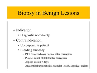
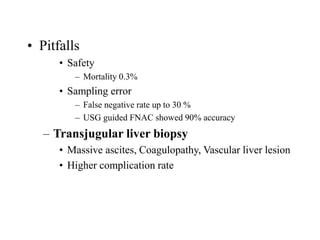
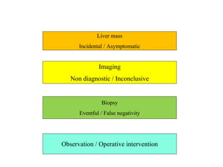
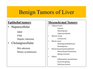
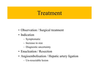
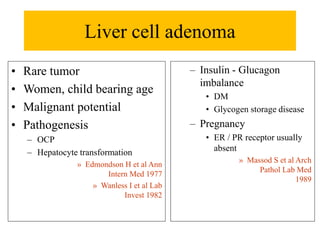
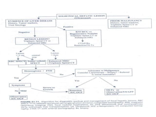
BENIGN LIVER
TUMORS
Department of Surgical Gastroenterology
King George’s Medical University
Lucknow
Background
• Unexpectedly or incidentally diagnosed
– Malignant to benign- 1: 2
• Different treatment and prognosis
• Diagnostic uncertainty- An acceptable indication for operative
intervention in 40 %
» Kammula US et al Int J Gatrointest Cancer 2001
Concerns
• Diagnostic confusion and management
• Imaging of choice
• Role of
– Biopsy
– Laparoscopy
• Observe or Treat
Liver mass
Incidental / Asymptomatic
Imaging
Non diagnostic / Inconclusive
Biopsy
Eventful / False negativity
Observation / Operative intervention
Benign Tumors of Liver
Mesenchymal Tumors
• Adipose tissue
Lipoma
Myelolipoma
Angiomyolipoma
• Muscle Tissue
Leiomyoma
• Blood vessel
Hemangioendothelioma
Hemangioma
• Mixed mesenchymal/epithelial
Mesenchymal hamartoma
Benign teratoma
• Others
Inflammatory pseudotumor
Focal fat sparing
Infectious lesions
Epithelial tumors
• Hepatocellular
NRH
FNH
Hepatic Adenoma
• Cholangiocellular
Bile adenoma
Biliary cystadenoma
John T et al Ann Surg 1994
Clinical Evaluation
Clinical context
History
Medications (eg OCPs)
Symptoms : Constitutional, GI
Complete physical
Stigmata of CLD
Rectal bleeding (hemoccult)
Breast and pelvic examination
Laboratories
CBC
LFTs
Hep B/C Serology
Tumor markers: AFP, CEA, CA19.9
Hemangioma
• Most common
• 0.4% - 20% (autopsy)
» Rubin R et al Med Clin North Am 1996
» Karhunen et al J Clin Path 1986
• Variable size
• Giant hemangioma - > 4cm
• Diagnosed more in female
• Third to fifth decade
• Pathogenesis
• Benign congenital hamartoma (Ectasia)
• Increase in growth or symptoms
– Pregnancy
– Oral contraceptive
– Estrogen may have role
» Nichols E et al SCNA 1989
• Pathology
• Compressible cystic lesion
• Histology
– Vascular space, endothelial lining, fibrous seta
• Clinical features
• Mostly asymptomatic
• Abdominal fullness, early satiety, anorexia
• Pain- Thrombosis. Infarcts, Necrosis, Pressure
• Size
– 40 % >4 cm & 90% >10cm are symptomatic
» Goodman Z et al Neoplasm of Liver 1987
• Spontaneous rupture- rare (Reported 28 case /100
year)
» Farge O et al Word J Surg 1995
• Physical findings unremarkable
• Biochemical analysis usually normal
• Consumption coagulopathy
(Kasaback Merrit Syndrome
– Giant hemangioma with feature of DIC
• Clinical course
• Benign
• May grow during pregnancy or OCP
• 8 out of 158 were symptomatic during 60 months
follow-up ( All > 5 cm)
» Gandolfi L et al Gut 1991
Imaging
• USG
• Well demarcated Homogenous Hypoechoic
• Posterior acoustic shadow
Plain
Hypodense mass,
Central scar
Arterial phase
Peripheral nodular enhancement
Portal venous phase
Centripetal filling
T1
Hypointense
T2
Hyperintense
Post contrast T1
Peripheral nodular enhancement
Post contrast T2
Centripetal filling with central scar
<2cm, multiple lesion, 90 % sensitivity
Tc 99 m Pertechnate / RBC
• Initial hypoperfusion with increase
tracer uptake in 30 minute
• False negative: Thrombosis / Fibrosis
• False Positivity: HCC /
Angiosarcoma
• Sensitivity - 80 %,
• Specificity – 100% for lesion >2
cm
» Kinnard M et al
Semin
Roentgenol
1995
SPECT
• Better spatial resolution
• Sensitivity & Specificity – 90 to 95%
Kudo M et al AJR 1989
• Lesion <2 cm & close to major vessels-
MRI > SPECT
Angiography
• Diagnostics confusion & Coexisting
pathology
– Cotton wool appearance
» Rapid filling of large
space, > 40 min)
– Absent AV shunt
– D/D: Vascular lesion
Combination USG, CT, SPECT has
86% sensitivity,
100 specificity,
91% accuracy
Treatment
Indication
• Symptomatic
• Complication
• ? Rupture
• Diagnostic doubt
Option
• Enucleation or Resection
Equally effective
Less complication in enucleation
Gedaly et al Arch Surg 1999
• Angioembolization/ Vascular
ligation
– Steroid & Radiation
– Interferon α 2a
Focal Nodular Hyperplasia
• Second most common
• 2.5- 8% in autopsy
• Female predominance
• 20-50 year
• Etiology: ? OCPs
• Clinical feature
Asymptomatic,
incidental
Pain, Rupture,
Hemorrhage – rare
Coexist
– Hemangioma
– Adenoma
– GSD
Pathogenesis
• ? Neoplastic / Hamartoma / Response to injury
• Response to hypo or hyper perfusion
Haber M et al Gastroenterology 1995
• Coexisting hemangioma ( Vascular Pathology)
Pathology
• Demarcated, well encapsulated
• Usually solitary
• Radial scar with central septa
• Preserved hepatic parenchyma with Kupfer cell
over activity
Imaging
Contrast enhanced USG
Spoke wheel pattern
Early phase
Isoechoic to normal liver
delayed phase
s/o Benign etiology
Precontrast
Isodense, central scar
Arterial Phase
Homogenous enhancement
Portal phase
No pooling
TI
Isointense to hypointense
T2 – Hyperintense,
Hyperintense central scar
Post contrast T1
Uniformly enhancing
• Tc 99m sulfur colloid scan
• Increased uptake with colloid concentration
• Angiography
• Central feeding artery
• Spoke wheel pattern
• Capillary blush
– FLC- Central scar with hypointense in T2
– Adenoma- No central scar
Treatment
• Observation / Surgical treatment
• Indication
– Symptomatic
– Increase in size
– Diagnostic uncertainty
• Enucleation / Resection
• Angioembolisation / Hepatic artery ligation
– Un-resectable lesion
Liver cell adenoma
• Rare tumor
• Women, child bearing age
• Malignant potential
• Pathogenesis
– OCP
– Hepatocyte transformation
» Edmondson H et al Ann
Intern Med 1977
» Wanless I et al Lab
Invest 1982
– Insulin - Glucagon
imbalance
• DM
• Glycogen storage disease
– Pregnancy
• ER / PR receptor usually
absent
» Massod S et al Arch
Pathol Lab Med
1989
• Pathology
• Large,
circumscribed,
partial
encapsulated
• Absent hepatic
architecture
• Reduced
Kupfer cell
activity
• Feeder -
Hepatic artery
• Peliosis
hepatitis
• Clinical feature
• Asymptomatic
• Pain – Most common
• Bleeding
• Mass
• Malignant
transformation
(2-7% years)
• Adenomatosis
– > 10 adenomas
– More potential
malignant
propensity
Unenhanced- Iso or hypodense
Arterial phase- Hyperdense,
irregular enhancement
Portal phase- Isodense
• USG
• Well defined lesion with variable echogenicity
• MRI
• T1
– Well defined hyper intense
• T2
– Heterogenous enhancement
• Sulfur colloid scan
• Reduced uptake (d/t reduced Kupffer cells)
Treatment
• Surgical removal
– Malignant transformation
– Bleeding and Necrosis
– Rapid increase in size in pregnancy
• Enucleation / Resection / Transplantation
• Angioembolization
• Follow-up
– USG & AFP
– Small, asymptomatic lesion after discontinuing OCP
Rare lesions
• Nodular regenerative hyperplasia
– Thoroughly distributed
– Usually <0.5 cm size
– Pathogenesis
• Vascular obstruction , atrophy and regenerative
hyperplasia
• Atrophy in central zone, regeneration around
periphery
» Altmann H et al 1989
• Association
• Chronic liver disease
(Cirrhosis, PHN)
• Chronic systemic disease
• Budd Chiari syndrome
• Felty syndrome
• Crest syndrome
• Collagen disorder
• Imaging
– USG
• Hypo or iso-echoic
– CT
• Hyperdense in
arterial phase while
imperceptible on
Portal phase
• Clinical feature
– Presentation as of
chronic liver disease
Adenomatous hyperplasia
• Compensatory lobular hyperplasia in response to
parenchyma collapse
• Macro-regenerative nodule
– In cirrhosis, lesion > 1 cm
– Type I- without atypia
– Type II- Associated with atypia
• Infantile bile duct adenoma
• Solitary subcapsular lesion
– D/D cholagiocarcinoma
• Non-neoplastic lesion
• Focal injury to bile duct
• Biliary hamartoma
– von Meyenberg Complex
• Anomalous bile duct proliferation
• Small nodular lesion, In elderly
• Precursor of cystic hepatic lesion of polycystic
kidney disease
• Infantile Hemangioendothelioma
– MC benign liver tumor in children and infancy
– Presentation
• Solitary or multiple mass with ill-defined margin
• Hepatomegaly, Vascular mass, Cardiac failure
• Consumption coagulopathy, Microangioathic
hemolytic anemia
• Coexisting cutaneous hemangioma, Cardiac defect,
Neural tube defect
• Imaging
• USG & CT s/o vascular lesion
• Treatment
– Management of CHF
– Operative intervention
• Progressive enlargement
• Symptomatic
• Resection / Enucleation
• Angioembolization / Hepatic artery ligation
• Inflammatory pseudotumor
• Acquired lesion, post inflammatory, >3cm size
• Solitary or multiple
• Show inflammatory infiltrates
• Presentation
– Jaundice, Pain, Fever, Hepatomegaly
• Imaging
– USG- Mosaic pattern
– CT- Non-enhancing irregular lesion
• Treatment
– Observation
– Resection if diagnostic dilemma
• Peliosis hepatitis
• Small multiple blood filled spaces
• Associated with steroid, OCP,
immunocompromised patient
D/D
Diffuse metastatic disease
Granulomatous lesion
(Sarcoidosis, extra intestinal
inflammatory bowel disease)
Diffuse coalescing hypodense lesion
Biopsy in Benign Lesions
– Indication
• Diagnostic uncertainty
– Contraindication
• Uncooperative patient
• Bleeding tendency
– PT > 3 second over normal after correction
– Platelet count <60,000 after correction
– Aspirin within 7 days
– Anatomical unsuitability, vascular lesion, Massive ascites
• Pitfalls
• Safety
– Mortality 0.3%
• Sampling error
– False negative rate up to 30 %
– USG guided FNAC showed 90% accuracy
– Transjugular liver biopsy
• Massive ascites, Coagulopathy, Vascular liver lesion
• Higher complication rate
Role of Laparoscopy
– Better surface tomography
– Guide biopsy at desired site
– Lap Ultrasound further increases sensitivity
(98%)
Approach to patient
Summary
Benign liver tumor represents a diagnostic dilemma -
both radiological and clinical
Significant overlap seen in lesion other than
hemangioma
Invasive approach reasonable
Biopsy / Laparoscopy
Predominant treatment is observation except in
adenoma
Diagnostic uncertainty is acceptable indication of
surgery

Topic Benign liver tumor

  • 1.
    BENIGN LIVER TUMORS Department ofSurgical Gastroenterology King George’s Medical University Lucknow
  • 2.
    Background • Unexpectedly orincidentally diagnosed – Malignant to benign- 1: 2 • Different treatment and prognosis • Diagnostic uncertainty- An acceptable indication for operative intervention in 40 % » Kammula US et al Int J Gatrointest Cancer 2001
  • 3.
    Concerns • Diagnostic confusionand management • Imaging of choice • Role of – Biopsy – Laparoscopy • Observe or Treat
  • 4.
    Liver mass Incidental /Asymptomatic Imaging Non diagnostic / Inconclusive Biopsy Eventful / False negativity Observation / Operative intervention
  • 5.
    Benign Tumors ofLiver Mesenchymal Tumors • Adipose tissue Lipoma Myelolipoma Angiomyolipoma • Muscle Tissue Leiomyoma • Blood vessel Hemangioendothelioma Hemangioma • Mixed mesenchymal/epithelial Mesenchymal hamartoma Benign teratoma • Others Inflammatory pseudotumor Focal fat sparing Infectious lesions Epithelial tumors • Hepatocellular NRH FNH Hepatic Adenoma • Cholangiocellular Bile adenoma Biliary cystadenoma
  • 6.
    John T etal Ann Surg 1994
  • 7.
    Clinical Evaluation Clinical context History Medications(eg OCPs) Symptoms : Constitutional, GI Complete physical Stigmata of CLD Rectal bleeding (hemoccult) Breast and pelvic examination Laboratories CBC LFTs Hep B/C Serology Tumor markers: AFP, CEA, CA19.9
  • 8.
    Hemangioma • Most common •0.4% - 20% (autopsy) » Rubin R et al Med Clin North Am 1996 » Karhunen et al J Clin Path 1986 • Variable size • Giant hemangioma - > 4cm • Diagnosed more in female • Third to fifth decade
  • 9.
    • Pathogenesis • Benigncongenital hamartoma (Ectasia) • Increase in growth or symptoms – Pregnancy – Oral contraceptive – Estrogen may have role » Nichols E et al SCNA 1989 • Pathology • Compressible cystic lesion • Histology – Vascular space, endothelial lining, fibrous seta
  • 10.
    • Clinical features •Mostly asymptomatic • Abdominal fullness, early satiety, anorexia • Pain- Thrombosis. Infarcts, Necrosis, Pressure • Size – 40 % >4 cm & 90% >10cm are symptomatic » Goodman Z et al Neoplasm of Liver 1987 • Spontaneous rupture- rare (Reported 28 case /100 year) » Farge O et al Word J Surg 1995 • Physical findings unremarkable • Biochemical analysis usually normal
  • 11.
    • Consumption coagulopathy (KasabackMerrit Syndrome – Giant hemangioma with feature of DIC • Clinical course • Benign • May grow during pregnancy or OCP • 8 out of 158 were symptomatic during 60 months follow-up ( All > 5 cm) » Gandolfi L et al Gut 1991
  • 12.
    Imaging • USG • Welldemarcated Homogenous Hypoechoic • Posterior acoustic shadow
  • 13.
    Plain Hypodense mass, Central scar Arterialphase Peripheral nodular enhancement Portal venous phase Centripetal filling
  • 14.
    T1 Hypointense T2 Hyperintense Post contrast T1 Peripheralnodular enhancement Post contrast T2 Centripetal filling with central scar <2cm, multiple lesion, 90 % sensitivity
  • 15.
    Tc 99 mPertechnate / RBC • Initial hypoperfusion with increase tracer uptake in 30 minute • False negative: Thrombosis / Fibrosis • False Positivity: HCC / Angiosarcoma • Sensitivity - 80 %, • Specificity – 100% for lesion >2 cm » Kinnard M et al Semin Roentgenol 1995 SPECT • Better spatial resolution • Sensitivity & Specificity – 90 to 95% Kudo M et al AJR 1989 • Lesion <2 cm & close to major vessels- MRI > SPECT Angiography • Diagnostics confusion & Coexisting pathology – Cotton wool appearance » Rapid filling of large space, > 40 min) – Absent AV shunt – D/D: Vascular lesion Combination USG, CT, SPECT has 86% sensitivity, 100 specificity, 91% accuracy
  • 16.
    Treatment Indication • Symptomatic • Complication •? Rupture • Diagnostic doubt Option • Enucleation or Resection Equally effective Less complication in enucleation Gedaly et al Arch Surg 1999 • Angioembolization/ Vascular ligation – Steroid & Radiation – Interferon α 2a
  • 17.
    Focal Nodular Hyperplasia •Second most common • 2.5- 8% in autopsy • Female predominance • 20-50 year • Etiology: ? OCPs • Clinical feature Asymptomatic, incidental Pain, Rupture, Hemorrhage – rare Coexist – Hemangioma – Adenoma – GSD Pathogenesis • ? Neoplastic / Hamartoma / Response to injury • Response to hypo or hyper perfusion Haber M et al Gastroenterology 1995 • Coexisting hemangioma ( Vascular Pathology) Pathology • Demarcated, well encapsulated • Usually solitary • Radial scar with central septa • Preserved hepatic parenchyma with Kupfer cell over activity
  • 18.
    Imaging Contrast enhanced USG Spokewheel pattern Early phase Isoechoic to normal liver delayed phase s/o Benign etiology
  • 19.
    Precontrast Isodense, central scar ArterialPhase Homogenous enhancement Portal phase No pooling
  • 20.
    TI Isointense to hypointense T2– Hyperintense, Hyperintense central scar Post contrast T1 Uniformly enhancing
  • 21.
    • Tc 99msulfur colloid scan • Increased uptake with colloid concentration • Angiography • Central feeding artery • Spoke wheel pattern • Capillary blush – FLC- Central scar with hypointense in T2 – Adenoma- No central scar
  • 22.
    Treatment • Observation /Surgical treatment • Indication – Symptomatic – Increase in size – Diagnostic uncertainty • Enucleation / Resection • Angioembolisation / Hepatic artery ligation – Un-resectable lesion
  • 23.
    Liver cell adenoma •Rare tumor • Women, child bearing age • Malignant potential • Pathogenesis – OCP – Hepatocyte transformation » Edmondson H et al Ann Intern Med 1977 » Wanless I et al Lab Invest 1982 – Insulin - Glucagon imbalance • DM • Glycogen storage disease – Pregnancy • ER / PR receptor usually absent » Massod S et al Arch Pathol Lab Med 1989
  • 24.
    • Pathology • Large, circumscribed, partial encapsulated •Absent hepatic architecture • Reduced Kupfer cell activity • Feeder - Hepatic artery • Peliosis hepatitis • Clinical feature • Asymptomatic • Pain – Most common • Bleeding • Mass • Malignant transformation (2-7% years) • Adenomatosis – > 10 adenomas – More potential malignant propensity
  • 25.
    Unenhanced- Iso orhypodense Arterial phase- Hyperdense, irregular enhancement Portal phase- Isodense
  • 26.
    • USG • Welldefined lesion with variable echogenicity • MRI • T1 – Well defined hyper intense • T2 – Heterogenous enhancement • Sulfur colloid scan • Reduced uptake (d/t reduced Kupffer cells)
  • 27.
    Treatment • Surgical removal –Malignant transformation – Bleeding and Necrosis – Rapid increase in size in pregnancy • Enucleation / Resection / Transplantation • Angioembolization • Follow-up – USG & AFP – Small, asymptomatic lesion after discontinuing OCP
  • 28.
    Rare lesions • Nodularregenerative hyperplasia – Thoroughly distributed – Usually <0.5 cm size – Pathogenesis • Vascular obstruction , atrophy and regenerative hyperplasia • Atrophy in central zone, regeneration around periphery » Altmann H et al 1989
  • 29.
    • Association • Chronicliver disease (Cirrhosis, PHN) • Chronic systemic disease • Budd Chiari syndrome • Felty syndrome • Crest syndrome • Collagen disorder • Imaging – USG • Hypo or iso-echoic – CT • Hyperdense in arterial phase while imperceptible on Portal phase • Clinical feature – Presentation as of chronic liver disease
  • 30.
    Adenomatous hyperplasia • Compensatorylobular hyperplasia in response to parenchyma collapse • Macro-regenerative nodule – In cirrhosis, lesion > 1 cm – Type I- without atypia – Type II- Associated with atypia
  • 31.
    • Infantile bileduct adenoma • Solitary subcapsular lesion – D/D cholagiocarcinoma • Non-neoplastic lesion • Focal injury to bile duct • Biliary hamartoma – von Meyenberg Complex • Anomalous bile duct proliferation • Small nodular lesion, In elderly • Precursor of cystic hepatic lesion of polycystic kidney disease
  • 32.
    • Infantile Hemangioendothelioma –MC benign liver tumor in children and infancy – Presentation • Solitary or multiple mass with ill-defined margin • Hepatomegaly, Vascular mass, Cardiac failure • Consumption coagulopathy, Microangioathic hemolytic anemia • Coexisting cutaneous hemangioma, Cardiac defect, Neural tube defect
  • 33.
    • Imaging • USG& CT s/o vascular lesion • Treatment – Management of CHF – Operative intervention • Progressive enlargement • Symptomatic • Resection / Enucleation • Angioembolization / Hepatic artery ligation
  • 34.
    • Inflammatory pseudotumor •Acquired lesion, post inflammatory, >3cm size • Solitary or multiple • Show inflammatory infiltrates • Presentation – Jaundice, Pain, Fever, Hepatomegaly • Imaging – USG- Mosaic pattern – CT- Non-enhancing irregular lesion • Treatment – Observation – Resection if diagnostic dilemma
  • 35.
    • Peliosis hepatitis •Small multiple blood filled spaces • Associated with steroid, OCP, immunocompromised patient D/D Diffuse metastatic disease Granulomatous lesion (Sarcoidosis, extra intestinal inflammatory bowel disease) Diffuse coalescing hypodense lesion
  • 36.
    Biopsy in BenignLesions – Indication • Diagnostic uncertainty – Contraindication • Uncooperative patient • Bleeding tendency – PT > 3 second over normal after correction – Platelet count <60,000 after correction – Aspirin within 7 days – Anatomical unsuitability, vascular lesion, Massive ascites
  • 37.
    • Pitfalls • Safety –Mortality 0.3% • Sampling error – False negative rate up to 30 % – USG guided FNAC showed 90% accuracy – Transjugular liver biopsy • Massive ascites, Coagulopathy, Vascular liver lesion • Higher complication rate
  • 38.
    Role of Laparoscopy –Better surface tomography – Guide biopsy at desired site – Lap Ultrasound further increases sensitivity (98%)
  • 39.
  • 40.
    Summary Benign liver tumorrepresents a diagnostic dilemma - both radiological and clinical Significant overlap seen in lesion other than hemangioma Invasive approach reasonable Biopsy / Laparoscopy Predominant treatment is observation except in adenoma Diagnostic uncertainty is acceptable indication of surgery