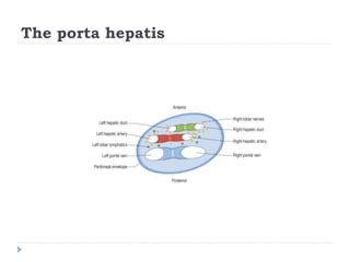
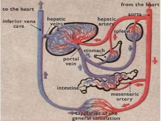
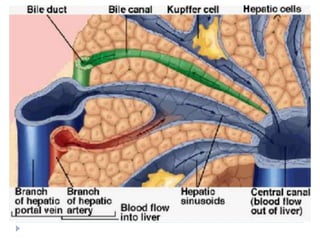
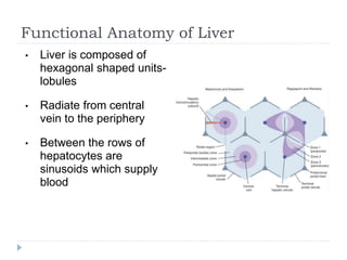
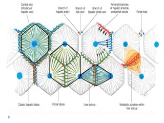
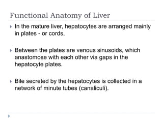
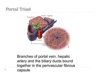
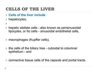
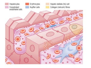
The liver is the largest organ in the abdominal cavity and performs many metabolic functions. It is composed of hepatocytes arranged in lobules around a central vein. Blood flows from the hepatic portal vein and hepatic artery into sinusoids between hepatocytes. Bile is secreted into bile canaliculi and flows through bile ducts. The liver regulates nutrients, metabolizes drugs and toxins, and synthesizes proteins. It is innervated by the hepatic plexus and refers pain to the epigastrium.
This is not a substitute for Books. Let it just help you understand some concepts in liver anatomy.
Continuation of this work will depend on your feedback. Stay Blessed.
anatomy of large intestine, its section, ceacum, ascending colon, transverse colon, descending colon, sigmoid colon, functions of large intestine , relations of each components of large intestine, carddinal siggns of large intestine, iliocecal junstion, difference between large and small intestine. abdominal angina, superior mesenteric and inferior mesenteric artery, lymphatic drainage, colonoscophy,
The human biliary system is composed of the liver, gallbladder, and bile duct. Understanding these complex systems will guide the students in training for better anatomical knowledge in the vein, artery, lymph nodes, and functional activities.
Portal Vein and portocaval Anastomosis. Anatomy of portal vein, tributaries, branches and course, formations and relations. Anatomy of portal vein and adjacent structures, their relation to liver and intestine, relation to IVC and Aorta, clinical and applied anatomy for both undergraduates and postgraduates. portal hypertension is an increase in blood pressure, however, rather than being systematic, it's localized to the portal system. Portal hypertension is most commonly caused by liver cirrhosis which in itself can be caused by alcoholism or other liver disease. It can also be caused by blood clots in the portal vein and schistosomiasis amongst other things. This increase in blood pressure can affect areas of anastomosis between the portal vasculature which we just discussed and the caval musculature which are classified as the vessels not relating to the portal system resulting in pressure pushing larger blood volumes into these anastomotic areas. This in turn can cause the vessels to dilate and form varicose veins which can result in potentially fatal hemorrhage. Some of these important porto-caval anastomotic areas are listed below – the first vein being the portal vein and the second vein being the caval vein – the superior rectal and inferior rectal veins, the left gastric and esophageal veins, the colonic veins and the retroperitoneal veins and the para-umbilical and epigastric veins.
In severe cases, the last anastomosis mentioned between the para-umbilical veins which are the small veins that run within the round ligament of the liver and the epigastric veins which are found in the anterior abdominal wall can form large dilations. These dilations can form the clinical presentation caput medusa or the head of the medusa as the dilated veins look like the snakes of the head of the medusa or Gorgon from Greek mythology. In this image on the right, we can only see the beginnings of a presentation of the caput medusa as in a true caput medusa, the veins would be raised and enlarged.
Hello everyone! This is Nicole from Kenhub, and today we're going to talk about the hepatic portal vein.
We are going to discuss the hepatic portal vein and to do so we'll be using this image here which is a ventral view of the portal hepatic vein with the central portion of the liver cut out so we can see the portal vein and other portal vessels. You can also see the aorta just here as well as the inferior vena cava just posterior to the portal hepatic vein. The portal venous system is an important system that has its own unique flow and we'll talk about how this works in tandem with the venous system in the coming slides.
The portal vein is one of the most important vessels in the body.
Its main functions are to direct blood to the liver from the gastrointestinal tract and receive nutrient rich blood from the intestines.
The portal hepatic vein also receives blood from the spleen, the pancreas and the gallbladder which are channels within the vessel.
Flu Vaccine Alert in Bangalore Karnatakaaddon Scans
As flu season approaches, health officials in Bangalore, Karnataka, are urging residents to get their flu vaccinations. The seasonal flu, while common, can lead to severe health complications, particularly for vulnerable populations such as young children, the elderly, and those with underlying health conditions.
Dr. Vidisha Kumari, a leading epidemiologist in Bangalore, emphasizes the importance of getting vaccinated. "The flu vaccine is our best defense against the influenza virus. It not only protects individuals but also helps prevent the spread of the virus in our communities," he says.
This year, the flu season is expected to coincide with a potential increase in other respiratory illnesses. The Karnataka Health Department has launched an awareness campaign highlighting the significance of flu vaccinations. They have set up multiple vaccination centers across Bangalore, making it convenient for residents to receive their shots.
To encourage widespread vaccination, the government is also collaborating with local schools, workplaces, and community centers to facilitate vaccination drives. Special attention is being given to ensuring that the vaccine is accessible to all, including marginalized communities who may have limited access to healthcare.
Residents are reminded that the flu vaccine is safe and effective. Common side effects are mild and may include soreness at the injection site, mild fever, or muscle aches. These side effects are generally short-lived and far less severe than the flu itself.
Healthcare providers are also stressing the importance of continuing COVID-19 precautions. Wearing masks, practicing good hand hygiene, and maintaining social distancing are still crucial, especially in crowded places.
Protect yourself and your loved ones by getting vaccinated. Together, we can help keep Bangalore healthy and safe this flu season. For more information on vaccination centers and schedules, residents can visit the Karnataka Health Department’s official website or follow their social media pages.
Stay informed, stay safe, and get your flu shot today!
Couples presenting to the infertility clinic- Do they really have infertility...Sujoy Dasgupta
Dr Sujoy Dasgupta presented the study on "Couples presenting to the infertility clinic- Do they really have infertility? – The unexplored stories of non-consummation" in the 13th Congress of the Asia Pacific Initiative on Reproduction (ASPIRE 2024) at Manila on 24 May, 2024.
TEST BANK for Operations Management, 14th Edition by William J. Stevenson, Ve...kevinkariuki227
TEST BANK for Operations Management, 14th Edition by William J. Stevenson, Verified Chapters 1 - 19, Complete Newest Version.pdf
MANAGEMENT OF ATRIOVENTRICULAR CONDUCTION BLOCK.pdfJim Jacob Roy
Cardiac conduction defects can occur due to various causes.
Atrioventricular conduction blocks ( AV blocks ) are classified into 3 types.
This document describes the acute management of AV block.
HOT NEW PRODUCT! BIG SALES FAST SHIPPING NOW FROM CHINA!! EU KU DB BK substit...GL Anaacs
Contact us if you are interested:
Email / Skype : kefaya1771@gmail.com
Threema: PXHY5PDH
New BATCH Ku !!! MUCH IN DEMAND FAST SALE EVERY BATCH HAPPY GOOD EFFECT BIG BATCH !
Contact me on Threema or skype to start big business!!
Hot-sale products:
NEW HOT EUTYLONE WHITE CRYSTAL!!
5cl-adba precursor (semi finished )
5cl-adba raw materials
ADBB precursor (semi finished )
ADBB raw materials
APVP powder
5fadb/4f-adb
Jwh018 / Jwh210
Eutylone crystal
Protonitazene (hydrochloride) CAS: 119276-01-6
Flubrotizolam CAS: 57801-95-3
Metonitazene CAS: 14680-51-4
Payment terms: Western Union,MoneyGram,Bitcoin or USDT.
Deliver Time: Usually 7-15days
Shipping method: FedEx, TNT, DHL,UPS etc.Our deliveries are 100% safe, fast, reliable and discreet.
Samples will be sent for your evaluation!If you are interested in, please contact me, let's talk details.
We specializes in exporting high quality Research chemical, medical intermediate, Pharmaceutical chemicals and so on. Products are exported to USA, Canada, France, Korea, Japan,Russia, Southeast Asia and other countries.
micro teaching on communication m.sc nursing.pdfAnurag Sharma
Microteaching is a unique model of practice teaching. It is a viable instrument for the. desired change in the teaching behavior or the behavior potential which, in specified types of real. classroom situations, tends to facilitate the achievement of specified types of objectives.
ARTIFICIAL INTELLIGENCE IN HEALTHCARE.pdfAnujkumaranit
Artificial intelligence (AI) refers to the simulation of human intelligence processes by machines, especially computer systems. It encompasses tasks such as learning, reasoning, problem-solving, perception, and language understanding. AI technologies are revolutionizing various fields, from healthcare to finance, by enabling machines to perform tasks that typically require human intelligence.
Factory Supply Best Quality Pmk Oil CAS 28578–16–7 PMK Powder in Stockrebeccabio
Factory Supply Best Quality Pmk Oil CAS 28578–16–7 PMK Powder in Stock
Telegram: bmksupplier
signal: +85264872720
threema: TUD4A6YC
You can contact me on Telegram or Threema
Communicate promptly and reply
Free of customs clearance, Double Clearance 100% pass delivery to USA, Canada, Spain, Germany, Netherland, Poland, Italy, Sweden, UK, Czech Republic, Australia, Mexico, Russia, Ukraine, Kazakhstan.Door to door service
Hot Selling Organic intermediates
2. Liver : Introduction
The liver is the largest of the abdominal viscera, occupying a
substantial portion of the upper abdominal cavity.
It performs a wide range of metabolic activities necessary for
homeostasis, nutrition and immune defence.
It is composed largely of epithelial cells (hepatocytes), which are
bathed in blood derived from the hepatic portal veins and hepatic
arteries.
Hepatocytes are also associated with an extensive system of minute
canals, which form the biliary system into which products are
secreted.
3. The liver lies in the upper right part of the abdominal
cavity.
It occupies most of the right hypochondrium and
epigastrium,
frequently extends into the left hypochondrium as far as
the left lateral line.
In adults the liver weighs 2% of body mass.
an overall wedge shape
4. The liver capsule plays an important part in maintaining
the integrity of its shape.
Once the capsule is lacerated, the liver tissue is
easily parted and provides only limited support for
surgical sutures.
These features, in combination with its exceptional
vascular supply, make the liver prone to potentially lethal
injuries if it is split open.
5. EXTERNAL FEATURES
The liver is attached to the anterior abdominal wall,
diaphragm and other viscera by several ligaments, which
are formed from condensations of the peritoneum :
Falciform ligament
Coronary ligament
Triangular ligaments
Lesser omentum
6.
7. Porta hepatis
The Porta hepatis is the area of the inferior surface
through which all the neurovascular and biliary
structures, except the hepatic veins, enter and leave the
liver.
It is situated between the quadrate lobe in front and the
caudate process behind.
Right and left hepatic bile ducts emerge from it.
All these structures are enveloped in the perivascular
fibrous capsule - hepatobiliary capsule of Glisson - a
sheath of loose connective tissue which surrounds the
vessels
9. LOBATION AND SEGMENTATION
The liver has four lobes or eight segments, depending
on whether it is defined by its gross anatomical
appearance or by its internal architecture.
10. Gross anatomical lobes
Right lobe
The right lobe is the largest in volume and
contributes to all surfaces
Quadrate lobe
The quadrate lobe is only visible from the inferior
surface. it is functionally related to the left lobe.
Caudate lobe
this lobe is said to arise from the right lobe, but it is
functionally separate
Left lobe
The left lobe is the smaller of the two 'main' lobes
12. Couinaud segments
The liver is divided by the 'principal plane' into two
halves of approximately equal size.
The principal plane is defined by an imaginary
parasagittal line from the gallbladder anteriorly to the
inferior vena cava posteriorly.
The usual functional division of the liver into right and left
lobes lies along this plane.
The liver is further subdivided into segments,
each supplied by a
principal branch of the hepatic artery,
portal vein and bile duct.
13. Segments I, II, III and IV make up the functional left lobe,
Segments V, VI, VII and VIII make up the functional right
lobe.
Segment I corresponds to the gross anatomical caudate
lobe and segment IV to the quadrate lobe.
14. VASCULAR SUPPLY
• Two sources provide blood to the liver
o Hepatic artery
Celiac trunk of aorta
o Portal vein
• Blood exits the liver via the central vein
• 25% of cardiac output
• Blood flow into the liver is controlled by number of
factors
• Muscular sphincters
• Number of different stimuli, including the autonomic nervous
system, circulating hormones, bile salts, and metabolites
15.
16. Portal vein
• Portal vein is a valveless
structure that is formed by
the confluence of the
superior mesenteric vein
and the splenic vein.
75% of the total liver
blood supply by volume
2 PV in liver
Left 2,3,4
Right 5,6,7,8
• Portal vein is formed by the
union of the superior
mesenteric vein and the
splenic vein at the level of the
second lumbar vertebra
behind the head of pancreas
• Portal vein is contained within
the hilum of the liver
17. Portal vein
• The portal vein gives rise to branching septal veins,.
• Blood from the septal veins enters directly into the
parenchymal sinusoids between hepatocytes.
•
• Sinusoids are lined by fenestrated and discontinuous
endothelial cells
o extrasinusoidal space of Disse, into which protrude abundant
microvilli of hepatocytes
Kupffer and presinusoidal stellate cells
18.
19. The portal vein and its tributaries (semi-diagrammatic). Portions of the stomach,
pancreas and left lobe of the liver and the transverse colon have been removed.
20. The portal vein supplies the liver with 5% of its resting
oxygen consumption but significantly more of its
metabolic nutrition.
Progressive occlusion of the hepatic artery rarely
results in complete necrosis of the liver, which is due
principally to the blood supply derived from the
portal vein.
21. Hepatic Artery
• Provides the remaining 25% of the blood flow to the liver
Large amount of variability
• Common hepatic artery arises from the celiac axis,
• ascends in the hepatoduodenal ligament, and gives rise to the right
gastric, gastroduodenal, and proper hepatic arteries;
• the proper hepatic artery then divides into the right and left hepatic
arterial branches in the liver hilum
• Right artery off SMA (#1 variant)
22.
23.
24. Hepatic veins
The liver has two venous systems.
The portal system conveys venous blood from the
majority of the gastrointestinal tract and its associated
organs to the liver.
The hepatic venous system drains blood from the liver
parenchyma into the inferior vena cava.
25. The hepatic veins convey blood from the liver to the
inferior vena cava.
The right hepatic vein drains segments V, VI, VII and VIII.
The left hepatic vein drains segments II and III with some
drainage from segment IV.
The hepatic veins have no valves.
26. Arrangement of the hepatic venous territories. Multiple lower group veins may be
present. Individual segments may drain into more than one hepatic venous territory.
27. INNERVATION
The liver has a dual innervation.
Hepatic nerves, which arise from the hepatic plexus and
contain sympathetic and parasympathetic (vagal) fibres
Supply the parenchyma.
They enter the liver at the porta hepatis and largely
accompany the hepatic arteries and bile ducts.
28. INNERVATION
The capsule is supplied by some fine branches of the
lower intercostal nerves, which also supply the parietal
peritoneum,
particularly in the area of the 'bare area' and superior
surface.
This is seen clinically when distension or disruption of the
liver capsule causes quite well localized sharp pain.
29. HEPATIC PLEXUS
The hepatic plexus is the largest derivative of the coeliac
plexus.
It also receives branches from the anterior and posterior
vagi.
These branches not only supply vasomotor fibres to the
hepatic vessels and biliary tree, but also innervate the
hepatocytes directly and are involved in the control of
some homeostatic mechanisms.
The vagal fibres are motor to the musculature of the
gallbladder and bile ducts and inhibitory to the sphincter
of the bile duct.
30. REFERRED PAIN
Pain arising from the parenchyma of the liver is poorly
localized.
In common with other structures of foregut origin, pain is
referred to the central epigastrium.
Stretch of or involvement of the liver capsule by
inflammatory or neoplastic processes rapidly produces
well-localized pain of a 'somatic' nature.
31. Functional Anatomy of Liver
• Liver is composed of
hexagonal shaped units-
lobules
• Radiate from central
vein to the periphery
• Between the rows of
hepatocytes are
sinusoids which supply
blood
32.
33. Functional Anatomy of Liver
In the mature liver, hepatocytes are arranged mainly
in plates - or cords,
Between the plates are venous sinusoids, which
anastomose with each other via gaps in the
hepatocyte plates.
Bile secreted by the hepatocytes is collected in a
network of minute tubes (canaliculi).
34. Portal Triad
Branches of portal vein, hepatic
artery and the biliary ducts bound
together in the perivascular fibrous
capsule
35. CELLS OF THE LIVER
Cells of the liver include
hepatocytes,
hepatic stellate cells - also known as perisinusoidal
lipocytes, or Ito cells - sinusoidal endothelial cells,
macrophages (Kupffer cells),
the cells of the biliary tree - cuboidal to columnar
epithelium - and
connective tissue cells of the capsule and portal tracts.
36.
37. Functions of the Liver
Formation and secretion of bile
Nutrient and vitamin metabolism
Glucose and other sugars
Amino acids
Lipids
Fatty acids
Cholesterol
Lipoproteins
Fat-soluble vitamins
Water-soluble vitamins
38. Inactivation of various substances
Toxins
Steroids
Other hormones
Synthesis of plasma proteins
Acute-phase proteins
Albumin
Clotting factors
Steroid-binding and other hormone-binding proteins
Immunity
Kupffer cells
39. Bile
About 500 mL is secreted per day.
Some of the components of the bile are reabsorbed in the
intestine and then excreted again by the liver
(enterohepatic circulation).
41. The glucuronides of the bile pigments, bilirubin and biliverdin,
are responsible for the golden yellow color of bile.
The bile salts are sodium and potassium salts of bile acids,
and all those secreted into the bile are conjugated to glycine or
taurine, a derivative of cysteine.
The bile acids are synthesized from cholesterol.
42. Primary bile acids
Formed in the liver are cholic acid and chenodeoxycholic acid.
Secondary bile acids
In the colon, bacteria convert cholic acid to deoxycholic acid
and chenodeoxycholic acid to lithocholic acid.
43. Enterohepatic Circulation
Enterohepatic circulation of bile salts. The solid lines entering the portal system represent bile salts of
hepatic origin, whereas the dashed lines represent bile salts resulting from bacterial action.
44. Functions of the Gallbladder
In normal individuals, bile flows into the gallbladder when the
sphincter of Oddi is closed.
In the gallbladder, the bile is concentrated by absorption of
water.
The degree of this concentration is shown by the increase in
the concentration of solids
liver bile is 97% water, whereas the average water content of
gallbladder bile is 89%.
45. When the bile duct and cystic duct are clamped, the
intrabiliary pressure rises to about 320 mm of bile in 30
minutes, and bile secretion stops.
However, when the bile duct is clamped and the cystic duct is
left open, water is reabsorbed in the gallbladder, and the
intrabiliary pressure rises only to about 100 mm of bile in
several hours.
Acidification of the bile is another function of the gallbladder.
47. Regulation of Biliary Secretion
Cholagogues:
Substances that cause contraction of the gallbladder
When food enters the mouth, the resistance of the sphincter
of Oddi decreases.
Fatty acids and amino acids in the duodenum release CCK,
which causes gallbladder contraction.
48. Effects of Cholecystectomy
The periodic discharge of bile from the gallbladder aids
digestion but is not essential for it.
Cholecystectomized patients maintain good health and
nutrition with a constant slow discharge of bile into the
duodenum.
Bile duct becomes somewhat dilated, and more bile tends to
enter the duodenum after meals than at other times.
Cholecystectomized patients can even tolerate fried foods,
although they generally must avoid foods that are particularly
high in fat content.
49. Choleretics:
Substances that increase the secretion of bile
The production of bile is increased by stimulation of the
vagus nerves and by the hormone secretin, which
increases the water and HCO3
– content of bile.
Bile salts themselves are among the most important
physiologic choleretics.