Downloaded 156 times


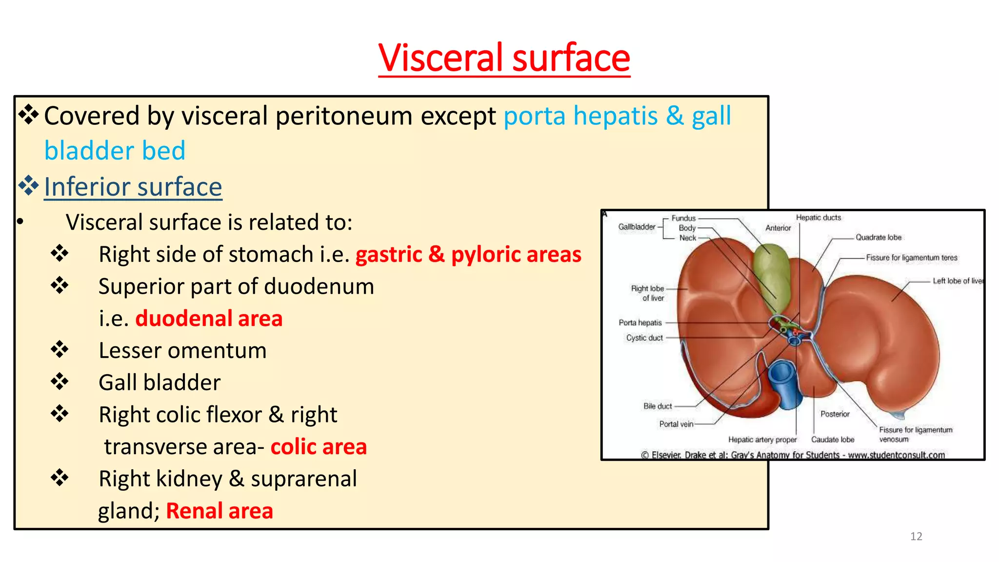


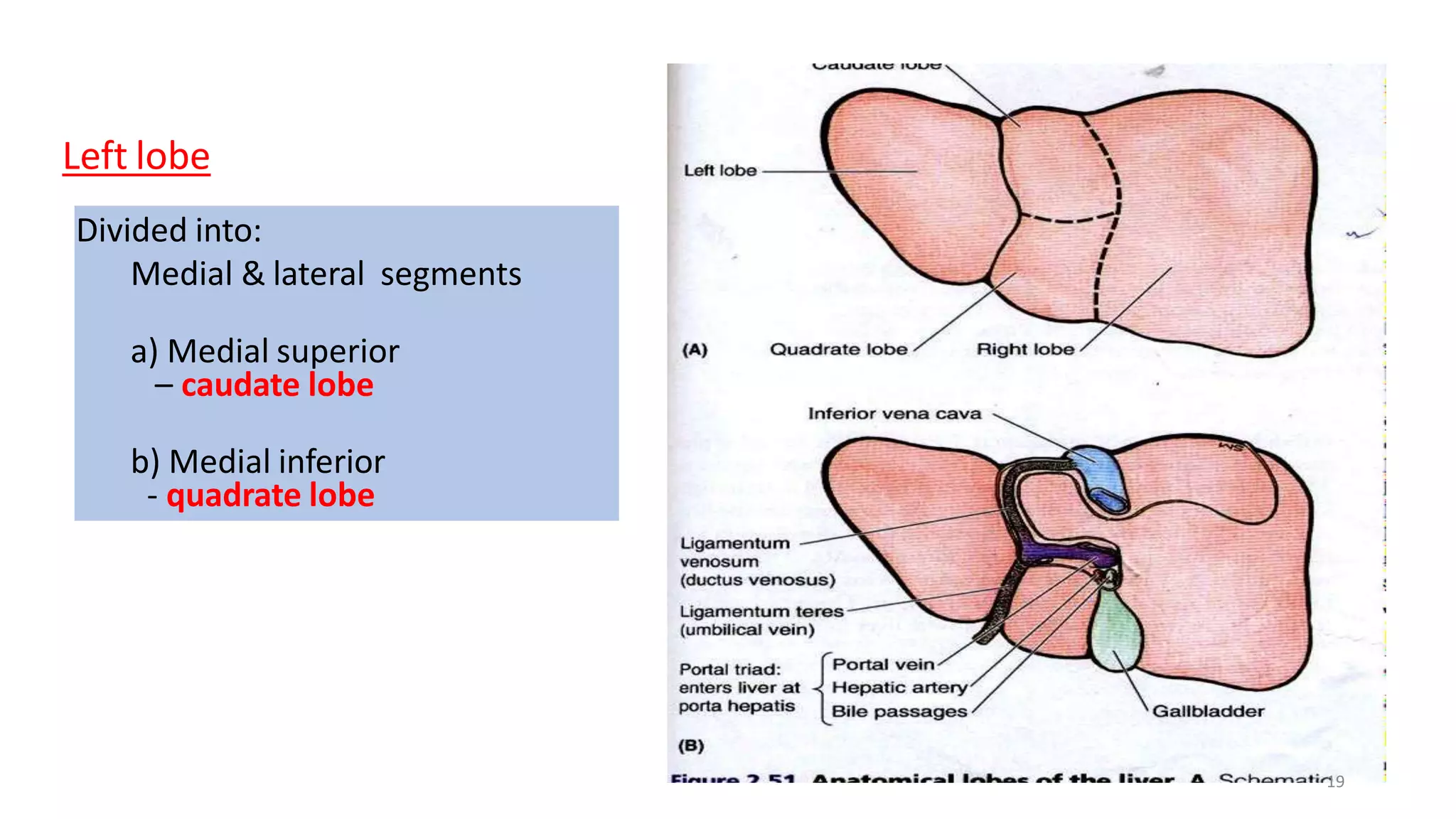


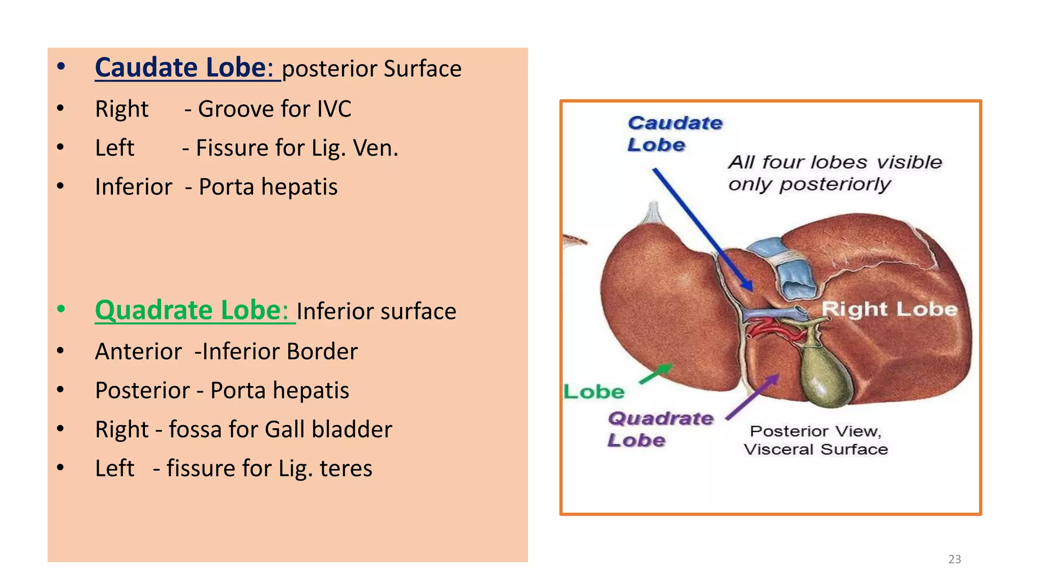


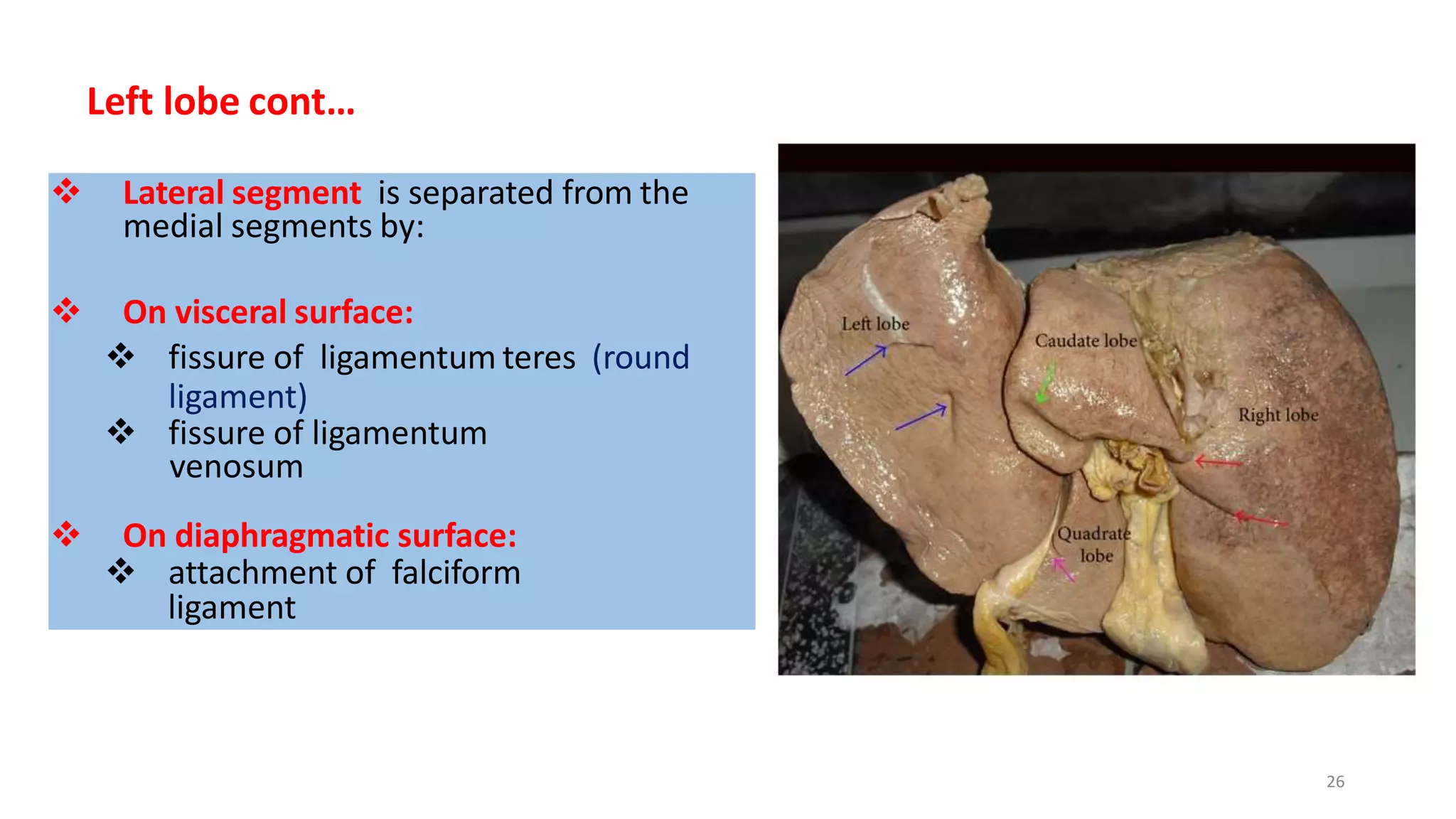


The document provides detailed information about the anatomy and features of the liver: 1. It describes the liver's location, lobes, ligaments, surfaces, segments, blood supply, nerve supply, lymphatic drainage and applied clinical aspects like hepatitis, cirrhosis and liver cancer. 2. Key points are that the liver has diaphragmatic and visceral surfaces, is divided into four lobes and eight segments, and receives dual blood supply from the hepatic artery and portal vein. 3. The bare area lacking peritoneal coverage is located on the posterior surface of the liver below the diaphragm.


