3. INTRODUCTION
Liposomes:
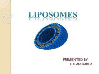
Liposomes are microscopic sealed structures in
which the aqueous compartment is enclosed by
one or more phospholipid bilayers. These carry
the drug and release it at the specific site of
action.
4.
5. These liposomes have proved to be excellent
carriers for drugs, as these are bio degradable,
inert and their lipid bilayers are similar in
composition to the biological memberanes.
6. Advantages
Liposomes are biodegradable, bio-compatible
They help in sustained drug release.
The drugs are delivered intact to their specific site of action.
The drug is not distributed to non- target sites, it reduces the
chances of drug toxicity.
The size, surface charge and other characteristics of the liposomes
can be altered depending on the drug and site of action.
7. Disadvantages
Expensive
Shorter half-lives in circulation.
Requires many modifications for deliver of
drugs to special organs.
Liposome's may undergo leakage during
their transit to the site of action.
8. TYPES OF LIPOSOMES
Based on their
size and number
of bilayers
liposomes are
classified into
three basic
types.
Multilamellar
Vesicles
(MLVs)
Small
Unilamellar
Vesicles
(SUVs)
Large
Unilamellar
Vesicles
(LUVs)
9. • Multilamellar vesicles of several lipid bilayers
separated one another by aqueous spaces.
• Ranging from few hundred to thousands of
nanometers in diameter.
Multilamellar
vesicles
• Single bilayer surrounding the entrapped
aqueous space.
• Size is larger than 100nm
Large
unilamellar
vesicles
• Sames as large unilamellar vesicles but size
is smaller than large unilamellar vesicles
• Size is less than 100nm
Small
unilamellar
vesicles
10.
11. Based on
composition and
mechanism of
intracellular
delivery of
liposomes
Long
circulating
liposomes
Immuno
liposomes
Cationic
liposomes
pH sensitive
liposomes
Conventional
liposomes
14. Mechanical dispersion
methods:
E.g., liquid film hydration, micro emulsification ( micro
fluidizer) sanitations dried reconstituted vesicle.
Lipid is solublised in organic solvent, drug to be
entrapped is solublised in aqueous solvent, the lipid
phase is hydrated at high speed stirring due to affinity
of aqueous phase to polar head it is entrapped in lipid
vesicles.
15. Solvent dispersion method:
e.g., ethanolinjection, ether injection, de- emulsification
In this method, lipid are first dissolved in organic
solvent, which then brought in to contact with aqueous
phase containing material which is to be entrapped in
liposomes under rapid dilution and evapouration of
organic solvent.
16. Detergent removal method:
In this methods, the phospholipids are brought into intimate contact with the
aqueous phase via detergent which associate with phospholipids molecules and
serve to screen the hydrophobic portions of the molecules from water.
Detergent removal from mixed micelles by
1. Dialysis
2. Column chromatography
3. Dilution
18. Hand Shaken MLVs
stand for 2 hours to get MLVs
Milky white dispersion formed
Rotate flask at room temperature, at 60 RPM for until lipid removes
from wall of RBF
Add 5ml buffer containing material to be entrapped
Till residues dry
Evaporate for 15 min above phase transition temperature
Lipids + solvent
(In 250ml
RBF)
(Flush with nitrogen)
19. Pro- Liposome
MLVs is formed
Flushed with nitrogen for drying properly
Dry the content using lyophilizer(freeze dryer)
Again add lipid solution
Add 5ml lipid solution (fitted to evaporator)
Sorbital/ Nacl (increase surface area of lipid film)
(Evaporation)
(Stand over night at room
temperature)
20. Micro emulsification liposomes(MEL)
MEL is prepared
by the “Micro
fluidizer”, which
pumps fluid at
very high
pressure
(10,000 psi)
through a 5nm
orifice.
Then, it is forced
along defined
micro channels,
which direct two
streams of fluid
to colloid
together at right
angle at very
high velocity.
After a single
pass, size
reduced to a
size 0.1 and 0.2
nm in diameter.
21. Sonicated unilamellar vesicles
MLV s in test tube
Sonicate for 5-10 min above
phase transition temperature
Filter & centrifuge at 10,000
rpm for 30min at 20ºc
Decant top layer to get
sonicated unilamellar vesicles
22. To increase size of liposomes : Freeze thaw
sonication
SUVs in
aqueous phase
+ solute
Freeze drying
FTS method,
(thawing
=melting)
Sonication (15-
30sec)
Solutes in
unilamellar
vesicles
23. pH induced vesiculation
SUV are formed
pH moves down to 7.5
Now add 0.1M Hcl
pH rises to 11
Add 1M NaOH (Less than 2 min)
MLVs or LUVs (pH 2.5-3)
24. EVALUATION
Evaluation tests are used used to ensure
their predictable in –vivo and in- vitro
performance.
Evaluation tests are classified into three
broad categories which include:
1. physical
2. chemical
3. biological tests
25. Physical
tests
• To ensure physical stability in
liposomes
Chemical
tests
• To establish purity and potency
of various liposomal constituents
Biological
tests
• To ensure safety and suitability of
the formulation
27. Particle size and particle size
distribution
Explains physical stability
These can be determined by the following
methods
1. Laser Light Scattering
2. Transmission Electron Microscopy
28. 1. LASER LIGHT SCATTERING
Laser light scattering is a very simple and rapid method which requires
expensive instrumentation.
In his method, a light of suitable wavelength is passed through a lipodial
suspension.
The intensity of light scattered by the liposomes is proportional to their
diameter.
Therefore, measuring the fluctuations in scattered light helps to indicate
the particle size of liposomes.
29. 2. Transmission Electron
Microscopy
The best method for determining the particle size of individual liposomes
is electron microscopy.
However, it requires high vaccum, is time consuming and may bring about
changes in the structure of the liposome.
Freeze fracture electron microscopy is found to be effective for evaluating
large- size vesicles.
This technique is also helpful in examining the morphological changes
that occur in liposomes when they undergo phase transition
30. Phase Behaviour
Liposomes at transition temperature undergo reversible
phase transition i.e., the polar head groups in gel state
become disordered to form the liquid crystalline state.
The phase behaviour of liposomes can be determined by
differential scanning calorimetry(DSC).
The transition temperature is indicative of stability.
31. Therapeutic applications
Liposomes as drug/ protein delivery vehicles.
Liposomes in antimicrobial, antifungal and antiviral therapy.
Liposomes in tumour therapy.
Liposomes in gene delivery.
Liposomes in immunology.
Liposomes as radiopharmaceutical and radio diagnostic carrier
Liposomes in cosmetic and dermatology.
Liposomes in enzyme immobilization and bioreactor.