
Recommended
More Related Content
Similar to Liposomes detail topic explanation notes
Similar to Liposomes detail topic explanation notes (20)
More from UVAS
More from UVAS (20)
Recently uploaded
Recently uploaded (20)
Liposomes detail topic explanation notes
- 1. LIPOSOMES
- 2. LIST OF CONTENTS Introduction Advantages with use of liposomes as drug delivery system. Classification Manufacturing of liposomes Liposome characterization and control Stability consideration for liposomal formulations Drug release from liposomes Applications Recent innovations Approved liposome products
- 3. INTRODUCTION The preparation of liposomes, with entrapped solutes, was demonstrated for the first time in 1965 by Prof. A.D. Bangham of the United Kingdom.
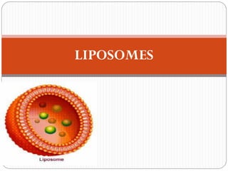
- 4. What is Liposomes “Are simple microscopic, concentric bilayered vesicles in which an aqueous material is entirely enclosed by a membranous lipid bilayer mainly composed of natural or synthetic phospholipids.” and range in size from 50 nanometers to several micrometers in diameter”
- 5. How Liposomes Formed? Phosphlipids(amphiphilic molecules) dispersed in water, they spontaneously form closed structure with internal aqueous enviornment bounded by phospholipid bilayer membranes, this vesicular system is called as Liposomes.
- 7. Structure Of Liposomes The structural main components of liposomes are phospholipids and cholesterol. Phospholipids Liposomes can be formed from a variety of phospholipids. The lipid most widely used is phosphatidyl choline, phosphatidyl ethanolamime and phosphatidlyl serine Cholesterol Condense the packing of phospholipids in bilayer, Thereby reducing their permeability to encapsulated compounds.
- 8. Phospholipid Bilayers are the core structure of liposome and cell membrane. Thus the structure of liposomes is similar to the structure of cell membranes. Liposome Cell Membrane
- 9. Advantages with Liposomes Suitable for delivery of hydrophobic, hydrophilic and amphipathic drugs and agents because its organized structure could hold drugs, depending on their solubility characteristics, in both the aqueous and lipid phases.
- 10. Chemically and physically well characterized entities Biocompatible and biologically inert in nature. Suitable for controlled release. Suitable to give localized action in particular tissues. Suitable to administer via various routes. Drug carrier for variety of small molecules, proteins, nucleotides and even plasmids.
- 11. Disadvantages of liposomes: Production cost is high. Leakage and fusion of encapsulated drug / molecules. Sometimes phospholipid undergoes oxidation and hydrolysis like reaction. Short half-life. Low solubility.
- 12. Cross-section of liposomes: H2O Layer Water Soluble ingredients (Drugs, Nutrients & vitamins) Polar Lipids (Phospholipid) Lipid Soluble ingredients (Drugs, Nutrients & vitamins)
- 13. Locus of drugs in liposomes: Hydrophilic (DOXORUBICIN) Low entrapment Low Leakage Hydrolytic degradation Lipophilic (CYCLOSPORINE) High entrapment Low leakage Chemical stability Amphiphilic (VINBLASTIN) High entrapment Rapid leakage Biphasic insoluble (ALLOPURINOL, 6- MERCAPTOPURINE) Poor loading & entrapment 43
- 14. Classification based on size of liposomes based on method of preparation based on composition and in vivo application
- 15. Classification based on structure & size 1. Small unilamellar vesicles 2. Medium sized unilamellar vesicles 3. Large unilamellar vesicles 4. Giant unilamellar vesicles 5. Unilamellar vesicles 6. Oligolamellar vesicles 7. Multilamellar large vesicles 8. Multivesicular vesicles
- 16. Lamella : Types of vesicles based on lamella
- 17. Unilamellar vesicles (UV) : UV refers to vesicles bounded by single bilayer membrane. Their further types are: SUV( Small Unilamellar vesicles): Size: 25-50nm No. of lipid bi layer: 1 MUV(Medium unilamellar vesicles): Size: >100nm No. of lipid bi layer: 1
- 18. LUV (Large unilamellar vesicles): Size: >1000nm No. of lipid bi layer: 1 GUV(Giant unilamellar vesicles): Size: >1um No. of lipid bi layer: 1
- 19. Advantages of Small unilamellar vesicles (SUV) Because of their small size, clearance from the systemic circulation is reduced, so they remain circulating for longer and thus have a better chance of exerting the desired therapeutic effect in tissues. Disadvantages of small unilamellar vesicles (SUV) The small size cause lower capacity for drug entrapment, less than 1% of the material available.
- 20. Advantages of Large unilamellar vesicles (LUV) There is a large space for incorporation of "drug.“ Disadvantages of Large unilamellar vesicles (LUV) They are more fragile than MLV and have increased Permeability to small solutes due to the absence of additional lamellae.
- 21. Multilamellar large vesicles (MLV): Refers to those vesicles bounded by two or more bilayer membranes. Size: > 0.5um No. of lipid bi layer: 5-20 As water added to the phosphholipid, the polar head groups at the surface of the exposed amphiphile become hydrated and start to reorganize into the lamellar form.
- 22. The water diffuses through this surface bilayer causing the underlying lipid to undergo a similar rearrangement, and the process is repeated until all of the lipid is organized into a series of parallel lamellae, each separated from the next by a layer of water. Mild agitation allows portions of close-packed, multilamellar lipid to break away resulting large spherical liposomes, each consisting of numerous concentric bilayers in close, alternating with layers of water, which are known as multilamellar vesicles (MLV).
- 23. Advantage of MLV: They are simple to make and have a relatively compact construction. Disadvantage of MLV: The volume available for solute incorporation is limited
- 24. Their large size is a drawback for many medical applications requiring parenteral administration, because it leads to rapid clearance from the bloodstream by the cells of the RES. On the other hand, this effect can be used for passive targeting of substances to the fixed macrophages of the liver and spleen.
- 25. Oligolamellar vesicles (OLV): Size: > 0.1-1um No. of lipid bi layer:~ 5 Multivesicular vesicles (MVV): Size: > 1um No. of lipid bi layer: Multi- compartmental structure
- 26. Liposome Function Depending on Size Large Multiple-layer liposome: Are liposomes within liposomes .They have a limited ability to penetrate narrow blood vessels or into the skin. The materials that are entrapped in the inner layers of these liposomes are practically less releasable . Large Unilamellar liposomes : Are easy to make by shaking phospholipids in water. These liposomes have very limited functions and are usually made of commercial lecithin (Phospotidyicholine PC), commonly found in food products.
- 27. Commercial lecithin’s main function is as an emulsifying agent, improving the ability of oil and water to remain mixed. Small Unilamellar liposomes (Nanosomes) Are constructed from the highest quality and high percentage of phosphatidylcholine (PC), one of the essential components of cell membranes. Thus, nanosomes can easily penetrate into small blood vessels by intravenous injection; and into the skin by topical application. Their entrapped material can be easily delivered to desired targets such as cells .
- 28. Classification based on composition and in vivo application Conventional Liposomes (CL) Long circulating Liposomes (LCL) Immuno Liposomes (Antibody targeted) (CL) (LCL) With attached monoclonal antibody or recognition sequence. Cationic Liposomes pH sensitive liposomes (Phospholipid such as PE or DOPE with either CHEMS or OA.
- 30. Conventional Liposomes Composed of only phospholipids (neutral or negatively charged) and/or cholesterol and used for passive targeting.
- 31. LONG CIRCULATING STEALTH LIPOSOMES These liposomes carry hydrophilic coatings(PEG coating) Also called sterically stabilized. Low permeability liquid matrix and internal aqueous buffer system Used to obtain prolonged circulation times.
- 32. Immunoliposomes (targeted liposomes) Either conventional or sterically stabilized. Used for active targeting purposes. Target specific ligands, such as antibodies, immuno globulins, lectins and oligosaccharides attached to the surface.
- 33. Cationic Liposomes Positively charged. Used for delivery of genetic material. Cationic lipid component interact with negatively- charged DNA, Results into Lipid –DNA Complexes.
- 34. Passive loading technique Active/remo te loading technique Loading of the entrapped agents before/ during the manufacture procedure. Certain types of compounds with ionizable groups & those with both lipid & water solubility can be Introduced into liposomes after the formationof intact vesicles. Methods of Liposome Preparation 21
- 35. General Method Of Liposome Preparation: 23
- 36. Methods of loading of liposome Passive loading techniques Active loading techniques Mechanical dispersion methods Solvent dispersion methods Detergentremoval technique 22
- 37. LOADING OF DRUG IN LIPOSOMES PASSIVE LOADINGTECHNIQUES Methods have been classified according to 3 basic modes of dispersion: 1. Physical(Mechanical)dispersion 2. Chemical dispersion 3. Detergent Solubilization
- 38. 1.PHYSICAL DISPERSION METHOD: In this method, aqueous volume enclosed in lipid membrane is 5- 10%. So water soluble drugs are more wasted while lipid soluble drugs encapsulated up to 100% efficiency. 4 methods of physical dispersion: 1. Hand shaking method. 2. Non shaking method. 3. Pro – liposomes . 4. Freeze drying .
- 39. Liposome Lipid spontaneously swell & Hydrate Solid lipid mixture is hydrated by using aqueous buffer Film deposition Remove organic solvent under vacuum Lipid dissolve in organic solvent/co-solvent 1. Physical( Mechanical )dispersion method: 24
- 40. 1. Hand Shaken Method PROCEDURE: 1. Prepare lipid mixture by dissolving phospholipids and charge components in chloroform: methanol mixture (2:1) ratio in 250ml round bottom flask. 2. Attach flask to rotary evaporator & rotate at 60 r.p.m to evaporate solvent at 30 ͦc. 3. Rotate for 15 min after dry residue appear. 4. Isolate evaporator from vacuum & introduce nitrogen till no pressure difference b/w inside and outside flask. 5. Remove evaporator and attach lyophilizer.
- 41. HYDRATION OF LIPIDS: After releasing the vacuum & Remove from lyophilizer, flush with nitrogen & 5ml of phosphate buffer containing solute(drug) to be entrapped is added. Attach flask again with evaporator at 60 r.p.m or less at room temp for 30 min until all lipid removed from walls of flask. Homogeneous white milky suspension formed. Allow milky suspension to stand for 2 hrs at room temp to allow swelling process which yields MLVs. At the end of this period, the loaded liposomes can be separated from nonencapsulated solute using a process such as centrifugation or dialysis. The possibility of lipid oxidation can be minimized by working in an inert atmosphere of nitrogen.
- 42. Lipids form stacks of film from organic solution Then film is treated with aqueous medium Upon hydration lipids swell and peel out from RB flask vesiculate to form Multi lamellar vesicles(MLVs) 26
- 43. 2. Non shaking method. In this method, LUVs formed with high entrapment volume. Use same procedure but instead of round bottom flask we will use flat bottom flask . More care over swelling process. Hydration and swelling process in two separate steps. Hydration: Dried film of lipid + stream of water saturated with nitrogen for 15 min. Swelling: In aqueous medium without shaking. Chloroform: methanol in ratio of 1:2 by volume is used.
- 45. 3. Pro-liposomes: To increase the surface area of dried lipid film & to facilitate instantaneous hydration. lipid Dried over lipid Finely divided particulate support like powdered NACL/ sorbital Pro - liposomes Pro- liposomes water Dispersion of MLV’S This Method overcome the stability problem. 27
- 46. In this method, the lipids are dried down to a finely divided particulate support, such as powdered sodium chloride, or sorbitol or other polysaccharides. The lipids are swelled upon adding water to dried lipid coated powder (pro-liposomes), where the support rapidly dissolves to give a suspension of MLVs in aqueous solution. This method overcomes the problems encountered when storing liposomes themselves in either liquid, dry or frozen form. This method is ideal for preparations where the material to be entrapped incorporates into lipid membrane.
- 47. 4. Freeze drying . Another method of dispersing lipid in finely divided form prior to the addition of aqueous media. In it freeze dry the lipid dissolved in suitable organic solvent (Tertiary butanol). After obtaining dry lipid, water or saline can be added with rapid mixing to give MLVs.
- 48. Processing of Liposomes 1. Microemulsification:
- 50. 2. Sonicated UnilamellarVesicles: a. Bath sonication b. Probe sonication Ultra sonicated sound( high frequency) waves travel through medium containing liposomes. Because sound waves can not pass through vaccum. Through any media when sound waves pass they pass in 2 region one is compressional region (air give it resistance) and this resistance region is called rear fractional region.This second region back convert these waves into smaller waves and spread in different regions.
- 52. 3.French Pressure Cell liposomes: The ultrasonic radiation can degrade the lipids, other sensitive compounds, macromolecules. For this extrusion of preformed larger liposomes in a French press under very high pressure (20,000 -40,000 psi) is done. This tech. yields unit or oligo lamellar liposomes of size (30-80 nm in dia) Includes high cost of press that consist of electric hydraulic press and pressure cell. Liposome prepare by this method are less likely to suffer from structural defects and instabilities as observed in sonication vesicles.
- 54. 4. Membrane Extrusion Method: Use to process LUVs as well as MLV. Liposomes prepare by this is called membrane filter extrusion liposomes. The 30% capture volume can be obtained using high lipid conc. The trapped volume in this process is 1-2 liter /mole of lipids. It is due to their ease of production, readily selectable vesicle diameter, batch to batch reproducibility and freedom from solvent or surfactant contamination is possible. All vesicle Form by this will of same size and lamellarity.
- 56. DRIED RECONSTITUEDVESICLE(DRV) and FREEZETHAW SONICATION:
- 58. 2. SOLVENT SOLVENT(CHEMICAL) DISPERSION METHODS: In this method, lipids are first dissolved in an organic solution, which is then brought in to contact with aqueous phase containing material to be entrapped within liposomes. At the interface between the organic and aqueous media, the phospholipids align themselves. Methods employing solvent dispersion fall in to one of three categories: Organic solvent miscible with aqueous phase. Organic solvent immiscible with aqueous phase and aq. Phase in excess. Organic solvent in excess and immiscible with aqueous phase.
- 59. Its further 5 types. A. Ethanol injection B. Ether injection C. Water in organic phase D. Double emulsion vesicles E. Reverse Phase evaporation vesicles.(REV)
- 60. Liposome Formation of monolayer and bilayer of phospholipids Lipids align at interface of aqueous and organic layer Excess addition of aqueous phase Lipid dissolve in organic solvent Solvent(Chemical) dispersion methods: 36
- 61. A:ETHANOL INJECTION: In this method, an ethanol solution of lipids is injected rapidly into an excess of saline or other aqueous medium (BUFFER), through fine needle. The force of injection is usually sufficient to achieve complete mixing, so that the ethanol is diluted almost instantaneously in water and phospholipid molecules are dispersed evenly throughout the medium. This procedure yields a high proportion of SUV (Dia. 25nm). Although lipid aggregates and larger vesicles may form if the mixing is not thorough enough. Disadvantages: Population is heterogeneous (30-110nm), liposomes are very dilute, the removal of ethanol is difficult because it forms into Azeotrope ( a mixture of two or more liquids whose components cannot be altered by simple distillation) with water.
- 62. B: ETHER INJECTION (SolventVaporization). This method is very similar to ethanol injection method. Ether injection method contrasts markedly with ethanol injection in many respects. It involve injecting the immiscible organic solution very slowly into an aqueous phase through a narrow needle at the temperature of vaporizing the organic solvent. This method may also treat sensitive lipids very gently and very little risk of causing oxidative degradation. Disadvantages of the technique are the long time taken to produce a batch of liposomes. The efficiency of encapsulation is relatively low.
- 63. Solvent dispersion methods: A:ETHANOL INJECTION/ B: ETHER INJECTION: 37
- 64. C. Reverse phase evaporation method: The novel key in this method is the removal of solvent from an emulsion by evaporation. The droplets are formed by bath sonication of mixture of the two phases, then the emulsion is dried down to a semi solid gel in a rotatory evaporator under reduced pressure. The next step is to bring about the collapse of a certain proportion of the water droplets by vigorous mechanical shaking with a vortex mixer. In these circumstances, the lipid monolayer which enclosed the collapsed vesicle, is contributed to adjacent intact vesicle to form the outer leaflet of the bilayer of a large unilamellar liposomes.
- 65. The aqueous content of the collapsed droplet provides the medium required for suspension of these newly formed liposomes. After conversion of the gel to a homogenous free flowing fluid , the suspension is dialyzed in order to remove the last traces of solvent. The vesicles formed are unilamellar and have a diameter of 0.5um. The encapsulation percentage is found to be not greater then 50%.
- 66. C. Reverse phase evaporation method: 39 Organic phase
- 67. Reverse phase evaporation technique. Lipid in solvent solution Two-phasesystem Water in oil emulsion Solvent removal Gel formation REV liposomes
- 68. D.WATER IN ORGANIC PHASE: The common feature of method is formation of “ water in oil emulsion’’ by introduction of a small quantity of aq. Medium containing the material to be entrapped, into large volume of immiscible organic solution of lipid followed by mechanical agitation to break up aq. Phase into microscopic droplets. These droplets are solubilized by the presence of phospholipid monolayer at the phase interface and form the central core of the final liposomes.
- 69. E. DOUBLE EMULSIONVESICLES In this method, firstly water is emulsified in organic phase, Which is followed by emulsifying w/o emulsion in aq. Phase resulting in a w/o/w system. The organic solvent is removed by evaporation and the product is centrifuged to remove the lipid aggregates. The organic solution which already contains water droplets is introduced into excess aqueous medium followed by mechanical dispersion, a multi compartment vesicle is obtained, which may be described as a w/o/w system (i.e double emulsion).
- 70. These vesicles are suspended in aqueous medium and have an aqueous core, the two aqueous compartments being separated from each other by a pair of phospholipid monolayers whose hydrophobic surfaces face each other across a thin film of organic solvent. Removal of this solvent clearly results in an intermediate size unilamellar vesicles.
- 71. Formation of Liposome by detergent removal By addition optimized concentration of detergent Phospholipids brought into intimate contact with aqueous phase Note:- Liposome size and shape depend on chemical nature of detergent, concentration and other lipid involved DETERGENT SOLUBILISATIOIN METHODS 40
- 73. 3.DETERGENT SOLUBILISATIOIN METHODS: Phospholipids can be solubilized in a aq. Medium by using detergent. The structures formed as a result of this association, are known as micelles. The concentration of detergent in water at which micelles just start to form is known as critical micelle concentration(CMC). Below CMC, detergent molecules exist in free soln. As the concentration is increased,micelles are formed. Production of liposomes is dependent on the removal of detergent molecule from aqueous dispersion of phospholipids / detergent mixed micelles. As detergent is removed, the micelles progressively richer in phospholipids and coalesce to form closed single bilayer vesicles (LUVs).
- 74. DETERGENTS: BILE SALTS such as sodium cholate and sodium deoxy cholate. SYNTHETIC such as octylglucoside. Liposome size and shape depend on chemical nature of detergent, concentration and other lipid involved. Sizes of liposomes can also control by controlling conditions of detergent removal. Not efficient method in terms of %age of solute entrapped.
- 76. PURIFICATION OF LIPOSOMES Purification of liposomes involved The removal of unbound drug from liposomes. Removal of detergents from liposomes. To fractionate heterogeneous liposomal dispersions. When lipophilic drugs of appropriate structure are encapsulated in the bilayer phase, the degree of "encapsulation" is dependent upon the saturation of the lipid phase. In this case degrees of encapsulation of over 90% is achieved. Thus it is unnecessary to remove the unbound drug.
- 77. However, in the case of water-soluble drugs, the encapsulated drug is only a fraction of the total drug used.Thus, it is required to remove the unbound drug from the drug-loaded liposomes in dispersion. Liposomes are purified by either: A. Dialysis B. Centrifugation C. Gel filtration or column chromatography.
- 78. A. Dialysis: Dialysis is the simplest procedure used for the removal of the unbound drug, except when macromolecular compounds are involved. Advantages: Dialysis Technique requiring no complicated or expensive equipment. Dialysis is effective in removing nearly all of the free drug with a sufficient number of changes of the dialyzing medium. For large scale liposomes, HOLLOW DIALYSIS CARTRIDGE may be used. Liposome dispersion
- 79. Disadvantages: • Dialysis is a slow process. • Removal of over 95% of the free drug require a minimum of 3 changes of the external medium over 10 to 24 hr at room temperature. • Care is taken to balance the osmotic strengths of the liposomal dispersion and the dialyzing medium to avoid leakage of the encapsulated drug.
- 80. B. Centrifugation: Centrifugation is an effective means of isolating liposomes from the free drug in the suspending medium. Separation of liposomes depend on size as well as composition of bilayer. SUVs Sediments by spinning at 200,000 g for 10-20 hrs in ultracentrifuge. MLVs Sediments rapidly at 100,000g in less than one hr.
- 81. C. Gel Filtration Gel permeation chromatographic technique is used extensively both to separate liposomes from unbound drug and also to fractionate heterogeneous liposomal dispersions. Advantages: The technique is very effective and rapid at the laboratory level.
- 83. Disadvantages: Gel filtration is expensive. Dilution of the liposomal dispersion with the eluting medium may necessitate another concentration step. Lipid losses on the column materials.
- 84. Active/remote loading technique: The lipid bilayer membrane is impermeable to ions & hydrophilic molecules. But, Permeation of hydrophobic molecules can be controlled by concentration gradients. Some weak acids or bases can be transported due to various transmembrane gradients Electrical gradients. Ionic(pH) gradients. Chemical potential gradients. Weak amphipathic bases accumulate in aq phase of lipid vesicles in response to difference in pH b/w Inside & outside of liposomes 41
- 85. pH gradient is createdby preparing liposomes withlow internal pH. [Basic compds ( lipophilic (non ionic) at high pH & hydrophilic(ionic) at low pH)] Lipophilic (UNPROTONATED) drug diffuse through the bilayer At low pH side, the molecules are predominantly protonated. . Weak bases like doxorubicine, adriamycin and vincristine are encapsulated. Solute bearing no chargeat neutral pH Liposomes with low internal pH Neutral solute passes easily through bilayer membrane by diffusion Charge aquired by solute inside liposomes makes them unable to exit 42
- 86. CHARACTERIZATION OF LIPOSOMES: PHYSICAL CHARACTERISATION → Vesicles size/shape/morphology → Surface -charge/electrical potential → Phase behaviour/ lamellarity → Drug release → % capture /free drug CHEMICA L CHARACTERISATION → Phospholipids /lipid concentration → Drug concentration → PH / Osmomolality →Phospholipids / cholesterols – peroxidation/oxidation/hydrolysis BIOLOGICAL CHARACTERISATION → Sterility → Pyrogenisity → Animal toxicity →Plasma Stability: 44
- 88. Characterization parameters Analytical method/Instrument 1. Vesicle shape and surfacemorphology Transmissionelectron microscopy, Freeze- fractureelectron microscopy 2.Meanvesicle size and size distribution (submicron and micron range) Electronmicroscopy, Photon correlation spectroscopy,laser light scattering,gel permeation and gel exclusion 3. Surfacecharge Free-flowelectrophoresis 4. Electricalsurfacepotential and surfacepH Zetapotential measurements 5. Lamellarity Small angle X-ray scattering,31 P-NMR, Freeze- fractureelectron microscopy 6. Phase behavior of Liposomes Freeze-fractureelectron microscopy, Differential scanning calorimetery 7. Percent of freedrug/ percent capture(entrapment) Minicolumn centrifugation,ion-exchange chromatography, radio labelling 8. Drug release Diffusioncell/ dialysis
- 94. Characterization parameters Analytical method/Instrument 1. Phospholipid concentration TLC & HPLC Barlett assay, stewart assay, 2. Cholesterol concentration Cholesterol oxidase assayand HPLC 3. Phopholipid oxidation UV absorbance 4. Phospholipid hydrolysis, Cholesterol auto-oxidation. HPLC and TLC 5. Osmolarity Osmometer
- 98. Characterization parameters Analytical method/Instrument 1. Sterility Aerobic or anaerobic cultures 2. Pyrogenicity Limulus Amebocyte Lysate (LAL) test 3. Animal toxicity Monitoring survival rates, histology and pathology
- 99. STABILITY OF LIPOSOMES: Liposomes stability problems are of course much more severe . ~ Perform Long term & accelerated stability Liposomal phospholipid can undergo chemical degradation such as ~ Lipid oxidation ~ Lipid Hydrolysis Either as a result of these changes or otherwise, liposomes maintained in aqueous suspension may aggregate, fuse or leak their contents.
- 100. PREVENTION OF CHEMICAL DEGRADATION: The following precautions may be taken to minimize chemical degradation. Start with freshly purified lipids and freshly distilled solvents. Avoid procedures which involves high temperature. Carry out manufacturing in the absence of oxygen. Deoxygenate aqueous solution with nitrogen. Store all liposome suspension in inert atmosphere. Include an anti-oxidant as a component of the lipid membrane.
- 102. 1. Endocytosis 2. Adsorption 3. fusion 4. Lipid transfer 48 5. Contact release
- 103. Liposomes can interact with cells by 5 different mechanims: Lipid Exchange Intermembrane Transfer Adsorption Endocytosis Fusion Contact Release
- 104. 1) Endocytosis by phagocytic cells of the reticuloendothelial system such as macrophages and neutrophils, that makes the liposomal content available to the cell, where lisosomes break , and phospholipids hydrolysed to fatty acids which can be incorporated into host phospholipids.
- 105. 2) Fusion with the cell membrane By insertion of the lipid bilayer of the liposome into the cell membrane to become its part, with simultaneous release of liposomal contents into the cytoplasm.
- 106. 3) Adsorption to the cell surface either by nonspecific weak hydrophobic or electrostatic forces, or by interactions of specific receptors on cell surface to ligands on the vesicle membrane. For water soluble components, vesicle contents are diffused through the lipids of the cell. For lipid soluble components, vesicle contents are exchanged with the cellular membrane along with the lipid of the vesicle.
- 107. 4) Inter-membrane Transfer (Lipid transfer): WithTransfer of liposomal lipids to cellular or sub cellular membranes, or vice versa.
- 108. 5) Contact-Release: This case can occur when the membranes of the cell and that of liposomes exert perturbation (agitation) which increase the permeability of liposomal membrane, and exposure of solute molecule to be entrapped by cell membrane.
- 109. CLINICAL APPLICATIONS In cancer chemotherapy In immunology (As carrier of vaccines, antigens) Liposomes as drug carrier in oral treatment As targeted delivery system Parental Liposomes Liposomes for topical application Liposomes for pulmonary delivery Liposomes for ophthalmic delivery of drugs Cell biological applications Advanced applications of Liposomes
- 110. Cancer chemotherapy Liposomes entrapped anticancer drugs and enhance the selectivity for tumor cells. Results in decreased toxicity to normal cells. Protects the metabolic degradation of drug. Increase circulation life time. EXAMPLE: (LipADM) LipADM is encapsulation of doxorubicin in liposome enhance the anti-tumor effects of drug and reduce side effects such as cardiac toxicity.
- 111. Liposomes could serve as tumor specific vehicles (even without special targeting) Liposomes better penetrate into tissues with disrupted endothelial lining 53
- 112. Liposomes in immunology As immunological adjuvant. Adjuvants may be incorporated with antigen into liposomes. Small amount of immunogen may be suitable as antigens. Hydrophobic antigen may be incorporated. Multiple antigens may be incorporated in to single liposomes. Toxicity or allergic reactions of antigens may be reduced.
- 113. Liposomes as drug carrier in oral treatment • Regular liposomes are typically unstable in intestine. Polymer coated liposomes have increased stability in GIT. Using Liposomes as a potential system for oral delivery of insulin has been extensively studied. As targeted delivery system: • The encapsulation of drugs with liposomes alters drug pharmacokinetics, and may be exploited to achieve targeted therapies by the flexibility in alteration of the liposome surface.
- 114. ARTHRITIS: Drugs used in arthritis specially steroids are destroyed by peripheral effects. This effect is minimized by local administration of drugs. Liposomes can be used in treatment of local diseases. Steroids can be entrapped in to large MLVs and administered locally in arthritis.
- 115. Parentral (I/V) Liposomes The closed pack of liposome structure can encapsulate aqueous soluble drugs within the central aqueous compartment or lipid soluble drugs within the bilayer membrane. ATRA-IV Liposomal retinoic acid: is a liposomes encapsulated, intravenous form of all trans retinoic acid (ATRA), a derivative of vitamin A.
- 116. Liposomes for topical application Liposomes have shown great potential as a topical drug delivery system. EXAMPLES: Hydrocortisone ointment Shows higher conc. Of drug in layers of human skin. Diclofenac gel Shows higher conc. Of drug and increase permeation through skin. Benzocaine gel Shows prolonged anesthesia as compared to plain benzocaine gel.
- 117. Liposomes for pulmonary delivery Liposomes are available in different sizes and provide the opportunity of a controlled drug delivery for administration to respiratory tract. Inhalation devices such as nebulizer will produce an aerosol of droplets containing liposomes. Liposomes encapsulated drugs for pulmonary administration are: Tobramycin Salbutamol Cytarabine
- 118. Cell biological applications • DNA delivery of Genes by Liposomes. • Cheaper than viruses. • No immune response. • Liposomes are also used to insert regulatory molecules such as AMP, CGMP.
- 119. Ophthalmic delivery of drugs Liposomes improve bioavailability of ophthalmic drugs after topical application due to lipophilisation of water soluble drugs which can not penetrate the lipophilic cornea. Liposomes have been used as vehicles for sub conjunctival inj. of cytotoxic drug and antibiotics. Dihydro-streptomycin Benzyl penicillin
- 120. Advanced applications of Liposomes The recent research is on use of liposomes to deliver hemoglobin act as red blood cell substitute. EXAMPLE: Liposomes encapsulated Hb (LEH)
- 121. DRUG ROUTE OF ADMINISTRATION APPLICATION TARGETED DISEASES Amphotericin B Oral delivery Ergosterol membrane Mycotic infection Insulin Oral,ocular,pulmonary And transdermal Decrease glucose level Diabetic mellitus Ketoprofen Ocular delivary Cyclooxygenase enzyme inhibitor Pain muscle condition Pentoxyfyllin Pulmonary delivery phosphodiesterase Asthama Tobramycin Pulmonary delivery Protein synthesis inhibitor Pseudomonas aeroginosai nfection, Salbutamol Pulmonary delivery ß2-adrenoceptor antagonist Asthama Cytarabin Pulmonary delivery DNA-polymerase inhibition Acute leukameias Benzocaine Transdermal Inhibition of nerve impulse from sensory nerves Ulcer on mucous surface with pain Ketaconazole Transdermal Inhibit ergosterol membrane Candida albicans Levanogesterol Transdermal Rhamnose receptor skin disorder hydroxyzine Transdermal H1-receptor antagonist Urtecaria,allergic skin disease Ibuprofen Oral delivery Chaemoceptor,free ending Rheumatoid arthritis triamcilonone Ocular delivery,Transdermal Inhibition of prostaglandin Anti-inflammatory Therapeutic application of liposomes: 54
- 122. NAME TRADE NAME COMPANY INDICATION Liposomal amphotericin B Abelcet Enzon Fungal infections Liposomal amphotericin B Ambisome Gilead Sciences Fungal and protozoal infections Liposomal cytarabine Depocyt Pacira (formerly SkyePharma) Malignant lymphomatous meningitis Liposomal daunorubicin DaunoXome Gilead Sciences HIV-related Kaposi’s sarcoma Liposomal doxorubicin Myocet Zeneus Combination therapy with cyclophosphamide in metastatic breast cancer Liposomal IRIV vaccine Epaxal Berna Biotech Hepatitis A Liposomal IRIV vaccine InflexalV Berna Biotech Influenza Liposomal morphine DepoDur SkyePharma, Endo Postsurgical analgesia Liposomal verteporfin Visudyne QLT, Novartis Age-related macular degeneration, pathologic myopia, ocular histoplasmosis Liposome-PEG doxorubicin Doxil/Caelyx Ortho Biotech, Schering-Plough HIV-related Kaposi’s sarcoma, metastatic breast cancer, metastatic ovarian cancer Micellular estradiol Estrasorb Novavax Menopausal therapy 55
- 123. THANKS
- 124. NANO REVIEW Open Access Liposome: classification, preparation, and applications Abolfazl Akbarzadeh1* , Rogaie Rezaei-Sadabady1,2 , Soodabeh Davaran1 , Sang Woo Joo5* , Nosratollah Zarghami1* , Younes Hanifehpour5 , Mohammad Samiei3 , Mohammad Kouhi4 and Kazem Nejati-Koshki1 Abstract Liposomes, sphere-shaped vesicles consisting of one or more phospholipid bilayers, were first described in the mid- 60s. Today, they are a very useful reproduction, reagent, and tool in various scientific disciplines, including mathematics and theoretical physics, biophysics, chemistry, colloid science, biochemistry, and biology. Since then, liposomes have made their way to the market. Among several talented new drug delivery systems, liposomes characterize an advanced technology to deliver active molecules to the site of action, and at present, several formulations are in clinical use. Research on liposome technology has progressed from conventional vesicles to ‘second-generation liposomes’, in which long-circulating liposomes are obtained by modulating the lipid composition, size, and charge of the vesicle. Liposomes with modified surfaces have also been developed using several molecules, such as glycolipids or sialic acid. This paper summarizes exclusively scalable techniques and focuses on strengths, respectively, limitations in respect to industrial applicability and regulatory requirements concerning liposomal drug formulations based on FDA and EMEA documents. Keywords: Liposomes, Glycolipids, Drug formulations, Drug delivery systems Review Introduction Liposomes are small artificial vesicles of spherical shape that can be created from cholesterol and natural non- toxic phospholipids. Due to their size and hydrophobic and hydrophilic character(besides biocompatibility), liposomes are promising systems for drug delivery. Lipo- some properties differ considerably with lipid compos- ition, surface charge, size, and the method of preparation (Table 1). Furthermore, the choice of bilayer components determines the ‘rigidity’ or ‘fluidity’ and the charge of the bilayer. For instance, unsaturated phos- phatidylcholine species from natural sources (egg or soybean phosphatidylcholine) give much more perme- able and less stable bilayers, whereas the satu- rated phospholipids with long acyl chains (for example, dipalmitoylphos phatidylcholine) form a rigid, rather im- permeable bilayer structure [1-3]. It has been displayed that phospholipids impulsively form closed structures when they are hydrated in aque- ous solutions. Such vesicles which have one or more phospholipid bilayer membranes can transport aqueous or lipid drugs, depending on the nature of those drugs. Because lipids are amphipathic (both hydrophobic and hydrophilic) in aqueous media, their thermodynamic phase properties and self assembling characteristics in- fluence entropically focused confiscation of their hydro- phobic sections into spherical bilayers. Those layers are referred to as lamellae [4]. Generally, liposomes are def- inite as spherical vesicles with particle sizes ranging from 30 nm to several micrometers. They consist of one or more lipid bilayers surrounding aqueous units, where the polar head groups are oriented in the pathway of the interior and exterior aqueous phases. On the other hand, self-aggregation of polar lipids is not limited to conven- tional bilayer structures which rely on molecular shape, temperature, and environmental and preparation con- ditions but may self-assemble into various types of col- loidal particles [5]. * Correspondence: akbarzadehab@tbzmed.ac.ir; swjoo@yu.ac.kr; zarghami@ tbzmed.ac.ir 1 Department of Medical Nanotechnology, Faculty of Advanced Medical Science, Tabriz University of Medical Sciences, Tabriz 51664, Iran 5 School of Mechanical Engineering, WCU Nanoresearch Center, Yeungnam University, Gyeongsan 712-749, South Korea Full list of author information is available at the end of the article © 2013 Akbarzadeh et al.; licensee Springer. This is an Open Access article distributed under the terms of the Creative Commons Attribution License (http://creativecommons.org/licenses/by/2.0), which permits unrestricted use, distribution, and reproduction in any medium, provided the original work is properly cited. Akbarzadeh et al. Nanoscale Research Letters 2013, 8:102 http://www.nanoscalereslett.com/content/8/1/102
- 125. Liposomes are extensively used as carriers for numer- ous molecules in cosmetic and pharmaceutical indus- tries. Additionally, food and farming industries have extensively studied the use of liposome encapsulation to grow delivery systems that can entrap unstable compounds (for example, antimicrobials, antioxidants, flavors and bioactive elements) and shield their func- tionality. Liposomes can trap both hydrophobic and hydrophilic compounds, avoid decomposition of the en- trapped combinations, and release the entrapped at designated targets [6-8]. Because of their biocompatibility, biodegradability, low toxicity, and aptitude to trap both hydrophilic and lipo- philic drugs [9] and simplify site-specific drug delivery to tumor tissues [10], liposomes have increased rate both as an investigational system and commercially as a drug- delivery system. Many studies have been conducted on liposomes with the goal of decreasing drug toxicity and/ or targeting specific cells [11-13]. Liposomal encapsulation technology (LET) is the newest delivery technique used by medical investigators to transmit drugs that act as curative promoters to the assured body organs. This form of delivery system pro- posal targeted the delivery of vital combinations to the body. LET is a method of generating sub-microscopic foams called liposomes, which encapsulate numerous materials. These ‘liposomes’ form a barrier around their contents, which is resistant to enzymes in the mouth and stomach, alkaline solutions, digestive juices, bile salts, and intestinal flora that are generated in the human body, as well as free radicals. The contents of the liposomes are, therefore, protected from oxidation and degradation. This protective phospholipid shield or bar- rier remains undamaged until the contents of the lipo- some are delivered to the exact target gland, organ, or system where the contents will be utilized [14]. Clinical medication keeps an enormously broad range of drug molecules at this time in use, and new drugs are added to the list every year. One of the main aims of any cure employing drug is to increase the therapeutic index of the drug while minimizing its side effects. The clinical usefulness of most conservative chemotherapeutics is restricted either by the incapability to deliver therapeutic drug concentrations to the target soft tissue or by Spar- tan and harmful toxic side effects on normal organs and tissues. Different approaches have been made to over- come these difficulties by providing the ‘selective’ deliv- ery to the target area; the ideal solution would be to target the drug alone to those cells, tissues, organs that are affected by the disease. Selected carriers, for instance colloidal particulates and molecular conjugates, can be appropriate for this determination. Colloidal particu- lates result from the physical incorporation of the drug into a particulate colloidal system, for instance reverse micelles, noisome, micro- and nano-spheres, erythro- cytes, and polymers and liposomes. Among these car- riers, liposomes have been most studied. Their attractiveness lies in their composition, which makes them biodegradable and biocompatible. Liposome involves an aqueous core entrapped by one or more bilayers composed of natural or synthetic lipids. They are composed of natural phospholipids that are biologic- ally inert and feebly immunogenic, and they have low in- herent toxicity. Furthermore, drugs with different lipophilicities can be encapsulated into liposomes: strongly lipophilic drugs are entrapped almost totally in the lipid bilayer, intensely hydrophilic drugs are located entirely in the aqueous compartment, and drugs with intermediary logP effortlessly partition between the lipid and aqueous phases, both in the bilayer and in the aque- ous core [15]. The present review will briefly explain the charac- teristics of liposomes and explore the related problems and solutions proposed, with a focus on liposome prep- aration, characterizations, affecting factors, advantages, and disadvantages. In particular, we return to the litera- ture relating to high-stability, long-circulating liposomes (stealth liposomes), and their field of application. Classification of liposomes The liposome size can vary from very small (0.025 μm) to large (2.5 μm) vesicles. Moreover, liposomes may have Table 1 Advantages and disadvantages of liposome [19] Advantages of liposome Disadvantages of liposome Liposomes increased efficacy and therapeutic index of drug (actinomycin-D) Low solubility Liposome increased stability via encapsulation Short half-life Liposomes are non-toxic, flexible, biocompatible, completely biodegradable, and non- immunogenic for systemic and non-systemic administrations Sometimes phospholipid undergoes oxidation and hydrolysis-like reaction Liposomes reduce the toxicity of the encapsulated agent (amphotericin B, Taxol) Leakage and fusion of encapsulated drug/ molecules Liposomes help reduce the exposure of sensitive tissues to toxic drugs Production cost is high Site avoidance effect Fewer stables Flexibility to couple with site-specific ligands to achieve active targeting Akbarzadeh et al. Nanoscale Research Letters 2013, 8:102 Page 2 of 9 http://www.nanoscalereslett.com/content/8/1/102
- 126. one or bilayer membranes. The vesicle size is an acute parameter in determining the circulation half-life of liposomes, and both size and number of bilayers affect the amount of drug encapsulation in the liposomes. On the basis of their size and number of bilayers, liposomes can also be classified into one of two categories: (1) multilamellar vesicles (MLV) and (2) unilamellar vesi- cles. Unilamellar vesicles can also be classified into two categories: (1) large unilamellar vesicles (LUV) and (2) small unilamellar vesicles (SUV) [16]. In unilamellar liposomes, the vesicle has a single phospholipid bilayer sphere enclosing the aqueous solution. In multilamellar liposomes, vesicles have an onion structure. Classically, several unilamellar vesicles will form on the inside of the other with smaller size, making a multilamellar structure of concentric phospholipid spheres separated by layers of water [17]. Methods of liposome preparation General methods of preparation All the methods of preparing the liposomes involve four basic stages: 1. Drying down lipids from organic solvent. 2. Dispersing the lipid in aqueous media. 3. Purifying the resultant liposome. 4. Analyzing the final product. Method of liposome preparation and drug loading The following methods are used for the preparation of liposome: 1. Passive loading techniques 2. Active loading technique. Passive loading techniques include three different methods: 1. Mechanical dispersion method. 2. Solvent dispersion method. 3. Detergent removal method (removal of non- encapsulated material) [18,19]. Mechanical dispersion method The following are types of mechanical dispersion methods: 1.1. Sonication. 1.2. French pressure cell: extrusion. 1.3. Freeze-thawed liposomes. 1.4. Lipid film hydration by hand shaking, non-hand. shaking or freeze drying. 1.5. Micro-emulsification. 1.6. Membrane extrusion. 1.7. Dried reconstituted vesicles [18,19]. Sonication Sonication is perhaps the most extensively used method for the preparation of SUV. Here, MLVs are sonicated either with a bath type sonicator or a probe sonicator under a passive atmosphere. The main disadvantages of this method are very low internal vol- ume/encapsulation efficacy, possible degradation of phospholipids and compounds to be encapsulated, elim- ination of large molecules, metal pollution from probe tip, and presence of MLV along with SUV [18]. There are two sonication techniques: a) Probe sonication. The tip of a sonicator is directly engrossed into the liposome dispersion. The energy input into lipid dispersion is very high in this method. The coupling of energy at the tip results in local hotness; therefore, the vessel must be engrossed into a water/ice bath. Throughout the sonication up to 1 h, more than 5% of the lipids can be de- esterified. Also, with the probe sonicator, titanium will slough off and pollute the solution. b) Bath sonication. The liposome dispersion in a cylinder is placed into a bath sonicator. Controlling the temperature of the lipid dispersion is usually easier in this method, in contrast to sonication by dispersal directly using the tip. The material being sonicated can be protected in a sterile vessel, dissimilar the probe units, or under an inert atmosphere [20]. French pressure cell: extrusion French pressure cell involves the extrusion of MLV through a small orifice [18]. An important feature of the French press vesicle method is that the proteins do not seem to be signifi- cantly pretentious during the procedure as they are in sonication [21]. An interesting comment is that French press vesicle appears to recall entrapped solutes signifi- cantly longer than SUVs do, produced by sonication or detergent removal [22-24]. The method involves gentle handling of unstable materials. The method has several advantages over son- ication method [25]. The resulting liposomes are rather larger than sonicated SUVs. The drawbacks of the method are that the high temperature is difficult to at- tain, and the working volumes are comparatively small (about 50 mL as the maximum) [18,19]. Freeze-thawed liposomes SUVs are rapidly frozen and thawed slowly. The short-lived sonication disper- ses aggregated materials to LUV. The creation of unilamellar vesicles is as a result of the fusion of SUV throughout the processes of freezing and thawing [26-28]. This type of synthesis is strongly inhibited by Akbarzadeh et al. Nanoscale Research Letters 2013, 8:102 Page 3 of 9 http://www.nanoscalereslett.com/content/8/1/102
- 127. increasing the phospholipid concentration and by in- creasing the ionic strength of the medium. The encapsu- lation efficacies from 20% to 30% were obtained [26]. Solvent dispersion method Ether injection (solvent vaporization) A solution of lipids dissolved in diethyl ether or ether-methanol mix- ture is gradually injected to an aqueous solution of the material to be encapsulated at 55°C to 65°C or under reduced pressure. The consequent removal of ether under vacuum leads to the creation of liposomes. The main disadvantages of the technique are that the popula- tion is heterogeneous (70 to 200 nm) and the exposure of compounds to be encapsulated to organic solvents at high temperature [29,30]. Ethanol injection A lipid solution of ethanol is rapidly injected to a huge excess of buffer. The MLVs are at once formed. The disadvantages of the method are that the population is heterogeneous (30 to 110 nm), liposomes are very dilute, the removal all ethanol is difficult because it forms into azeotrope with water, and the probability of the various biologically active macromolecules to inactivate in the presence of even low amounts of ethanol is high [31]. Reverse phase evaporation method This method provided a progress in liposome technology, since it allowed for the first time the preparation of liposomes with a high aqueous space-to-lipid ratio and a capability to entrap a large percentage of the aqueous material presented. Reverse-phase evaporation is based on the creation of inverted micelles. These inverted micelles are shaped upon sonication of a mixture of a buffered aque- ous phase, which contains the water-soluble molecules to be encapsulated into the liposomes and an organic phase in which the amphiphilic molecules are solubilized. The slow elimination of the organic solvent leads to the conversion of these inverted micelles into viscous state and gel form. At a critical point in this process, the gel state collapses, and some of the inverted micelles were disturbed. The excess of phospholipids in the environment donates to the formation of a complete bilayer around the residual micelles, which results in the creation of liposomes. Liposomes made by reverse phase evaporation method can be made from numerous lipid formulations and have aqueous volume-to-lipid ratios that are four times higher than hand-shaken liposomes or multilamellar liposomes [19,20]. Briefly, first, the water-in-oil emulsion is shaped by brief sonication of a two-phase system, containing phospholipids in organic solvent such as isopropyl ether or diethyl ether or a mixture of isopropyl ether and chloroform with aqueous buffer. The organic solvents are detached under reduced pressure, resulting in the creation of a viscous gel. The liposomes are shaped when residual solvent is detached during continued ro- tary evaporation under reduced pressure. With this method, high encapsulation efficiency up to 65% can be obtained in a medium of low ionic strength for example 0.01 M NaCl. The method has been used to encapsulate small, large, and macromolecules. The main drawback of the technique is the contact of the materials to be encapsulated to organic solvents and to brief periods of sonication. These conditions may possibly result in the breakage of DNA strands or the denaturation of some proteins [32]. Modified reverse phase evaporation method was presented by Handa et al., and the main benefit of the method is that the liposomes had high en- capsulation efficiency (about 80%) [33]. Detergent removal method (removal of non-encapsulated material) Dialysis The detergents at their critical micelle concentrations (CMC) have been used to solubilize lipids. As the detergent is detached, the micelles become increasingly better-off in phospholipid and lastly com- bine to form LUVs. The detergents were removed by dialysis [34-36]. A commercial device called LipoPrep (Diachema AG, Switzerland), which is a version of dialysis system, is obtainable for the elimination of detergents. The dialysis can be performed in dialysis bags engrossed in large detergent free buffers (equilib- rium dialysis) [17]. Detergent (cholate, alkyl glycoside, Triton X-100) re- moval of mixed micelles (absorption) Detergent absorption is attained by shaking mixed micelle solution with beaded organic polystyrene adsorbers such as XAD-2 beads (SERVA Electrophoresis GmbH, Heidelberg, Germany) and Bio-beads SM2 (Bio-Rad Laboratories, Inc., Hercules, USA). The great benefit of using detergent adsorbers is that they can eliminate detergents with a very low CMC, which are not entirely depleted. Gel-permeation chromatography In this method, the detergent is depleted by size special chromatography. Sephadex G-50, Sephadex G-l 00 (Sigma-Aldrich, MO, USA), Sepharose 2B-6B, and Sephacryl S200-S1000 (General Electric Company, Tehran, Iran) can be used for gel filtration. The liposomes do not penetrate into the pores of the beads packed in a column. They perco- late through the inter-bead spaces. At slow flow rates, the separation of liposomes from detergent monomers is very good. The swollen polysaccharide beads adsorb Akbarzadeh et al. Nanoscale Research Letters 2013, 8:102 Page 4 of 9 http://www.nanoscalereslett.com/content/8/1/102
- 128. substantial amounts of amphiphilic lipids; therefore, pre- treatment is necessary. The pre-treatment is done by pre-saturation of the gel filtration column by lipids using empty liposome suspensions. Dilution Upon dilution of aqueous mixed micellar solution of de- tergent and phospholipids with buffer, the micellar size and the polydispersity increase fundamentally, and as the system is diluted beyond the mixed micellar phase boundary, a spontaneous transition from poly-dispersed micelles to vesicles occurs. Stealth liposomes and conventional liposomes Although liposomes are like biomembranes, they are still foreign objects of the body. Therefore, liposomes are known by the mononuclear phagocytic system (MPS) after contact with plasma proteins. Accordingly, lipo- somes are cleared from the blood stream. These stability difficulties are solved through the use of synthetic phospholipids, particle coated with amphi- pathic polyethylene glycol, coating liposomes with chitin derivatives, freeze drying, polymerization, micro-encap- sulation of gangliosides [17]. Coating liposomes with PEG reduces the percentage of uptake by macrophages and leads to a prolonged pres- ence of liposomes in the circulation and, therefore, make available abundant time for these liposomes to leak from the circulation through leaky endothelium. A stealth liposome is a sphere-shaped vesicle with a membrane composed of phospholipid bilayer used to de- liver drugs or genetic material into a cell. A liposome can be composed of naturally derived phospholipids with mixed lipid chains coated or steadied by polymers of PEG and colloidal in nature. Stealth liposomes are attained and grown in new drug delivery and in con- trolled release. This stealth principle has been used to develop the successful doxorubicin-loaded liposome product that is presently marketed as Doxil (Janssen Biotech, Inc., Horsham, USA) or Caelyx (Schering- Plough Corporation, Kenilworth, USA) for the treatment of solid tumors. Recently impressive therapeutic impro- vements were described with the useof corticosteroid- loaded liposome in experimental arthritic models. The concerning on the application of stealth liposomes has been on their potential to escape from the blood circula- tion. However, long circulating liposome may also act as a reservoir for prolonged release of a therapeutic agent. Pharmacological action of vasopressin is formulated in long circulating liposome [37,38]. Drug loading in liposomes Drug loading can be attained either passively (i.e., the drug is encapsulated during liposome formation) or actively (i.e., after liposome formation). Hydrophobic drugs, for example amphotericin B taxol or annamycin, can be directly combined into liposomes during vesicle formation, and the amount of uptake and retention is governed by drug-lipid interactions. Trapping effective- ness of 100% is often achievable, but this is dependent on the solubility of the drug in the liposome membrane. Passive encapsulation of water-soluble drugs depends on the ability of liposomes to trap aqueous buffer containing a dissolved drug during vesicle formation. Trapping effectiveness (generally <30%) is limited by the trapped volume delimited in the liposomes and drug solubility. On the other hand, water-soluble drugs that have protonizable amine functions can be actively en- trapped by employing pH gradients [39], which can re- sult in trapping effectiveness approaching 100% [40]. Freeze-protectant for liposomes (lyophilization) Natural excerpts are usually degraded because of oxida- tion and other chemical reactions before they are delivered to the target site. Freeze-drying has been a standard practice employed to the production of many pharmaceutical products. The overwhelming majority of these products are lyophilized from simple aqueous solutions. Classically, water is the only solvent that must be detached from the solution using the freeze-drying process, but there are still many examples where pharmaceutical products are manufactured via a process that requires freeze-drying from organic co-solvent systems [14]. Freeze-drying (lyophilization) involves the removal of water from products in the frozen state at tremendously low pressures. The process is normally used to dry products that are thermo-labile and would be demo- lished by heat-drying. The technique has too much potential as a method to solve long-term stability diffi- culties with admiration to liposomal stability. Studies showed that leakage of entrapped materials may take place during the process of freeze-drying and on recon- stitution. Newly, it was shown that liposomes when freeze-dried in the presence of adequate amounts of tre- halose (a carbohydrate commonly found at high concentrations in organism) retained as much as 100% of their original substances. It shows that trehalose is an excellent cryoprotectant (freeze-protectant) for lipo- somes. Freeze-driers range in size from small laboratory models to large industrial units available from pharma- ceutical equipment suppliers [41]. Mechanism of transportation through liposome The limitations and benefits of liposome drug carriers lie critically on the interaction of liposomes with cells and their destiny in vivo after administration. In vivo and in vitro studies of the contacts with cells have shown that the main interaction of liposomes with cells Akbarzadeh et al. Nanoscale Research Letters 2013, 8:102 Page 5 of 9 http://www.nanoscalereslett.com/content/8/1/102
- 129. is either simple adsorption (by specific interactions with cell-surface components, electrostatic forces, or by non- specific weak hydrophobic) or following endocytosis (by phagocytic cells of the reticuloendothelial system, for ex- ample macrophages and neutrophils). Fusion with the plasma cell membrane by insertion of the lipid bilayer of the liposome into the plasma mem- brane, with simultaneous release of liposomal content into the cytoplasm, is much rare. The fourth possible inter- action is the exchange of bilayer components, for instance cholesterol, lipids, and membrane-bound molecules with components of cell membranes. It is often difficult to de- termine what mechanism is functioning, and more than one may function at the same time [42-44]. Fusogenic liposomes and antibody-mediated liposomes in cancer therapy It has been infrequently well-known that a powerful anticancer drug, especially one that targets the cyto- plasm or cell nucleus, does not work due to the low per- meability across a plasma membrane, degradation by lysosomal enzymes through an endocytosis-dependent pathway, and other reasons. Thus, much attention on the use of drug delivery systems is focused on overcom- ing these problems, ultimately leading to the induction of maximal ability of anti-cancer drug. In this respect, a new model for cancer therapy using a novel drug deliv- ery system, fusogenic liposome [45], was developed. Fusogenic liposomes are poised of the ultraviolet- inactivated Sendai virus and conventional liposomes. Fusogenic liposomes effectively and directly deliver their encapsulated contents into the cytoplasm using a fusion mechanism of the Sendai virus, whereas conventional liposomes are taken up by endocytosis by phagocytic cells of the reticuloendothelial system, for example macrophages and neutrophils. Thus, fusogenic liposome is a good candidate as a vehicle to deliver drugs into the cytoplasm in an endocytosis-independent manner [45]. Liposomal drug delivery systems provide steady formulation, provide better pharmacokinetics, and make a degree of ‘passive’ or ‘physiological’ targeting to tumor tissue available. However, these transporters do not directly target tumor cells. The design modifications that protect liposomes from unwanted interactions with plasma proteins and cell membranes which differed them with reactive carriers, for example cationic liposomes, also prevent interactions with tumor cells. As an alternative, after extravasation into tumor tissue, liposomes remain within tumor stroma as a drug-loaded depot. Liposomes ultimately become subject to enzym- atic degradation and/or phagocytic attack, leading to re- lease of drug for subsequent diffusion to tumor cells. The next generation of drug carriers under development features directs molecular targeting of cancer cells via antibody-mediated or other ligand-mediated interactions [17,45]. Applications of liposomes in medicine and pharmacology Applications of liposomes in medicine and pharmacol- ogy can be divided into diagnostic and therapeutic applications of liposomes containing various markers or drugs, and their use as a tool, a model, or reagent in the basic studies of cell interactions, recognition processes, and mode of action of certain substances [43]. Unfortunately, many drugs have a very narrow thera- peutic window, meaning that the therapeutic concentra- tion is not much lower than the toxic one. In several cases, the toxicity can be reduced or the efficacy can be enhanced by the use of a suitable drug carrier which alters the temporal and spatial delivery of the drug, i.e., its biodistribution and pharmacokinetics. It is clear from many pre-clinical and clinical studies that drugs, for instance antitumor drugs, parceled in liposome demon- stration reduced toxicities, while retentive enhanced efficacy. Advances in liposome design are leading to new applications for the delivery of new biotechnology products, for example antisense oligonucleotides, cloned genes, and recombinant proteins. A vast literature define the viability of formulating wide range of conservative drugs in liposomes, frequently resultant in improved therapeutic activity and/or reduced toxicity compared with the free drug. As a whole, changed pharmacokinet- ics for liposomal drugs can lead to improved drug bio- availability to particular target cells that live in the circulation, or more prominently, to extravascular dis- ease sites, for example, tumors. Recent improvements include liposomal formulations of all-trans-retinoic acid [46,47] and daunorubicin [48-51], which has received Food and Drug Administration consent as a first-line treatment of AIDS-related advanced Kaposi's sarcoma. Distinguished examples are vincristine, doxorubicin, and amphotericin B [38]. The benefits of drug load in liposomes, which can be applied as (colloidal) solution, aerosol, or in (semi) solid forms, such as creams and gels, can be summarized into seven categories [44] (Table 2): Liposomes in parasitic diseases and infections From the time when conventional liposomes are digested by phagocytic cells in the body after intraven- ous management, they are ideal vehicles for the targeting drug molecules into these macrophages. The best known instances of this ‘Trojan horse-like’ mechanism are sev- eral parasitic diseases which normally exist in the cell of MPS. They comprise leishmaniasis and several fungal infections. Akbarzadeh et al. Nanoscale Research Letters 2013, 8:102 Page 6 of 9 http://www.nanoscalereslett.com/content/8/1/102
- 130. Leishmaniasis is a parasitic infection of macrophages which affects over 100 million people in tropical regions and is often deadly. The effectual dose of drugs, mostly different antimonials, is not much lower than the toxic one. Liposomes accumulate in the very same cell popu- lation which is infected, and so an ideal drug delivery ve- hicle was proposed [52]. Certainly, the therapeutic index was increased in rodents as much as several hundred times upon administration of the drug in various liposomes. Unexpectedly, and unfortunately, there was not much interest to scale up the formulations and clin- ically approve them after several very encouraging stud- ies dating back to 1978. Only now, there are several continuing studies with various anti-parasitic liposome formulations in humans. These formulations use mostly ionosphere amphotericin B and are transplanted from very successful and prolific area of liposome formu- lations in antifungal therapy. The best results reported so far in human therapy are probably liposomes as carriers foramphotericin B in antifungal therapies. This is the drug of choice in dispersed fungal infections which often in parallel work together with chemotherapy, immune system, or AIDS, and is frequently fatal. Unfortunately, the drug itself is very toxic and its dosage is limited due to its ionosphere and neurotoxicity. These toxicities are normally related with the size of the drug molecule or its complex. Obvi- ously, liposome encapsulation inhibits the accumulation of drug in these organs and radically reduces toxicity [53]. Furthermore, often, the fungus exists in the cells of the mononuclear phagocytic system; therefore, the encapsula- tion results in reduced toxicity and passive targeting. These benefits, however, can be associated with any colloidal drug carrier. Certainly, similar improvements in therapy were observed with stable mixed micellar formulations and micro-emulsions [54]. Additionally, it seems that many of the early liposomal preparations were in actual fact liquid crystalline colloidal particles rather than self-closed MLV. Since the lives of the first terminally ill patients (who did not rely to all the conventional therapies) were saved [53], many patients were very effect- ively treated with diverse of amphotericin B formulations. Comparable methods can be achieved in antiviral and antibacterial therapies [55]. Most of the antibiotics, how- ever, are orally available; liposome encapsulation can be considered only in the case of very potent and toxic ones which are administered parenterally. The preparation of antibiotic-loaded liposomes at sensibly high drug-to-lipid ratios may not be easy because of the interactions of these molecules with bilayers and high densities of their aqueous solutions which often force liposomes to float as a creamy layer on the top of the tube. Several other ways, for instance, topical or pulmonary (by inhalation) administration are being considered also. Liposome- encapsulated antivirals (for example ribavirin, azidothy- midine, or acyclovir) have also shown to reduce toxicity; currently, more detailed experiments are being per- formed in relation to their efficacy. Liposomes in anticancer therapy Numerous different liposome formulations of numerous anticancer agents were shown to be less toxic than the free drug [56-59]. Anthracyclines are drugs which stop the growth of dividing cells by intercalating into the DNA and, thus, kill mainly rapidly dividing cells. These cells are not only in tumors but are also in hair, gastro- intestinal mucosa, and blood cells; therefore, this class of drug is very toxic. The most used and studied is Adriamycin (commercial name for doxorubicin HCl; Ben Venue Laboratories, Bedford, Ohio). In addition to the above-mentioned acute toxicities, its dosage is limited by its increasing cardio toxicity. Numerous di- verse formulations were tried. In most cases, the toxicity was reduced to about 50%. These include both acute and chronic toxicities because liposome encapsulation reduces the delivery of the drug molecules towards those tissues. For the same reason, the efficiency was in many cases compromised due to the reduced bioavailability of the drug, especially if the tumor was not phagocytic or located in the organs of mononuclear phagocytic system. Table 2 Benefits of drug load in liposomes Benefits of drug load in liposome Examples 1. Improved solubility of lipophilic and amphiphilic drugs Amphotericin B, porphyrins, minoxidil, some peptides, and anthracyclines, respectively; hydrophilic drugs, such as anticancer agent doxorubicin or acyclovir 2. Passive targeting to the cells of the immune system, especially cells of the mononuclear phagocytic system Antimonials, amphotericin B, porphyrins, vaccines, immunomodulators 3. Sustained release system of systemically or locally administered liposomes Doxorubicin, cytosine arabinoside, cortisones, biological proteins or peptides such as vasopressin 4. Site-avoidance mechanism Doxorubicin andamphotericin B 5. Site-specific targeting Anti-inflammatory drugs, anti-cancer, anti-infection 6. Improved transfer of hydrophilic, charged molecules Antibiotics, chelators, plasmids, and genes 7. Improved penetration into tissues Corticosteroids, anesthetics, and insulin Akbarzadeh et al. Nanoscale Research Letters 2013, 8:102 Page 7 of 9 http://www.nanoscalereslett.com/content/8/1/102
- 131. In some cases, such as systemic lymphoma, the effect of liposome encapsulation showed enhanced efficacy due to the continued release effect, i.e., longer presence of therapeutic concentrations in the circulation [60-62], while in several other cases, the sequestration of the drug into tissues of mononuclear phagocytic system ac- tually reduced its efficacy. Applications in man showed, in general, reduced tox- icity and better tolerability of administration with not too encouraging efficacy. Several different formulations are in different phases of clinical studies and show mixed results. Conclusions Liposomes have been used in a broad range of pharmaceut- ical applications. Liposomes are showing particular promise as intracellular delivery systems for anti-sense molecules, ribosomes, proteins/peptides, and DNA. Liposomes with enhanced drug delivery to disease locations, by ability of long circulation residence times, are now achieving clinical acceptance. Also, liposomes promote targeting of particular diseased cells within the disease site. Finally, liposomal drugs exhibit reduced toxicities and retain enhanced effi- cacy compared with free complements. Only time will tell which of the above applications and speculations will prove to be successful. However, based on the pharmaceutical applications and available products, we can say that liposomes have definitely established their position in mod- ern delivery systems. Competing interests The authors declare that they have no competing interests. Authors’ contributions SWJ conceived the study and participated in its design and coordination. NZ participated in the sequence alignment and drafted the manuscript. AA, RRS, SD, YH, MS, MK, and KNK helped in drafting the manuscript. All authors read and approved the final manuscript. Acknowledgments The authors thank the Department of Medical Nanotechnology, Faculty of Advanced Medical Science of Tabriz University for all the support provided. This work is funded by the 2012 Yeungnam University Research Grant. Author details 1 Department of Medical Nanotechnology, Faculty of Advanced Medical Science, Tabriz University of Medical Sciences, Tabriz 51664, Iran. 2 Department of Biology, Science and Research Branch, Islamic Azad University, Tehran, Iran. 3 Department of Endodontics, Dental School, Tabriz University of Medical Sciences, Tabriz, Iran. 4 Department of Physics, Tabriz Branch, Islamic Azad University, Tabriz, Iran. 5 School of Mechanical Engineering, WCU Nanoresearch Center, Yeungnam University, Gyeongsan 712-749, South Korea. Received: 5 January 2013 Accepted: 22 January 2013 Published: 22 February 2013 References 1. Sahoo SK, Labhasetwar V: Nanotech approaches to drug delivery and imaging. DDT 2003, 8:24. 2. Gabizon A, Goren D, Cohen R, Barenholz Y: Development of liposomal anthracyclines: from basics to clinical applications. J Control Release 1998, 53:275–279. 3. Allen TM: Liposomes. Opportunities in drug delivery. Drugs 1997, 54(Suppl 4):8–14. 4. Chrai SS, Murari R, Imran A: Liposomes: a review. Bio Pharm 2001, 14(11):10–14. 5. Andreas W, Karola VU: Liposome technology for industrial purposes. J Drug Deliv 2011, 2011:9. 6. Atrooz OM: Effects of alkylresorcinolic lipids obtained from acetonic extract of Jordanian wheat grains on liposome properties. Int J Biol Chem 2011, 5(5):314–321. 7. Benech RO, Kheadr EE, Laridi R, Lacroix C, Fliss I: Inhibition of Listeria innocua in cheddar cheese by addition of nisin Z in liposomes or by in situ production in mixed culture. Applied Environ Microbiol 2002, 68:3683–3690. 8. Shehata T, Ogawara K, Higaki K, Kimura T: Prolongation of residence time of liposome by surface-modification with mixture of hydrophilic polymers. Int J Pharm 2008, 359:272–279. 9. Johnston MJ, Semple SC, Klimuk SK, Ansell S, Maurer N, Cullis PR: Characterization of the drug retention and pharmacokinetic properties of liposomal nanoparticles containing dihydrosphingomyelin. Biochim Biophys Acta 2007, 1768:1121–1127. 10. Hofheinz RD, Gnad-Vogt SU, Beyer U, Hochhaus A: Liposomal encapsulated anti-cancer drugs. Anticancer Drugs 2005, 16:691–707. 11. Omri A, Suntres ZE, Shek PN: Enhanced activity of liposomal polymyxin B against Pseudomonas aeruginosa in a rat model of lung infection. Biochem Pharmacol 2002, 64:1407–1413. 12. Schiffelers RM, Storm G, Bakker-Woudenberg IA: Host factors influencing the preferential localization of sterically stabilized liposomes in Klebsiella pneumoniae-infected rat lung tissue. Pharm Res 2001, 18:780–787. 13. Stano P, Bufali S, Pisano C, Bucci F, Barbarino M, Santaniello M, Carminati P, Luisi PL: Novel camptothecin analogue (gimatecan)-containing liposomes prepared by the ethanol injection method. J Liposome Res 2004, 14:87–109. 14. Hemanthkumar M, Spandana V: Liposomal encapsulation technology a novel drug delivery system designed for ayurvedic drug preparation. IRJP 2011, 2(10):4–7. 15. Maria Laura I, Franco D, Cattel L: Stealth liposomes: review of the basic science, rationale, and clinical applications, existing and potential. Int J Nanomedicine 2006, 1(3):297–315. 16. Amarnath S, Sharma US: Liposomes in drug delivery: progress and limitations. Int J Pharm 1997, 154:123–140. 17. Shaheen SM, Shakil Ahmed FR, Hossen MN, Ahmed M, Amran MS, Ul-Islam MA: Liposome as a carrier for advanced drug delivery. Pak J Biol Sci 2006, 9(6):1181–1191. 18. Riaz M: Liposome preparation method. Pak J Pharm Sci 1996, 9(1):65–77. 19. Himanshu A, Sitasharan P, Singhai AK: Liposomes as drug carriers. IJPLS 2011, 2(7):945–951. 20. Kataria S, Sandhu P, Bilandi A, Akanksha M, Kapoor B, Seth GL, Bihani SD: Stealth liposomes: a review. IJRAP 2011, 2(5):1534–1538. 21. Mayer LD, Bally MB, Hope MJ, Cullis PR: Techniques for encapsulating bioactive agents in to liposomes. Chem Phys Lipids 1986, 40:333–345. 22. Song H, Geng HQ, Ruan J, Wang K, Bao CC, Wang J, Peng X, Zhang XQ, Cui DX: Development of polysorbate 80/phospholipid mixed micellar formation for docetaxel and assessment of its in vivo distribution in animal models. Nanoscale Res Lett 2011, 6:354. 23. Zhang Y: Relations between size and function of substance particles. Nano Biomed Eng 2011, 3(1):1–16. 24. Mozafari MR: Liposomes: an overview of manufacturing techniques. Cell Mol Biol Lett 2005, 10(4):711–719. 25. Hamilton RL, Guo LSS: Liposomes preparation methods. J Clin Biochem Nut 1984, 7:175. 26. Pick U: Liposomes with a large trapping capacity prepared by freezing and thawing of sonicated phospholipid mixtures. Arch Biochem Biophys 1981, 212:186–194. 27. Ohsawa T, Miura H, Harada K: Improvement of encapsulation efficiency of water-soluble drugs in liposomes formed by the freeze-thawing method. Chem Pharm Bull 1985, 33(9):3945–3952. 28. Liu L, Yonetaini T: Preparation and characterization of liposome- encapsulated haemoglobin by a freeze-thaw method. J Microencapsulation 1994, 11(4):409–421. 29. Deamer D, Bangham AD: Large volume liposomes by an ether vaporization method. Biochim Biophys Acta 1976, 443(3):629–634. Akbarzadeh et al. Nanoscale Research Letters 2013, 8:102 Page 8 of 9 http://www.nanoscalereslett.com/content/8/1/102
- 132. 30. Schieren H, Rudolph S, Findelstein M, Coleman P, Weissmann G: Comparison of large unilamellar vesicles prepared by a petroleum ether vaporization method with multilamellar vesicles: ESR, diffusion and entrapment analyses. Biochim Biophys Acta 1978, 542(1):137–153. 31. Batzri S, Korn ED: Single bilayer liposomes prepared without sonication. Biochim Biophy Acta 1973, 298(4):1015–1019. 32. Szoka F Jr, Papahadjopoulos D: Procedure for preparation of liposomes with large internal aqueous space and high capture by reverse-phase evaporation. Proc Natl Acad Sci USA 1978, 75(9):4194–4198. 33. Handa T, Naito S, Hiramatsu M, Tsuboi M: Thermal SiO and H13 CO+ line observations of the dense molecular cloud G0.11-0.11 in the Galactic Center Region. Astrophys J 2006, 636:261–266. 34. Daemen T, Hofstede G, Ten Kate MT, Bakker-Woudenberg IAJM, Scherphof GL: Liposomal doxorubicin induced toxicity: depletion and impairment of phagocytic activity of liver macrophages. Int Cancer 1995, 61:761–721. 35. Kirby CJ, Gregoriadis G: A simple procedure for preparing liposomes capable of high encapsulation efficiency under mild conditions. In Liposome Technology. 1st edition. Edited by Gregoriadis G. Boca Raton: CRC; 1984:19–27. 36. Alpes H, Allmann K, Plattner H, Reichert J, Rick R, Schulz S: Formation of large unilamellar vesicles using alkyl maltoside detergents. Biochim Biophys Acta 1986, 862:294. 37. Gabizon AA: Stealth liposomes and tumor targeting: one step further in the quest for the magic bullet. Clin Cancer Res 2001, 7:223. 38. Romberg B, Hennink WE, Storm G: Sheddable coatings for long-circulating nanoparticles. Pharm Res 2008, 25(1):55–71. 39. Mayer ID, Madden TM, Bally MU, Cullis PR: pH gradient-mediated drug entrapment in liposomes. In Liposome Technology. 2nd edition. Edited by Gregoriadis G. Boca Raton: CRC Press; 1993:27–44. 40. Arcadio C, Cullis PR: Recent advances in liposomal drug-delivery systems. Curr Opin Biotechnol 1995, 6:698–708. 41. Awada A, Gil T, Sales F, Dubuisson M, Vereecken P, Klastersky J, Moerman C, de Valeriola D, Piccart MJ: Prolonged schedule of temozolomide (Temodal) plus liposomal doxorubicin (Caelyx) in advanced solid cancers. Anticancer Drugs 2004, 15:499–502. 42. Babai I, Samira S, Barenholz Y, Zakay-Rones Z, Kedar E: A novel influenza subunit vaccine composed of liposome-encapsulated haemagglutinin/ neuraminidase and IL-2 or GM-CSF. I. Vaccine characterization and efficacy studies in mice. Vaccine 1999, 17:1223–1238. 43. Banerjee R, Tyagi P, Li S, Huang L: Anisamide-targeted stealth liposomes: a potent carrier for targeting doxorubicin to human prostate cancer cells. Int J Cancer 2004, 112:693–700. 44. Baselga J, Metselaar JM: Monoclonal antibodies: clinical applications: monoclonal antibodies directed against growth factor receptors. In Principles and Practice of Biological Therapy of Cancer. Edited by Rosenburg SA. Philadelphia: Lippincott; 2000:475–489. 45. Kunisawa J, Mayumi T: Fusogenic liposome delivers encapsulated nanoparticles for cytosolic controlled gene release. J Control Release 2005, 105:344–353. 46. Parthasarathy R, Sacks PC, Harris D, Brock H, Mehta K: Interaction of liposorae-associated all-trans-retinoic acid with squamous carcinoma cells. Cancer Chemother Pharmacol 1994, 34:527–534. 47. Mehta K, Sadeghi T, McQueen T, Lopez-Berestein G: Liposome encapsulation circumvents the hepatic clearance mechanisms of all- trans-retinoic acid. Leuk Res 1994, 18:587–596. 48. Gill PS, Espina M, Muggia F, Cabriales S, Tulpule A, Esplin JA, Liebman HA, Forssen E, Ross ME, Levine AM: Phase I/II clinical and pharmacokinetic evaluation of liposomal daunorubicin. J Clin Oncol 1995, 13:996–1003. 49. Gauglianone P, Chan K, DelaFfor-Weiss E, Hanixh R, Jeffers S, Sharma D, Muggia F: Phase I and pharmacologic study of liposomal daunorubicin (DaunoXome). Invest New Drugs 1994, 12:103–110. 50. Eckardt JR, Campbell E, Burris HA, Weiss CR, Rodriguez CI, Fields SM, Thurman AM, Peacock NW, Cobb P, Rothenbeig ML: A phase II trial of DaunoXome, liposome-encapsulated daunorubicin, in patients with metastatic adenocarcinoma of the colon. Am Clin Oncol 1994, 17:498–501. 51. Schurmann D, Dormann A, Grunewald T, Ruf B: Successful treatment of AIDS-related pulmonary Kaposi's sarcoma with liposomal daunorubicin. Eur Respir J 1994, 7:824–825. 52. New RRC, Chance SM, Thomas SC, Peters W: Nature antileishmanial activity of antimonials entrapped in liposomes. Nature 1978, 272:55–58. 53. Lopez-Berestein G, Fainstein V, Hopter R, Mehta KR, Sullivan M, Keating M, Luna M, Hersh EM: Liposomal amphotericin B for the treatment of systemic fungal infections in patients with cancer. J Infect Diseases 1985, 151:704–710. 54. Lasic DD: Mixed micelles in drug delivery. Nature 1992, 355:279–280. 55. Svenson CE, Popescu MC, Ginsberg RC: Liposome treatments of viral, bact and protozoal infections. Crit Rev Microbiol 1988, 15:S1–S31. 56. Gabizon A: Liposomes as a drug delivery system in cancer therapy. In Drug Carrier Systems. Edited by Roerdink FHD, Kron AM. Chichester: Wiley; 1989:185–211. 57. Storm G, Roerdink FH, Steerenberg PA, de Jong WH, Crommelin DJA: Influence of lipid composition on the antitumor activity exerted by doxorubicin containing liposomes in a rat solid tumor model. Cancer Res 1987, 47:3366–3372. 58. Akbarzadeh A, Asgari D, Zarghami N, Mohammad R, Davaran S: Preparation and in vitro evaluation of doxorubicin-loaded Fe3O4 magnetic nanoparticles modified with biocompatible co-polymers. Int J Nanomedicine 2012, 7:511–526. 59. Akbarzadeh A, Zarghami N, Mikaeili H, Asgari D, Goganian AM, Khiabani HK, Samiei M, Davaran S: Synthesis, characterization, and in vitro evaluation of novel polymer-coated magnetic nanoparticles for controlled delivery of doxorubicin. Nanotechnol Sci Appl 2012, 5:13–25. 60. Akbarzadeh A, Samiei M, Davaran S: Magnetic nanoparticles: preparation, physical properties, and applications in biomedicine. Nanoscale Res Lett 2012, 7:144. 61. Valizadeh A, Mikaeili H, Samiei M, Mussa Farkhani S, Zarghami N, Kouhi M, Akbarzadeh A, Davaran S: Quantum dots: synthesis, bioapplications, and toxicity. Nanoscale Res Lett 2012, 7:480. 62. Akbarzadeh A, Samiei M, Joo SW, Anzaby M, Hanifehpour Y, Nasrabadi HT, Davaran S: Synthesis, characterization and in vitro studies of doxorubicin- loaded magnetic nanoparticles grafted to smart copolymers on A549 lung cancer cell line. J Nanobiotechnology 2012, 10:46. doi:10.1186/1556-276X-8-102 Cite this article as: Akbarzadeh et al.: Liposome: classification, preparation, and applications. Nanoscale Research Letters 2013 8:102. Submit your manuscript to a journal and benefit from: 7 Convenient online submission 7 Rigorous peer review 7 Immediate publication on acceptance 7 Open access: articles freely available online 7 High visibility within the field 7 Retaining the copyright to your article Submit your next manuscript at 7 springeropen.com Akbarzadeh et al. Nanoscale Research Letters 2013, 8:102 Page 9 of 9 http://www.nanoscalereslett.com/content/8/1/102
- 133. Medical nanoparticles for next generation drug delivery to the lungs Sabine H. van Rijt1 , Thomas Bein2 and Silke Meiners1 Affiliations: 1 Comprehensive Pneumology Center (CPC), University Hospital, Ludwig-Maximilians University, Helmholtz Zentrum München, Member of the German Center for Lung Research (DZL), Munich, Germany. 2 Dept of Chemistry, Center for NanoScience (CeNS), Ludwig-Maximilians University, Munich, Germany. Correspondence: Sabine H. van Rijt, Comprehensive Pneumology Center, Ludwig-Maximilians University, Helmholtz Zentrum München, Max-Lebsche-Platz 31, 81377 Munich, Germany. E-mail: sabine.vanrijt@helmholtz-muenchen.de ABSTRACT Respiratory diseases are an increasing burden for the ageing population. Although our understanding of these diseases has improved significantly over the past decades, diagnostic and therapeutic options for treating lung diseases, such as chronic obstructive pulmonary disease, idiopathic pulmonary fibrosis and lung cancer, remain limited. Multidisciplinary approaches that bridge the gap between medicinal and materials sciences will likely contribute to promising new therapeutic and diagnostic solutions. One such multidisciplinary approach is the use of nanoparticles as carriers for the delivery of drugs. The advantages of using nanoparticles to deliver drugs include: increased drug concentration at the disease site; minimised drug degradation and loss; ease of creating inhalable formulations; and the possibility of specific cell targeting. This article gives a brief overview on the emerging field of nanocarriers as drug delivery vehicles for the treatment of lung diseases. @ERSpublications This article provides an overview on the emerging field of nanocarriers as drug delivery vehicles for lung therapy http://ow.ly/vkNCx Received: Dec 06 2013 | Accepted after revision: March 26 2014 | First published online: May 2 2014 Support statement: The Alexander von Humboldt foundation (Bonn, Germany) provided funding for S.H. van Rijt. Conflict of interest: None declared. Copyright ßERS 2014 | BACK TO BASICS MEDICAL NANOPARTICLES Eur Respir J 2014; 44: 765–774 | DOI: 10.1183/09031936.00212813 765
- 134. Introduction The lung is a unique organ for drug delivery. Its large surface area, thin epithelium layer and rich blood supply ensure fast drug absorption. These properties offer not only the possibility to treat lung disease, but also allow for the systemic application of drugs. Levels of metabolism in the lung are much lower compared to the gastrointestinal tract and the liver. Therefore, drugs largely bypass metabolism when directly delivered into the lung. Administration of drugs via the lung is made even more attractive by its noninvasiveness and possibility for self-administration. Even though inhaled drugs have been popular for over 50 years, drawbacks such as difficulties with formulation, administration, dosing, compliance and stability of inhaled drugs have limited their clinical success. Only one inhalable therapeutic protein is currently on the market for the treatment of cystic fibrosis (CF) (pulmozyme; Genentech Inc., San Francisco, CA, USA). Inhalable insulin (exubera; Pfizer, New York, NY, USA) for systemic drug delivery was approved in 2006 but has already been withdrawn from the market due to disappointing sales and adverse effects. Another inhalable insulin product (AFREZZA; MannKind Corporation, Valencia, CA, USA) is currently under review by the US Food and Drug Administration (FDA) [1]. Exactly where it is best to deposit drugs in the lungs is not completely clear and depends on the disease type. In asthma, for example, drug delivery to the upper airways is needed while for alveolar lung diseases, such as chronic obstructive pulmonary disease (COPD) and idiopathic pulmonary fibrosis, it is important to have drug deposition in the deep lung rather than the upper airways. Depositing drugs in the lung sounds straightforward but in reality it’s quite challenging, for example, due to the efficient clearance mechanisms of the lung (fig. 1). Inhaled particles can be cleared out of the lungs by two systems. In the conducting zone of the lung, particles can efficiently be removed by moving patches of mucus. This mechanism hinders drug delivery to the underlying epithelium, a difficulty that is worsened in lung diseases such as CF and COPD, which are characterised by increased mucus production and thicker mucus. In the deep lung, the monolayer of the alveolar epithelium is extremely thin and the surface liquid lining the alveoli is much thinner than in the bronchial tree and therefore represents less of a hindrance for particle uptake. However, the air side of the alveolar cells is patrolled by macrophages, which engulf and digest insoluble particles that are deposited there quite rapidly [7]. The deposition and distribution of inhaled particles in the lungs is very complex and depends on many factors, such as breathing rate and lung volume of the individual. Additionally, depending on particle size, air flow and desired location in the lung, particle deposition occurs via several different mechanisms. A detailed summary of this is beyond the scope of this article and has been excellently reviewed elsewhere [2, 3]. However, many studies suggest that the size of the inhaled particles is an important factor in dictating their distribution and deposition site in the lung. There is some consensus in the literature that particles in the range of 1 mm to 5 mm are deposited in the deep lungs, while particles .10 mm are generally deposited in the oropharyngeal region [4, 5]. Only a few studies have examined deposition of nanosized particles in lungs and these data suggest that nanosized particles are deposited to a larger extent and more evenly through the lung compared to micron-sized particles [6]. In the alveolar region, the size of the particles appears to dominate the clearance rate. Although many aspects concerning the interactions of particles with macrophages remain unknown, several studies agree on the following statements. 1) Particles at the nanoscale (f200 nm) can cross the cellular barrier independent of energy and are phagocytosed by macrophages to a minor extent. 2) Micron scale particles between 1 mm and 5 mm are efficiently taken up by macrophages. 3) Particles .6 mm are generally not phagocytosed but rather exhaled [2]. This suggests that particles that are small enough can evade macrophage clearance when deposited in the deep lung. As a result, nanosized particles have ideal deposition characteristics for delivery to alveolar regions of the lung (fig. 1). The drawbacks associated with generating inhalable drugs, namely formulation and deposition difficulties, can mostly be circumvented by using nanoparticles as almost any drug can be encapsulated. In addition, formulating drugs in nanosized particles, rather than the currently used micron-sized particles, offers additional benefits. Unlike the micron-sized particles, nanoparticles penetrate the lung more deeply and are able to enter the alveolar region. In addition, as mentioned previously, nanosized particles evade macrophage clearance more easily and penetrate the epithelium. At the same time, nanoparticles can be modified on the surface to increase bioavailability or help in penetrating the mucus layers, as well as targeting functionalities to aid drug delivery (discussed in the following sections). Nano-sized drug carriers Over the past decades, the application of nanotechnology in medicine has received much attention. In particular, the use of nanoparticles as carriers for drug delivery is a rapidly growing field of research. The advantages of using nanoscale drug carriers are numerous and include: increased drug circulation times; MEDICAL NANOPARTICLES | S.H. VAN RIJT ET AL. DOI: 10.1183/09031936.00212813 766
- 135. increased drug concentration at the disease site; minimised drug degradation and loss; and ease of administration [8]. It is also important that the use of nanoparticles allows for controlled drug release at the disease site, as well as the possibility for specific cell targeting. Nanocarriers are solid colloidal particles with dimensions usually between 10 nm and 200 nm. Depending on the nanocarrier, the drug can be captured inside the particle using different techniques and can be retained with different interactions (e.g. covalent and electrostatic interactions). As such not every drug is compatible with every type of nanoparticle. To date, there are many different kinds of known nanoparticles such as polymeric nanoparticles, liposomes, carbon nanotubes, mesoporous silica, protein nanoassemblies, dendrimers, micelles and many more (fig. 2). Depending on the material used and modifications on the surface, nanocarriers can have different properties and drug release characteristics. Nanoparticles have sizes similar to biological entities such as proteins and viruses (fig. 3) and can, therefore, interact with biomolecules on the cell surface and within the cell. Biodegradable polymers have received a lot of attention due to their biocompatibility, possibility for surface modifications and sustained release of the drug [8]. Polymeric particles have also been extensively studied for the delivery of pulmonary medicines; these include asthmatic drugs [9, 10], anti-tuberculosis drugs [11] and anti-cancer drugs [12, 13]. Many types of liposomal nano-formulations have been investigated for their drug delivery properties as well. Currently, there are several US FDA-approved liposomal drug delivery systems on the market and some liposomal drug formulations are in clinical trials [14]. Liposomes seem particularly appropriate for delivery to the lung since they can be prepared from compounds endogenous to the lungs (e.g. lung surfactants). Encouragingly, some liposomal particles made from surfactants (e.g. exosurf (GlaxosSmithKline, Brentford, UK) and alveofact (Lyomark Pharma, Oberhaching, Germany)) are currently on the market for the treatment of acute respiratory distress syndrome [15]. Polymeric and liposomal nanoparticles can be classified as ‘‘soft’’ organic particles. Recently, ‘‘hard’’, often inorganic-based, nanocarriers have received much attention for their drug delivery properties, for example carbon nanotubes and mesoporous silica nanoparticles. Many exciting new nanocarriers with different properties are being reported in this rapidly evolving field of research, making it impossible to highlight all novel findings. However, several properties that are important for all nanocarriers designed for drug delivery are highlighted in figure 4 and discussed in the following sections. Surface modifications Most particles allow for surface modifications that can positively affect their biodistribution and circulation times. Interestingly, nanocarrier hydrophobicity determines, to a large degree, the level of particle binding to blood components. Indeed, nonsurface-modified hydrophobic particles are known to be cleared rapidly [16]. To increase the blood circulation time of these particles, they can be coated with hydrophilic polymers or surfactants. For example, low molecular weight poly(ethylene glycol) (PEG) chains that decorate the particle in a brush-like fashion (PEGylation) are known to reduce phagocytosis [17]. Importantly for chronic obstructive lung diseases, PEGylation of nanoparticles has also been shown to increase mucous Largely phagocytosed >1–5 µm <1 µm >5 µm Exhaled Deposition and uptake in alveoli FIGURE 1 Schematic diagram showing the size-dependent deposition of particles in the lungs [2–6]. MEDICAL NANOPARTICLES | S.H. VAN RIJT ET AL. DOI: 10.1183/09031936.00212813 767