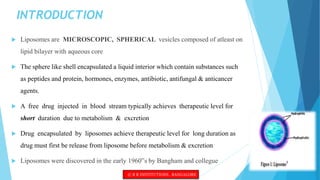
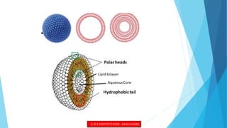
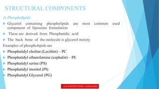
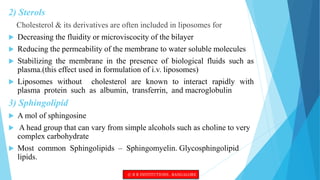
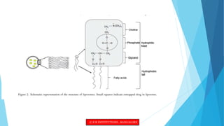
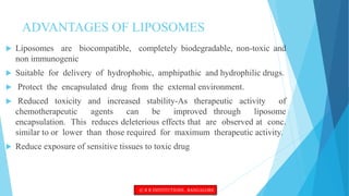
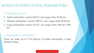
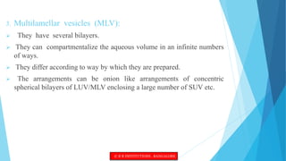
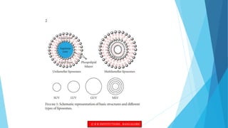
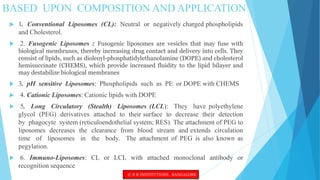
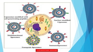
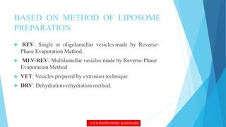
RR COLLEGE OF PHARMACY
OCCULAR LIPOSOMES
SUBMITTED BY: SUBMITTED TO:
SUPRITH D. Dr. A. GEETHA LAKSHMI
1ST SEM , M.PHARMACY PROF. & HOD
DEPARTMENT OF PHARMACEUTICS
© R R INSTITUTIONS , BANGALORE
CONTENTS
 INTRODUCTION
 STRUCTURAL COMPONENTS
 ADVANTAGES
 DIS-ADVANTAGES
 TYPES OF LIPOSOMES
 METHODS OF LIPOSOMES FORMATION
 MODES OF LIPOSOME ACTION
 APPLICATION OF LIPOSOMES IN OPTHALMIC DRUG DELIVERY
SYSTEM
 MECHANISM OF PERMIATION OF LIPOSOMES THROUGH
OCULAR SURFACE
 EVALUATION OF LIPOSOMES
 REFRENCE © R R INSTITUTIONS , BANGALORE
INTRODUCTION
 Liposomes are MICROSCOPIC, SPHERICAL vesicles composed of atleast on
lipid bilayer with aqueous core
 The sphere like shell encapsulated a liquid interior which contain substances such
as peptides and protein, hormones, enzymes, antibiotic, antifungal & anticancer
agents.
 A free drug injected in blood stream typically achieves therapeutic level for
short duration due to metabolism & excretion
 Drug encapsulated by liposomes achieve therapeutic level for long duration as
drug must first be release from liposome before metabolism & excretion
 Liposomes were discovered in the early 1960‟s by Bangham and collegue
© R R INSTITUTIONS , BANGALORE
STRUCTURAL COMPONENTS
1) Phospholipids
 Glycerol containing phospholipids are most common used
component of liposome formulation
 These are derived from Phosphatidic acid
 The back bone of the molecule is glycerol moiety
Examples of phospholipids are
 Phosphatidyl choline (Lecithin) – PC
 Phosphatidyl ethanolamine (cephalin) – PE
 Phosphatidyl serine (PS)
 Phosphatidyl inositol (PI)
 Phosphatidyl Glycerol (PG)
© R R INSTITUTIONS , BANGALORE
2) Sterols
Cholesterol & its derivatives are often included in liposomes for
 Decreasing the fluidity or microviscocity of the bilayer
 Reducing the permeability of the membrane to water soluble molecules
 Stabilizing the membrane in the presence of biological fluids such as
plasma.(this effect used in formulation of i.v. liposomes)
 Liposomes without cholesterol are known to interact rapidly with
plasma protein such as albumin, transferrin, and macroglobulin
3) Sphingolipid
 A mol of sphingosine
 A head group that can vary from simple alcohols such as choline to very
complex carbohydrate
 Most common Sphingolipids – Sphingomyelin. Glycosphingolipid
lipids.
© R R INSTITUTIONS , BANGALORE
ADVANTAGES OF LIPOSOMES
 Liposomes are biocompatible, completely biodegradable, non-toxic and
non immunogenic
 Suitable for delivery of hydrophobic, amphipathic and hydrophilic drugs.
 Protect the encapsulated drug from the external environment.
 Reduced toxicity and increased stability-As therapeutic activity of
chemotherapeutic agents can be improved through liposome
encapsulation. This reduces deleterious effects that are observed at conc.
similar to or lower than those required for maximum therapeutic activity.
 Reduce exposure of sensitive tissues to toxic drug
© R R INSTITUTIONS , BANGALORE
DISADVANTAGES OF LIPOSOMES
 Production cost is high
 Leakage and fusion of encapsulated drug/molecules
 Short half-life
 Sometimes phospholipid undergoes oxidation and hydrolysis-like
reaction
© R R INSTITUTIONS , BANGALORE
TYPES OF LIPOSOMES
Liposomes are classified on the basis of
A) BASED ON STRUCTURAL PARAMETERS
B) BASED UPON COMPOSITION AND APPLICATION
C) BASED ON METHOD OF LIPOSOME PREPARATION
© R R INSTITUTIONS , BANGALORE
BASED ON STRUCTURAL PARAMETERS
1. Unilamellar vesicles:
 Small unilamellar vesicles (SUV): size ranges from 20-40 nm
 Medium unilamellar vesicles (MUV}: size ranges from 40-80 nm.
 Large unilamellar vesicles (LUV): size ranges from 100 nm-1,000
nm
2. Oligolamellar vesicles (OLV):
These are made up of 2-10 bilayers of lipids surrounding a large
internal volume
© R R INSTITUTIONS , BANGALORE
3. Multilamellar vesicles (MLV):
 They have several bilayers.
 They can compartmentalize the aqueous volume in an infinite numbers
of ways.
 They differ according to way by which they are prepared.
 The arrangements can be onion like arrangements of concentric
spherical bilayers of LUV/MLV enclosing a large number of SUV etc.
© R R INSTITUTIONS , BANGALORE
BASED UPON COMPOSITION AND APPLICATION
 1. Conventional Liposomes (CL): Neutral or negatively charged phospholipids
and Cholesterol.
 2. Fusogenic Liposomes : Fusogenic liposomes are vesicles that may fuse with
biological membranes, thereby increasing drug contact and delivery into cells. They
consist of lipids, such as dioleoyl-phosphatidylethanolamine (DOPE) and cholesterol
hemisuccinate (CHEMS), which provide increased fluidity to the lipid bilayer and
may destabilize biological membranes
 3. pH sensitive Liposomes: Phospholipids such as PE or DOPE with CHEMS
 4. Cationic Liposomes: Cationic lipids with DOPE
 5. Long Circulatory (Stealth) Liposomes (LCL): They have polyethylene
glycol (PEG) derivatives attached to their surface to decrease their detection
by phagocyte system (reticuloendothelial system; RES). The attachment of PEG to
liposomes decreases the clearance from blood stream and extends circulation
time of liposomes in the body. The attachment of PEG is also known as
pegylation.
 6. Immuno-Liposomes: CL or LCL with attached monoclonal antibody or
recognition sequence
© R R INSTITUTIONS , BANGALORE
BASED ON METHOD OF LIPOSOME
PREPARATION
 REV: Single or oligolamellar vesicles made by Reverse-
Phase Evaporation Method.
 MLV-REV: Multilamellar vesicles made by Reverse-Phase
Evaporation Method
 VET: Vesicles prepared by extrusion technique
 DRV: Dehydration-rehydration method.
© R R INSTITUTIONS , BANGALORE
METHODS OF LIPOSOME FORMATION
 Convectional method
 Sonication method
 High-pressure extrusion method
 Solubilization and detergent removal method
 Reverse phase evaporation technique
© R R INSTITUTIONS , BANGALORE
Convectional method
 The phospholipids are dissolved in an organic solvent (usually a
chloroform/methanol mixture) and deposited from the solvents as a thin film on the
wall of a round bottom flask by use of rotary evaporation under reduced pressure.
MLVs form spontaneously when an excess volume of aqueous buffer containing
the drug is added to the dried lipid film. Drug containing liposomes can be
separated from non sequestered drug by centrifugation of the liposomes or by gel
filtration. The time allowed for hydration of the dried film and conditions of
agitation are critical in determining the amount of the aqueous buffer (or drug
solution) that will be entrapped within the internal compartments of the MLVs.
© R R INSTITUTIONS , BANGALORE
Sonication method
 This method is used in the preparation of SUVs and it involves the subsequent
sonication of MLVs prepared by the conventional method either with a bath type or
a probe type sonicator under an inert atmosphere, usually nitrogen or argon. The
principle of sonication involves the use of pulsed, high frequency sound waves
(sonic energy) to agitate a suspension of the MLVs. Such disruption of the MLVs
produces SUVs with diameter in the range of 15–50nm. The purpose of sonication,
therefore, is to produce a homogenous dispersion of small vesicles with a potential
for greater tissue penetration. The commonly used sonicators are of the bath and
probe tip type. The major drawbacks in the preparation of liposomes by sonication
include oxidation of unsaturated bonds in the fatty acid chains of phospholipids and
hydrolysis to lyso phospholipids and free fatty acids. Another drawback is the
denaturation or inactivation of some thermolabile substances (e.g., DNA, certain
proteins, etc.) to be entrapped © R R INSTITUTIONS , BANGALORE
High-pressure extrusion method
 This is another method for converting MLV to SUV suspensions. By this method,
suspensions of MLVs prepared by the convectional method are repeatedly passed
through filters polycarbonate membranes with very small pore diameter (0.8–
1.0μm) under high pressure up to 250psi. By choosing filters with appropriate pore
sizes, liposomes of desirable diameters can be produced. The mechanism of action
of the high pressure extrusion method appears to be much like peeling an onion. As
the MLVs are forced through the small pores, successive layers are peeled off until
only one remains. Besides reducing the liposome size, the extrusion method
produces liposomes of homogeneous size distributions. A variety of different lipids
can be used to form stable liposomes by this method. Extrusion at low pressures
© R R INSTITUTIONS , BANGALORE
Solubilization and detergent removal method
 This method is used in the preparation of LUVs and it involves the use of detergent
(surfactant) for the solubilization of the lipids. Detergents used include the non-
ionic surfactants [e.g., n-octyl-beta-D-glucopyranose (octyl gluside), anionic
surfactants (e.g., dodecyl sulphate) and cationic surfactants (e.g., hexadecyl
trimethyl ammonium bromide). The procedure involves the solubilization of the
lipids in an aqueous solution of the detergent and the protein(s) to be encapsulated.
The detergent should have a high critical micelle concentration (CMC), so that it is
easily removed. The detergent is subsequently removed by dialysis or column
chromatography. During detergent removal, LUVs of diameter 0.08–0.2μm are
produced. This detergent removal method has been found suitable for the
encapsulation of proteins of biomedical importance.
© R R INSTITUTIONS , BANGALORE
Reverse phase evaporation technique
 It consists of a rapid injection of aqueous solution of the drug into an organic solvent,
which contains the lipid dissolved with simultaneous bath sonication of the mixture
leading to the formation of water droplets in the organic solvent (i.e., a “water-in-oil”
emulsion). The resulting emulsion is dried down to a semi solid gel in a rotary evaporator.
The next step is to subject the gel to vigorous mechanical agitation to induce a phase
reversal from water-in-oil to oil-in-water dispersion (i.e., an aqueous suspension of the
vesicles). During the agitation, some of the water droplets collapse to form the external
phase while the remaining portion forms the entrapped aqueous volume. Large unilamellar
vesicles (diameter 0.1–1μm) are formed in the process. This method has been used to
encapsulate both small and macromolecules such as RNA and various enzymes without
loss of activity. The expected limitation of this method is the exposure of the material to
be encapsulated to organic solvents and mechanical agitation, which can lead to the
denaturation of some proteins or breakage of DNA strands.
Modes of liposome action
Liposomes as drug delivery systems can offer several advantages over
conventional dosage forms especially for parenteral (i.e. local or systemic
injection or infusion), topical, and pulmonary route of administration.
1. Passive targeting to the cells of the immune system, especially cells of the
mononuclear phagocytic system (in older literature reticuloendothelial
system). Examples are antimonials, Amphotericin B, porphyrins and also
vaccines, immunomodulators or (immuno)suppressor’s
2. Sustained release system of systemically or locally administered
liposomes. Examples are doxorubicin, cytosine arabinose, cortisones,
biological proteins or peptides such as vasopressin;
3. Site-avoidance mechanism: liposomes do not dispose in certain organs, such
as heart, kidneys, brain, and nervous system and this reduces cardio-,
nephro-, and neuro-toxicity. Typical examples are reduced nephrotoxicity of
Amphotericin B, and reduced cardiotoxicity of Doxorubicin liposomes;
© R R INSTITUTIONS , BANGALORE
4) Site specific targeting: in certain cases liposomes with surface attached ligands
can bind to target cells („key and lock‟ mechanism), or can be delivered into the
target tissue by local anatomical conditions such as leaky and badly formed blood
vessels, their basal lamina, and capillaries.
Examples include anticancer, antiinfection and anti-inflammatory drugs;
5) Improved transfer of hydrophilic, charged molecules such as chelators,
antibiotics, plasmids, and genes into cells
© R R INSTITUTIONS , BANGALORE
Application of Liposomes in Ophthalmic
Drug Delivery
 Liposomes have been investigated for ophthalmic drug delivery since it offers advantages as
a carrier system
 It is a biodegradable and biocompatible nanocarrier
 It can enhance the permeation of poorly absorbed drug molecules by binding to the corneal
surface and improving residence time
 It can encapsulate both the hydrophilic and hydrophobic drug molecules. In addition,
liposomes can improve pharmacokinetic profile, enhance therapeutic effect, and reduce
toxicity associated with higher dose
 Current approaches for the anterior segment drug delivery are focused on improving corneal
adhesion and permeation by incorporating various bioadhesive and penetration enhancing
polymers.
© R R INSTITUTIONS , BANGALORE
Mechanisms of permeation of liposomes
through ocular surface
 The mechanisms of interaction of liposomes with cell membranes that
result into intracellular drug delivery have been studied
(1) Adsorption:
Adsorption of liposomes to cell membrane is one of the
important mechanisms of intracellular drug delivery. The adsorbed
liposomes, in the presence of cell surface proteins, become leaky and
release their contents in the vicinity of cell membrane. This results in a
higher concentration of drug dose to cell membrane and facilitates
cellular uptake of drug by passive diffusion or transport
© R R INSTITUTIONS , BANGALORE
(2) Endocytosis:
Adsorption of liposomes on the surface of cell membrane is followed by
their engulfment and internalization into endosomes. Endosomes transport
liposomes to lysosomes. Subsequently, lysosomal enzymes degrade the lipids
and release the entrapped drug into the cytoplasm
(3) Fusion:
Fusion of lipid bilayer of liposomes with lipoidal cell membrane by
intermixing and lateral diffusion of lipids results in direct delivery of
liposomal contents into the cytoplasm
(4) Lipid exchange:
Due to the similarity of liposomal membrane lipids with the cell membrane
phospholipids, lipid transfer proteins in the cell membrane recognize
liposomes and consequently cause lipid exchange. This results in the
destabilization of liposomal membranes and intracellular release of drug
© R R INSTITUTIONS , BANGALORE
EVALUATION OF LIPOSOMES
Vesicle shape and lamellarity:
Vesicle shape can be assessed using Electron Microscopic
Techniques. Lamellarity of vesicles i.e. number of bilayers presents in liposomes is
determined using Freeze-Fracture Electron Microscopy and Nuclear Magnetic Resonance
Analysis
Optical Microscopy:
The microscopic method includes use of Bright-Field, Phase Contrast
Microscope and Fluorescent Microscope and is useful in evaluating vesicle size of large
vesicle.
Negative Stain TEM :
Electron Microscopic Techniques used to assess liposome shape and size are mainly
negative-stain TEM and Scanning Electron Microscopy. The latter technique is less
preferred. Negative Stain Electron Microscopy visualizes bright areas against dark
background (hence termed as negative stain) The negative stains used in TEM analysis are
ammonium molybdate or Phosphotungstic acid (PTA) or uranyl acetate. Both PTA and
ammonium molybdate are anionic in nature while uranyl acetate are cationic in nature
REFERENCES
 INTERNATIONAL RESEARCH JOURNAL OF PHARMACY www.irjponline.com
ISSN 2230 – 8407
 Taylor and Francis ISSN: 1071-7544 (Print) 1521-0464 (Online) Journal homepage:
https://www.tandfonline.com/loi/idrd20
 Hindawi Publishing Corporation Journal of Drug Delivery Volume 2011, Article ID
863734, 14 pages doi:10.1155/2011/863734
 Akbarzadeh et al. Nanoscale Research Letters 2013, 8:102
http://www.nanoscalereslett.com/content/8/1/102
 Abdelbary G. (2011). Ocular ciprofloxacin hydrochloride mucoadhesive chitosan-
coated liposomes. Pharm Dev Technol 16:44–56.
 Abdel-Rhaman MS, Soliman W, Habib F, Fathalla D. (2012). A new long-acting
liposomal topical antifungal formula: human clinical study. Cornea 31:126–9.
 Abdul Nasir NA, Agarwal R, Tripathy M, et al. (2013). Tocotrienol delays onset and
progression of galactose-induced cataract in rat. Abstracts of the 12th Meeting of the
Asia Pacific Federation of Pharmacologists. Shanghai, China, July 9–13, 2013. Acta
Pharmocol Sin 34:147
© R R INSTITUTIONS , BANGALORE

Liposomes.pptx

  • 1.
    RR COLLEGE OFPHARMACY OCCULAR LIPOSOMES SUBMITTED BY: SUBMITTED TO: SUPRITH D. Dr. A. GEETHA LAKSHMI 1ST SEM , M.PHARMACY PROF. & HOD DEPARTMENT OF PHARMACEUTICS © R R INSTITUTIONS , BANGALORE
  • 2.
    CONTENTS  INTRODUCTION  STRUCTURALCOMPONENTS  ADVANTAGES  DIS-ADVANTAGES  TYPES OF LIPOSOMES  METHODS OF LIPOSOMES FORMATION  MODES OF LIPOSOME ACTION  APPLICATION OF LIPOSOMES IN OPTHALMIC DRUG DELIVERY SYSTEM  MECHANISM OF PERMIATION OF LIPOSOMES THROUGH OCULAR SURFACE  EVALUATION OF LIPOSOMES  REFRENCE © R R INSTITUTIONS , BANGALORE
  • 3.
    INTRODUCTION  Liposomes areMICROSCOPIC, SPHERICAL vesicles composed of atleast on lipid bilayer with aqueous core  The sphere like shell encapsulated a liquid interior which contain substances such as peptides and protein, hormones, enzymes, antibiotic, antifungal & anticancer agents.  A free drug injected in blood stream typically achieves therapeutic level for short duration due to metabolism & excretion  Drug encapsulated by liposomes achieve therapeutic level for long duration as drug must first be release from liposome before metabolism & excretion  Liposomes were discovered in the early 1960‟s by Bangham and collegue © R R INSTITUTIONS , BANGALORE
  • 4.
    © R RINSTITUTIONS , BANGALORE
  • 5.
    STRUCTURAL COMPONENTS 1) Phospholipids Glycerol containing phospholipids are most common used component of liposome formulation  These are derived from Phosphatidic acid  The back bone of the molecule is glycerol moiety Examples of phospholipids are  Phosphatidyl choline (Lecithin) – PC  Phosphatidyl ethanolamine (cephalin) – PE  Phosphatidyl serine (PS)  Phosphatidyl inositol (PI)  Phosphatidyl Glycerol (PG) © R R INSTITUTIONS , BANGALORE
  • 6.
    2) Sterols Cholesterol &its derivatives are often included in liposomes for  Decreasing the fluidity or microviscocity of the bilayer  Reducing the permeability of the membrane to water soluble molecules  Stabilizing the membrane in the presence of biological fluids such as plasma.(this effect used in formulation of i.v. liposomes)  Liposomes without cholesterol are known to interact rapidly with plasma protein such as albumin, transferrin, and macroglobulin 3) Sphingolipid  A mol of sphingosine  A head group that can vary from simple alcohols such as choline to very complex carbohydrate  Most common Sphingolipids – Sphingomyelin. Glycosphingolipid lipids. © R R INSTITUTIONS , BANGALORE
  • 7.
    © R RINSTITUTIONS , BANGALORE
  • 8.
    ADVANTAGES OF LIPOSOMES Liposomes are biocompatible, completely biodegradable, non-toxic and non immunogenic  Suitable for delivery of hydrophobic, amphipathic and hydrophilic drugs.  Protect the encapsulated drug from the external environment.  Reduced toxicity and increased stability-As therapeutic activity of chemotherapeutic agents can be improved through liposome encapsulation. This reduces deleterious effects that are observed at conc. similar to or lower than those required for maximum therapeutic activity.  Reduce exposure of sensitive tissues to toxic drug © R R INSTITUTIONS , BANGALORE
  • 9.
    DISADVANTAGES OF LIPOSOMES Production cost is high  Leakage and fusion of encapsulated drug/molecules  Short half-life  Sometimes phospholipid undergoes oxidation and hydrolysis-like reaction © R R INSTITUTIONS , BANGALORE
  • 10.
    TYPES OF LIPOSOMES Liposomesare classified on the basis of A) BASED ON STRUCTURAL PARAMETERS B) BASED UPON COMPOSITION AND APPLICATION C) BASED ON METHOD OF LIPOSOME PREPARATION © R R INSTITUTIONS , BANGALORE
  • 11.
    BASED ON STRUCTURALPARAMETERS 1. Unilamellar vesicles:  Small unilamellar vesicles (SUV): size ranges from 20-40 nm  Medium unilamellar vesicles (MUV}: size ranges from 40-80 nm.  Large unilamellar vesicles (LUV): size ranges from 100 nm-1,000 nm 2. Oligolamellar vesicles (OLV): These are made up of 2-10 bilayers of lipids surrounding a large internal volume © R R INSTITUTIONS , BANGALORE
  • 12.
    3. Multilamellar vesicles(MLV):  They have several bilayers.  They can compartmentalize the aqueous volume in an infinite numbers of ways.  They differ according to way by which they are prepared.  The arrangements can be onion like arrangements of concentric spherical bilayers of LUV/MLV enclosing a large number of SUV etc. © R R INSTITUTIONS , BANGALORE
  • 13.
    © R RINSTITUTIONS , BANGALORE
  • 14.
    BASED UPON COMPOSITIONAND APPLICATION  1. Conventional Liposomes (CL): Neutral or negatively charged phospholipids and Cholesterol.  2. Fusogenic Liposomes : Fusogenic liposomes are vesicles that may fuse with biological membranes, thereby increasing drug contact and delivery into cells. They consist of lipids, such as dioleoyl-phosphatidylethanolamine (DOPE) and cholesterol hemisuccinate (CHEMS), which provide increased fluidity to the lipid bilayer and may destabilize biological membranes  3. pH sensitive Liposomes: Phospholipids such as PE or DOPE with CHEMS  4. Cationic Liposomes: Cationic lipids with DOPE  5. Long Circulatory (Stealth) Liposomes (LCL): They have polyethylene glycol (PEG) derivatives attached to their surface to decrease their detection by phagocyte system (reticuloendothelial system; RES). The attachment of PEG to liposomes decreases the clearance from blood stream and extends circulation time of liposomes in the body. The attachment of PEG is also known as pegylation.  6. Immuno-Liposomes: CL or LCL with attached monoclonal antibody or recognition sequence © R R INSTITUTIONS , BANGALORE
  • 15.
    © R RINSTITUTIONS , BANGALORE
  • 16.
    BASED ON METHODOF LIPOSOME PREPARATION  REV: Single or oligolamellar vesicles made by Reverse- Phase Evaporation Method.  MLV-REV: Multilamellar vesicles made by Reverse-Phase Evaporation Method  VET: Vesicles prepared by extrusion technique  DRV: Dehydration-rehydration method. © R R INSTITUTIONS , BANGALORE
  • 17.
    METHODS OF LIPOSOMEFORMATION  Convectional method  Sonication method  High-pressure extrusion method  Solubilization and detergent removal method  Reverse phase evaporation technique © R R INSTITUTIONS , BANGALORE
  • 18.
    Convectional method  Thephospholipids are dissolved in an organic solvent (usually a chloroform/methanol mixture) and deposited from the solvents as a thin film on the wall of a round bottom flask by use of rotary evaporation under reduced pressure. MLVs form spontaneously when an excess volume of aqueous buffer containing the drug is added to the dried lipid film. Drug containing liposomes can be separated from non sequestered drug by centrifugation of the liposomes or by gel filtration. The time allowed for hydration of the dried film and conditions of agitation are critical in determining the amount of the aqueous buffer (or drug solution) that will be entrapped within the internal compartments of the MLVs. © R R INSTITUTIONS , BANGALORE
  • 19.
    Sonication method  Thismethod is used in the preparation of SUVs and it involves the subsequent sonication of MLVs prepared by the conventional method either with a bath type or a probe type sonicator under an inert atmosphere, usually nitrogen or argon. The principle of sonication involves the use of pulsed, high frequency sound waves (sonic energy) to agitate a suspension of the MLVs. Such disruption of the MLVs produces SUVs with diameter in the range of 15–50nm. The purpose of sonication, therefore, is to produce a homogenous dispersion of small vesicles with a potential for greater tissue penetration. The commonly used sonicators are of the bath and probe tip type. The major drawbacks in the preparation of liposomes by sonication include oxidation of unsaturated bonds in the fatty acid chains of phospholipids and hydrolysis to lyso phospholipids and free fatty acids. Another drawback is the denaturation or inactivation of some thermolabile substances (e.g., DNA, certain proteins, etc.) to be entrapped © R R INSTITUTIONS , BANGALORE
  • 20.
    High-pressure extrusion method This is another method for converting MLV to SUV suspensions. By this method, suspensions of MLVs prepared by the convectional method are repeatedly passed through filters polycarbonate membranes with very small pore diameter (0.8– 1.0μm) under high pressure up to 250psi. By choosing filters with appropriate pore sizes, liposomes of desirable diameters can be produced. The mechanism of action of the high pressure extrusion method appears to be much like peeling an onion. As the MLVs are forced through the small pores, successive layers are peeled off until only one remains. Besides reducing the liposome size, the extrusion method produces liposomes of homogeneous size distributions. A variety of different lipids can be used to form stable liposomes by this method. Extrusion at low pressures © R R INSTITUTIONS , BANGALORE
  • 21.
    Solubilization and detergentremoval method  This method is used in the preparation of LUVs and it involves the use of detergent (surfactant) for the solubilization of the lipids. Detergents used include the non- ionic surfactants [e.g., n-octyl-beta-D-glucopyranose (octyl gluside), anionic surfactants (e.g., dodecyl sulphate) and cationic surfactants (e.g., hexadecyl trimethyl ammonium bromide). The procedure involves the solubilization of the lipids in an aqueous solution of the detergent and the protein(s) to be encapsulated. The detergent should have a high critical micelle concentration (CMC), so that it is easily removed. The detergent is subsequently removed by dialysis or column chromatography. During detergent removal, LUVs of diameter 0.08–0.2μm are produced. This detergent removal method has been found suitable for the encapsulation of proteins of biomedical importance. © R R INSTITUTIONS , BANGALORE
  • 22.
    Reverse phase evaporationtechnique  It consists of a rapid injection of aqueous solution of the drug into an organic solvent, which contains the lipid dissolved with simultaneous bath sonication of the mixture leading to the formation of water droplets in the organic solvent (i.e., a “water-in-oil” emulsion). The resulting emulsion is dried down to a semi solid gel in a rotary evaporator. The next step is to subject the gel to vigorous mechanical agitation to induce a phase reversal from water-in-oil to oil-in-water dispersion (i.e., an aqueous suspension of the vesicles). During the agitation, some of the water droplets collapse to form the external phase while the remaining portion forms the entrapped aqueous volume. Large unilamellar vesicles (diameter 0.1–1μm) are formed in the process. This method has been used to encapsulate both small and macromolecules such as RNA and various enzymes without loss of activity. The expected limitation of this method is the exposure of the material to be encapsulated to organic solvents and mechanical agitation, which can lead to the denaturation of some proteins or breakage of DNA strands.
  • 23.
    Modes of liposomeaction Liposomes as drug delivery systems can offer several advantages over conventional dosage forms especially for parenteral (i.e. local or systemic injection or infusion), topical, and pulmonary route of administration. 1. Passive targeting to the cells of the immune system, especially cells of the mononuclear phagocytic system (in older literature reticuloendothelial system). Examples are antimonials, Amphotericin B, porphyrins and also vaccines, immunomodulators or (immuno)suppressor’s 2. Sustained release system of systemically or locally administered liposomes. Examples are doxorubicin, cytosine arabinose, cortisones, biological proteins or peptides such as vasopressin; 3. Site-avoidance mechanism: liposomes do not dispose in certain organs, such as heart, kidneys, brain, and nervous system and this reduces cardio-, nephro-, and neuro-toxicity. Typical examples are reduced nephrotoxicity of Amphotericin B, and reduced cardiotoxicity of Doxorubicin liposomes; © R R INSTITUTIONS , BANGALORE
  • 24.
    4) Site specifictargeting: in certain cases liposomes with surface attached ligands can bind to target cells („key and lock‟ mechanism), or can be delivered into the target tissue by local anatomical conditions such as leaky and badly formed blood vessels, their basal lamina, and capillaries. Examples include anticancer, antiinfection and anti-inflammatory drugs; 5) Improved transfer of hydrophilic, charged molecules such as chelators, antibiotics, plasmids, and genes into cells © R R INSTITUTIONS , BANGALORE
  • 25.
    Application of Liposomesin Ophthalmic Drug Delivery  Liposomes have been investigated for ophthalmic drug delivery since it offers advantages as a carrier system  It is a biodegradable and biocompatible nanocarrier  It can enhance the permeation of poorly absorbed drug molecules by binding to the corneal surface and improving residence time  It can encapsulate both the hydrophilic and hydrophobic drug molecules. In addition, liposomes can improve pharmacokinetic profile, enhance therapeutic effect, and reduce toxicity associated with higher dose  Current approaches for the anterior segment drug delivery are focused on improving corneal adhesion and permeation by incorporating various bioadhesive and penetration enhancing polymers. © R R INSTITUTIONS , BANGALORE
  • 26.
    Mechanisms of permeationof liposomes through ocular surface  The mechanisms of interaction of liposomes with cell membranes that result into intracellular drug delivery have been studied (1) Adsorption: Adsorption of liposomes to cell membrane is one of the important mechanisms of intracellular drug delivery. The adsorbed liposomes, in the presence of cell surface proteins, become leaky and release their contents in the vicinity of cell membrane. This results in a higher concentration of drug dose to cell membrane and facilitates cellular uptake of drug by passive diffusion or transport © R R INSTITUTIONS , BANGALORE
  • 27.
    (2) Endocytosis: Adsorption ofliposomes on the surface of cell membrane is followed by their engulfment and internalization into endosomes. Endosomes transport liposomes to lysosomes. Subsequently, lysosomal enzymes degrade the lipids and release the entrapped drug into the cytoplasm (3) Fusion: Fusion of lipid bilayer of liposomes with lipoidal cell membrane by intermixing and lateral diffusion of lipids results in direct delivery of liposomal contents into the cytoplasm (4) Lipid exchange: Due to the similarity of liposomal membrane lipids with the cell membrane phospholipids, lipid transfer proteins in the cell membrane recognize liposomes and consequently cause lipid exchange. This results in the destabilization of liposomal membranes and intracellular release of drug © R R INSTITUTIONS , BANGALORE
  • 28.
    © R RINSTITUTIONS , BANGALORE
  • 29.
    EVALUATION OF LIPOSOMES Vesicleshape and lamellarity: Vesicle shape can be assessed using Electron Microscopic Techniques. Lamellarity of vesicles i.e. number of bilayers presents in liposomes is determined using Freeze-Fracture Electron Microscopy and Nuclear Magnetic Resonance Analysis Optical Microscopy: The microscopic method includes use of Bright-Field, Phase Contrast Microscope and Fluorescent Microscope and is useful in evaluating vesicle size of large vesicle. Negative Stain TEM : Electron Microscopic Techniques used to assess liposome shape and size are mainly negative-stain TEM and Scanning Electron Microscopy. The latter technique is less preferred. Negative Stain Electron Microscopy visualizes bright areas against dark background (hence termed as negative stain) The negative stains used in TEM analysis are ammonium molybdate or Phosphotungstic acid (PTA) or uranyl acetate. Both PTA and ammonium molybdate are anionic in nature while uranyl acetate are cationic in nature
  • 30.
    REFERENCES  INTERNATIONAL RESEARCHJOURNAL OF PHARMACY www.irjponline.com ISSN 2230 – 8407  Taylor and Francis ISSN: 1071-7544 (Print) 1521-0464 (Online) Journal homepage: https://www.tandfonline.com/loi/idrd20  Hindawi Publishing Corporation Journal of Drug Delivery Volume 2011, Article ID 863734, 14 pages doi:10.1155/2011/863734  Akbarzadeh et al. Nanoscale Research Letters 2013, 8:102 http://www.nanoscalereslett.com/content/8/1/102  Abdelbary G. (2011). Ocular ciprofloxacin hydrochloride mucoadhesive chitosan- coated liposomes. Pharm Dev Technol 16:44–56.  Abdel-Rhaman MS, Soliman W, Habib F, Fathalla D. (2012). A new long-acting liposomal topical antifungal formula: human clinical study. Cornea 31:126–9.  Abdul Nasir NA, Agarwal R, Tripathy M, et al. (2013). Tocotrienol delays onset and progression of galactose-induced cataract in rat. Abstracts of the 12th Meeting of the Asia Pacific Federation of Pharmacologists. Shanghai, China, July 9–13, 2013. Acta Pharmocol Sin 34:147 © R R INSTITUTIONS , BANGALORE
  • 31.
    © R RINSTITUTIONS , BANGALORE