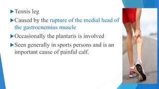
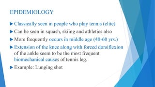
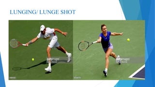
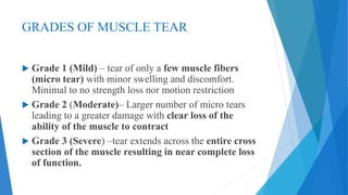
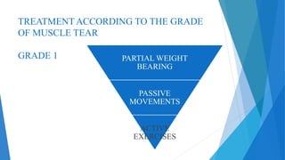
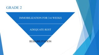
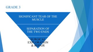
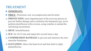
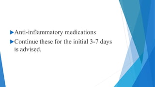
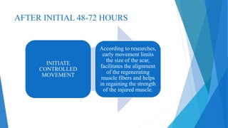
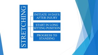
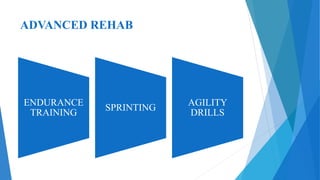
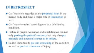
TENNIS LEG
(BPT 4th year – Sports physiotherapy topic)
PRESENTED BY:
Dr. LAKSHMI PAVANI P. (PT)
OBJECTIVES
TO KNOW:
1. Epidemiology
2. Clinical presentation
3. Causes
4. Radiographic features
5. Treatment according to the grade of injury
6. Rehabilitation
Tennis leg
Caused by the rupture of the medial head of
the gastrocnemius muscle
Occasionally the plantaris is involved
Seen generally in sports persons and is an
important cause of painful calf.
EPIDEMIOLOGY
 Classically seen in people who play tennis (elite)
 Can be seen in squash, skiing and athletics also
 More frequently occurs in middle age (40-60 yrs.)
 Extension of the knee along with forced dorsiflexion
of the ankle seem to be the most frequent
biomechanical causes of tennis leg.
 Example: Lunging shot
LUNGING/ LUNGE SHOT
CLINICAL PRESENTATION
 Audible pop when the injury occurred is usually
reported.
 Sensation of something “snapping” within the
calf.
 Sudden onset of severe calf pain with significant
disability.
 Tenderness and swelling present
 May be associated with extensive bruising
 A focal ‘gap’ can be felt sometimes on early
palpation, prior to the onset of swelling at the site
of tear
 Moderate to severe pain is present – passive
ankle dorsiflexion( due to stretching of the torn
muscle fibers)
 Active resistance to plantar flexion is painful (
due to the activity in torn muscle fibers)
CAUSES
Deconditioned / un-stretched muscles
of recreational athletes - “weekend
warriors”
Previous injuries/ recurrent calf strains
DD: DVT, ruptured baker’s cyst
GRADES OF MUSCLE TEAR
 Grade 1 (Mild) – tear of only a few muscle fibers
(micro tear) with minor swelling and discomfort.
Minimal to no strength loss nor motion restriction
 Grade 2 (Moderate)– Larger number of micro tears
leading to a greater damage with clear loss of the
ability of the muscle to contract
 Grade 3 (Severe) –tear extends across the entire cross
section of the muscle resulting in near complete loss
of function.
RADIOGRAPHIC FEATURES
 Ultrasound : A tear in the deep surface of
gastrocnemius may be seen as a disruption in contour
and echogenicity of muscle fibres.
 MRI : focal area of disruption of muscle continuity
noted along the deep aspect of the medial head of the
gastrocnemius, with associated edema of the muscle
 Plantaris tendon may be identified either torn or intact.
TREATMENT ACCORDING TO THE GRADE
OF MUSCLE TEAR
GRADE 1 PARTIAL WEIGHT
BEARING
PASSIVE
MOVEMENTS
ACTIVE
EXERCISES
GRADE 2
IMMOBILIZATION FOR 3-6 WEEKS
ADEQUATE REST
REHABILITATION
GRADE 3
SIGNIFICANT TEAR OF THE
MUSCLE
SEPARATION OF
THE TWO ENDS
SURGICAL
EXPLORATION
& REPAIR
TREATMENT
 IMMEDIATE:
 PRICE : Protection, rest, ice,compression and elevation
1) PROTECTION: most important part of the recovery process to
prevent further damage and to minimise the hematoma (eg: not to
perform dorsiflexion with eversion, using crutches to unload the
calf during locomotion)
2) REST: Immobilization
3) ICE: for 10-15 min and repeat this several times a day
4) COMPRESSION BANDAGE to prevent and minimize the intra
muscular hematoma
5) ELEVATION: Above the heart level and foot held in slight
plantarflexion.
Anti-inflammatory medications
Continue these for the initial 3-7 days
is advised.
AFTER INITIAL 48-72 HOURS
INITIATE
CONTROLLED
MOVEMENT
According to researches,
early movement limits
the size of the scar,
facilitates the alignment
of the regenerating
muscle fibers and helps
in regaining the strength
of the injured muscle.
STRETCHING
INITIATE 10 DAYS
AFTER INJURY
START IN LONG
SITTING POSITION
PROGRESS TO
STANDING
STRETCHING
STRENGTHENING EXERCISES
INTRINSIC FOOT MUSCLES: Eg:
GRASPING A TOWEL WITH TOES etc.,
SWIMMING & CYCLING- ENHANCES
BLOOD FLOW & HELPS IN RECOVERY
GRADUATED RETURN TO WEIGHT
BEARING
WALKING
STRENGTHENING
RUNNING
• BRISK WALKING
• EASY JOGS
• BILATERAL CALF RAISES
• UNILATERAL CALF RAISES
• ECCENTRIC LOWERING
EXERCISES
PRE REQUISITES TO RUNNING
Symmetrical flexibility of the calf muscle in
comparison to the non-involved side.
No residual signs of swelling/inflammation
Patient Should tolerate ADL’s Without
hindrances/issues.
ADVANCED REHAB
ENDURANCE
TRAINING
SPRINTING
AGILITY
DRILLS
IN RETROSPECT
 Calf muscle is regarded as the peripheral heart in the
human body and plays a major role in locomotion as
well.
 Calf muscle strains/ tennis leg can be a debilitating
condition.
 Failure in proper evaluation and rehabilitation can not
only prolong the patient’s recovery but may also pre
maturely end a person’s career as an athlete.
 So it is important to prevent worsening of the condition
as well as prevent recurrence of injury.
REFERENCES
 Clinical sports medicine, Peter Brukner and Karim Khan; Third edition, Tata Mc Graw
Hill.
 http://radsource.us/tennis-leg-plantaris-tendon-rupture/
 http://webcache.googleusercontent.com/search?q=cache:3AW3e16wtr0J:www.chsm.net
/tennisleg.html+&cd=12&hl=en&ct=clnk&gl=in
 http://www.sportsmedicineofatlanta.com/reference/tennis_leg_common_sports_injury.h
tml
Tennis leg

Tennis leg

  • 1.
    TENNIS LEG (BPT 4thyear – Sports physiotherapy topic) PRESENTED BY: Dr. LAKSHMI PAVANI P. (PT)
  • 2.
    OBJECTIVES TO KNOW: 1. Epidemiology 2.Clinical presentation 3. Causes 4. Radiographic features 5. Treatment according to the grade of injury 6. Rehabilitation
  • 3.
    Tennis leg Caused bythe rupture of the medial head of the gastrocnemius muscle Occasionally the plantaris is involved Seen generally in sports persons and is an important cause of painful calf.
  • 4.
    EPIDEMIOLOGY  Classically seenin people who play tennis (elite)  Can be seen in squash, skiing and athletics also  More frequently occurs in middle age (40-60 yrs.)  Extension of the knee along with forced dorsiflexion of the ankle seem to be the most frequent biomechanical causes of tennis leg.  Example: Lunging shot
  • 5.
  • 6.
    CLINICAL PRESENTATION  Audiblepop when the injury occurred is usually reported.  Sensation of something “snapping” within the calf.  Sudden onset of severe calf pain with significant disability.  Tenderness and swelling present  May be associated with extensive bruising
  • 7.
     A focal‘gap’ can be felt sometimes on early palpation, prior to the onset of swelling at the site of tear  Moderate to severe pain is present – passive ankle dorsiflexion( due to stretching of the torn muscle fibers)  Active resistance to plantar flexion is painful ( due to the activity in torn muscle fibers)
  • 8.
    CAUSES Deconditioned / un-stretchedmuscles of recreational athletes - “weekend warriors” Previous injuries/ recurrent calf strains DD: DVT, ruptured baker’s cyst
  • 9.
    GRADES OF MUSCLETEAR  Grade 1 (Mild) – tear of only a few muscle fibers (micro tear) with minor swelling and discomfort. Minimal to no strength loss nor motion restriction  Grade 2 (Moderate)– Larger number of micro tears leading to a greater damage with clear loss of the ability of the muscle to contract  Grade 3 (Severe) –tear extends across the entire cross section of the muscle resulting in near complete loss of function.
  • 10.
    RADIOGRAPHIC FEATURES  Ultrasound: A tear in the deep surface of gastrocnemius may be seen as a disruption in contour and echogenicity of muscle fibres.  MRI : focal area of disruption of muscle continuity noted along the deep aspect of the medial head of the gastrocnemius, with associated edema of the muscle  Plantaris tendon may be identified either torn or intact.
  • 11.
    TREATMENT ACCORDING TOTHE GRADE OF MUSCLE TEAR GRADE 1 PARTIAL WEIGHT BEARING PASSIVE MOVEMENTS ACTIVE EXERCISES
  • 12.
    GRADE 2 IMMOBILIZATION FOR3-6 WEEKS ADEQUATE REST REHABILITATION
  • 13.
    GRADE 3 SIGNIFICANT TEAROF THE MUSCLE SEPARATION OF THE TWO ENDS SURGICAL EXPLORATION & REPAIR
  • 14.
    TREATMENT  IMMEDIATE:  PRICE: Protection, rest, ice,compression and elevation 1) PROTECTION: most important part of the recovery process to prevent further damage and to minimise the hematoma (eg: not to perform dorsiflexion with eversion, using crutches to unload the calf during locomotion) 2) REST: Immobilization 3) ICE: for 10-15 min and repeat this several times a day 4) COMPRESSION BANDAGE to prevent and minimize the intra muscular hematoma 5) ELEVATION: Above the heart level and foot held in slight plantarflexion.
  • 15.
    Anti-inflammatory medications Continue thesefor the initial 3-7 days is advised.
  • 16.
    AFTER INITIAL 48-72HOURS INITIATE CONTROLLED MOVEMENT According to researches, early movement limits the size of the scar, facilitates the alignment of the regenerating muscle fibers and helps in regaining the strength of the injured muscle.
  • 17.
    STRETCHING INITIATE 10 DAYS AFTERINJURY START IN LONG SITTING POSITION PROGRESS TO STANDING
  • 18.
  • 19.
    STRENGTHENING EXERCISES INTRINSIC FOOTMUSCLES: Eg: GRASPING A TOWEL WITH TOES etc., SWIMMING & CYCLING- ENHANCES BLOOD FLOW & HELPS IN RECOVERY GRADUATED RETURN TO WEIGHT BEARING
  • 20.
    WALKING STRENGTHENING RUNNING • BRISK WALKING •EASY JOGS • BILATERAL CALF RAISES • UNILATERAL CALF RAISES • ECCENTRIC LOWERING EXERCISES
  • 21.
    PRE REQUISITES TORUNNING Symmetrical flexibility of the calf muscle in comparison to the non-involved side. No residual signs of swelling/inflammation Patient Should tolerate ADL’s Without hindrances/issues.
  • 22.
  • 23.
    IN RETROSPECT  Calfmuscle is regarded as the peripheral heart in the human body and plays a major role in locomotion as well.  Calf muscle strains/ tennis leg can be a debilitating condition.  Failure in proper evaluation and rehabilitation can not only prolong the patient’s recovery but may also pre maturely end a person’s career as an athlete.  So it is important to prevent worsening of the condition as well as prevent recurrence of injury.
  • 24.
    REFERENCES  Clinical sportsmedicine, Peter Brukner and Karim Khan; Third edition, Tata Mc Graw Hill.  http://radsource.us/tennis-leg-plantaris-tendon-rupture/  http://webcache.googleusercontent.com/search?q=cache:3AW3e16wtr0J:www.chsm.net /tennisleg.html+&cd=12&hl=en&ct=clnk&gl=in  http://www.sportsmedicineofatlanta.com/reference/tennis_leg_common_sports_injury.h tml