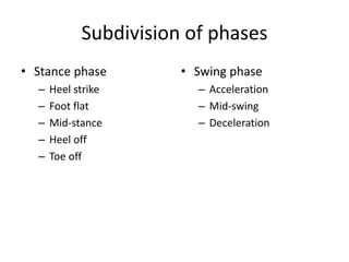
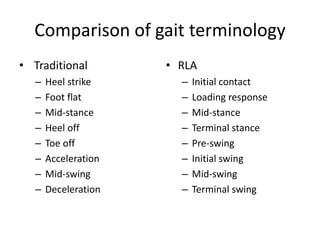
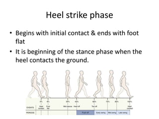
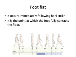
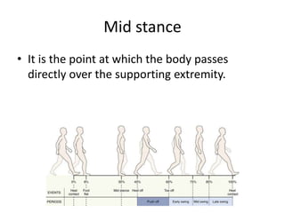
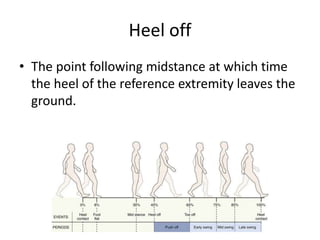
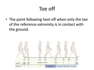
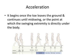
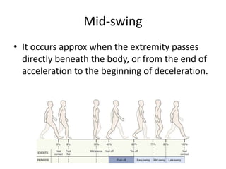
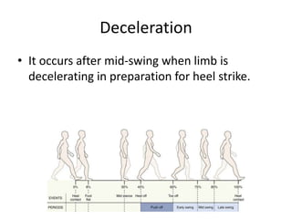
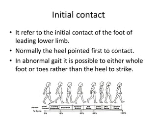
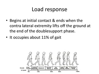
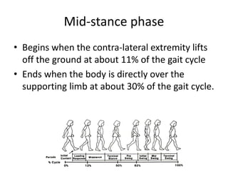
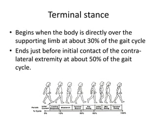
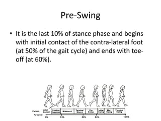
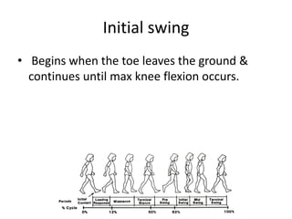
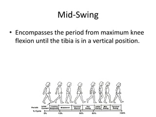
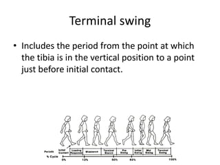
Gait Cycle
Dr. Sairamakrishnan S
Defenition
• Locomotion or gait
– It is defined as a translatory progression of the
body as a whole produce by coordinated, rotatory
movements of body segments.
• Normal gait
– It is a rhythmic & characterized by alternating
propulsive & retropulsive motions of the lower
extremities.
Task involved in walking
• According to “Rancho Los Amigos” (RLA),
California
– Weight acceptance
– Single limb support
– Swing limb advance
Gait initiation
• A series of events occur from the initiation of
body movement to beginning of gait cycle.
• It is stereotyped activity in all people.
• Total duration of this phase is about – 0.60sec
Phases of gait
• Stance phase
• Swing phase
Stance phase
• It begins at the instant that one extremity
contacts the ground & continues as long as
some portion of the foot is in contact with the
ground.
• It is approx 60% of normal gait duration
Swing phase
• It begins as soon as the toe of one extremity
leaves the ground & ceases just before heel
strike or contact of the same extremity.
• It makes up 40% of normal gait cycle.
Double support
• Lower limb of one side of body is beginning
its stance phase & the opposite side is ending
its stance phase.
• During double support both the lower limb
are in contact with the ground at the same
time.
• It account approx 22% of gait cycle.
• This phase is absent in running
Subdivision of phases
• Stance phase
– Heel strike
– Foot flat
– Mid-stance
– Heel off
– Toe off
• Swing phase
– Acceleration
– Mid-swing
– Deceleration
Comparison of gait terminology
• Traditional
– Heel strike
– Foot flat
– Mid-stance
– Heel off
– Toe off
– Acceleration
– Mid-swing
– Deceleration
• RLA
– Initial contact
– Loading response
– Mid-stance
– Terminal stance
– Pre-swing
– Initial swing
– Mid-swing
– Terminal swing
TRADITIONAL PHASES OF GAIT
Heel strike phase
• Begins with initial contact & ends with foot
flat
• It is beginning of the stance phase when the
heel contacts the ground.
Foot flat
• It occurs immediately following heel strike
• It is the point at which the foot fully contacts
the floor.
Mid stance
• It is the point at which the body passes
directly over the supporting extremity.
Heel off
• The point following midstance at which time
the heel of the reference extremity leaves the
ground.
Toe off
• The point following heel off when only the toe
of the reference extremity is in contact with
the ground.
Acceleration
• It begins once the toe leaves the ground &
continues until midswing, or the point at
which the swinging extremity is directly under
the body.
Mid-swing
• It occurs approx when the extremity passes
directly beneath the body, or from the end of
acceleration to the beginning of deceleration.
Deceleration
• It occurs after mid-swing when limb is
decelerating in preparation for heel strike.
RLA PHASES OF GAIT
Initial contact
• It refer to the initial contact of the foot of
leading lower limb.
• Normally the heel pointed first to contact.
• In abnormal gait it is possible to either whole
foot or toes rather than the heel to strike.
Load response
• Begins at initial contact & ends when the
contra lateral extremity lifts off the ground at
the end of the doublesupport phase.
• It occupies about 11% of gait
Mid-stance phase
• Begins when the contra-lateral extremity lifts
off the ground at about 11% of the gait cycle
• Ends when the body is directly over the
supporting limb at about 30% of the gait cycle.
Terminal stance
• Begins when the body is directly over the
supporting limb at about 30% of the gait cycle
• Ends just before initial contact of the contra-
lateral extremity at about 50% of the gait
cycle.
Pre-Swing
• It is the last 10% of stance phase and begins
with initial contact of the contra-lateral foot
(at 50% of the gait cycle) and ends with toe-
off (at 60%).
Initial swing
• Begins when the toe leaves the ground &
continues until max knee flexion occurs.
Mid-Swing
• Encompasses the period from maximum knee
flexion until the tibia is in a vertical position.
Terminal swing
• Includes the period from the point at which
the tibia is in the vertical position to a point
just before initial contact.
Variables of gait
• There are two basic variables which provide a
basic description of human gait.
– Time/ Temporal variable
– Distance variables
• Provide essential quantitative information
about gait
Factors affecting variables
• Age
• Gender
• Height
• Size & shape of bony
components
• Distribution of mass in
body segments
• Joint mobility
• Muscle strength
• Type of clothing &
footwear
• Habit
• Psychological status
Variables
• Temporal variable
– Stance time
– Single-limb & double
support time
– Swing time
– Stride and step time
– Cadence
– Speed
• Distance variable
– Stride length
– Step length and
width
– Degree of toe-out
Stance time
• It is the amount of time that elapses during
the stance phase of one extremity in a gait
cycle.
Single-support time
• It is the amount of time that elapses during
the period when only one extremity is on the
supporting surface in a gait cycle
Double-support time
• It is the amount of time spent with both feet
on the ground during one gait cycle.
• The % of time spent increased in elderly
persons and in those with balance disorders.
• The percentage of time spent decreases as the
speed of walking increases
Stride length
• It is the linear distance from the heel strike of
one lower limb to the next heel strike of the
same limb.
Step length
• It is the linear distance from the heel strike of
one lower limb to the next heel strike of
opposite limb.
Stride duration
• It refers to amount of time taken to
accomplish one stride.
• Stride duration and gait cycle duration are
synonymous.
• One stride, for a normal adult, lasts approx 1
sec
Step duration
• It refers to the amount of time spent during a
single step.
• Measurement usually is expressed as sec/step.
• When weakness or pain in limb, step duration
may be decreased on the affected side and
increased on the unaffected side
Cadence
• It is the no of steps taken by a person per unit
of time.
• It is measured as the no of steps / sec or per
minute.
• Cadence = Number of steps / Time
Walking velocity
• It is the rate of linear forward motion of the
body, which can be measured in meters or
cm/second, meters/minute, or miles/hour.
Speed of gait
• It is referred to as slow, free, and fast.
• Free speed of gait refers to a person‟s normal
walking speed
• Slow & fast speeds of gait refer to speeds
slower or faster than the person’s normal
comfortable walking speed.
Step width
• It is the measure of linear distance between
the midpoint of the heel of one foot and the
same point on the other foot
Degree of toe-out
• It represents the angle of foot formed by each
foot’s line of progression and a line
intersecting the centre of the heel and the
second toe.
• The normal angle is about 7 degree from the
line of progression of each foot at free speed
walking.
• The Degree of toe-out decreases as the speed
of walking increases.
Path of Center of Gravity
• Center of Gravity:
– Midway between the hips
– Few cm in front of S2
• Least energy consumption if Center of Gravity
travels in a straight line
Vertical displacement
• Rhythmic up & down
movement
• Highest point: midstance
• Lowest point: double
support
• Average displacement:
5cm
• Path: extremely smooth
sinusoidal curve
Lateral displacement
• Rhythmic side-to-side movement
• Lateral limit: mid-stance
• Average displacement: 5cm
• Path: extremely smooth sinusoidal curve
Overall displacement
• Sum of vertical & horizontal displacement
• Figure ‘8’ movement of CG as seen from AP
view
Saunders’ Determinants of gait
• Gait “determinants” was first described by
“Saunders & Coworkers” in 1953.
• Six optimizations used to minimize excursion
of CG in vertical & horizontal planes.
• The “determinants” represent adjustments
made by the pelvis, hips, knees, and ankles
that help to keep movement of the body’s
Center of Gravity to a minimum.
• By decreasing the vertical & lateral excursions
of the body’s Center of Gravity it was thought
that energy expenditure would be less & gait
be more efficient.
Pelvic rotation
• Forward rotation of the pelvis in the
horizontal plane is approx. 8o on the swing-
phase side.
• It reduces the angle of hip flexion & extension
• It enables a slightly longer step-length
Pelvic tilt
• 5 degree dip of the swinging side
• It reduces the height of the apex of the curve
of Center of Gravity
• If this occurs in standing it is a +ve
Trendelenberg test
Knee flexion
• Approx. 20 degree
• It shortens the leg in the middle of stance
phase
• It reduces the height of the apex of Center of
Gravity curve
Ankle mechanism
• It lengthens the leg at heel contact
• It helps in smoothening the curve of Center of
Gravity
• It reduces the lowering of Center of Gravity
Foot mechanism
• Lengthens the leg at toe-off as ankle moves
from dorsiflexion to plantarflexion
• It helps in smoothening the curve of Center of
Gravity
• It reduces the lowering of Center of Gravity
Physiological knee valgus
• Physiologic valgus of the knee reduce side-
toside movement of the COM in frontal plane.
• The normally narrow width of the walking
base minimizes the lateral displacement of CG
• Reduced muscular energy consumption due to
reduced lateral acceleration & deceleration
ABNORMAL (PATHOLOGICAL) GAIT
Antalgic gait
• This is a compensatory gait pattern adopted in
order to remove or diminish the discomfort
caused by pain in the LL or pelvis.
• Characteristic features
– Decreased in duration of stance phase of the affected
limb
– There is a lack of weight shift laterally over the stance
limb and also to keep weight off the involved limb
– Decrease in stance phase in affected side will result in
a decrease in swing phase of sound limb.
Psoatic gait
• Psoas bursa may be inflamed & edematous,
which cause limitation of movement due to
pain & produce a atypical gait.
– Hip externally rotated
– Hip adducted
– Knee in slight flexion
• This process seems to relieve tension of the
muscle & hence relieve the inflamed
structures.
Gluteus maximus gait
• The gluteus maximus act as a restraint for
forward progression.
• The trunk quickly shifts posteriorly at heel strike
(initial contact).
• This will shift the body’s COG posteriorly over the
gluteus maximus, moving the line of force
posterior to the hip joints.
• This shifting is referred to as a “Rocking Horse
Gait” because of the extreme backward-forward
movement of the trunk.
Gluteus medius gait
• It is also known as “Trendelenberg gait” or
“Lurching Gait” when one side affected.
• The individual shifts the trunk over the affected
side during stance phase.
• When right gluteus medius or hip abductor is
weak it causes two thing
– The body leans over the left leg during stance phase
of the left leg
– Right side of the pelvis will drop when the right leg
leaves the ground & begins swing phase.
• Shifting the trunk over the affected side is an
attempt to reduce the amount of strength
required of the gluteus medius to stabilize the
pelvis.
• Bilateral paralysis, waddling or duck gait.
• The patient lurch to both sides while walking.
• The body sways from side to side on a wide
base with excessive shoulder swing
Quadriceps gait
• Quadriceps action is needed during heel
strike & foot flat when there is a flexion force
acting at the knee.
• Quadriceps weakness/ paralysis will lead to
buckling of the knee during gait & thus loss of
balance.
• Patient can compensate this if he has normal
hip extensor & plantar flexors
• Compensation:
– With quadriceps weakness, the individual may lean
forward over the quadriceps at the early part of
stance phase, as weight is being shifted on to the
stance leg.
– Normally, the line of force falls behind the knee,
requiring quadriceps action to keep the knee from
buckling.
– By leaning forward at the hip, the COG is shifted
forward & the line of force now falls in front.
– This will force the knee backward into extension
• Another compensatory manoeuvre to use is
the hip extensors & ankle plantar flexors in a
closed chain action to pull the knee into
extension at heel strike (initial contact).
• In addition, the person may physically push on
the anterior thigh during stance phase,
holding the knee in extension.
Hemiplegic gait
• With spastic pattern of hemiplegic leg
– Hip into extension, adduction & medial rotation
– Knee in extension, though often unstable
– Ankle in drop foot with ankle plantar flexion and
inversion (equinovarus), which is present during
both stance and swing phases.
• In order to clear the foot from the ground the
hip & knee should flex
• But the spastic muscles won‟t allow the hip &
knee to flex for the floor clearance.
• So the patient hikes hip & bring the affected leg
by making a half circle i.e. circumducting the leg.
• Hence the gait is known as “Circumductory Gait”.
• Usually, there will be no reciprocal arm swing.
• Step length tends to be lengthened on the
involved side & shortened on the uninvolved side
Scissoring gait
• It results from spasticity of bilateral adductor
muscle of hip.
• One leg crosses directly over the other with
each step like crossing the blades of a scissor.
Cerebellar ataxic gait
• Abnormal function of cerebellum result in a
disturbance of normal mechanism controlling
balance & therefore patient walks with wider
BOS.
• The wider Step width creates a larger side to
side deviation of COG.
• This result in irregularly swinging sideways to
a tendency to fall with each steps.
• Hence it is known as “Drunkard’s Gait”.
Sensory ataxic gait
• This is a typical gait pattern seen in patients
affected by tabes dorsalis.
• It is a degenerative disease affecting the
posterior horn cells & posterior column of the
spinal cord.
• Because of lesion, the proprioceptive impulse
won‟t reach the cerebellum.
• The patient will loss his joint sense & position
for his limb on space.
• Because of loss of joint sense, the patient
abnormally raises his leg (high step) jerks it
forward to strike the ground with a stamp.
• So it is also called as “Stamping Gait”.
• The patient compensated this loss of joint
position sense by vision.
• So his head will be down while he is walking
Short shuffling or festinate gait
• Normal function at basal ganglia are
– Control of muscle tone
– Planning & programming of normal movements.
– Control of associated movements like reciprocal
arm swing.
• Typical example for basal ganglia leision is
parkinsonism.
• Because of rigidity, all the joint will go for a
flexion position with spine stooping forward.
• This posture displaces the COG anteriorly.
• So in order to keep the COG within the BOS,
the patient will no of small shuffling steps.
• Due to loss of voluntary control over the
movement, they loses balance & walks faster
as if he is chasing the COG.
• So it is called as “Festinate Gait”.
Foot drop gait
• This is due to dorsiflexor weakness caused by
paralysis of common peroneal nerve.
• There won‟t be normal heel strike, instead the
foot comes in contact with ground as a whole
with a slapping sound.
• So it is also known as “Slapping gait”.
• Due to plantarflexion of the ankle, there will
be relatively lengthening at the leading
extremity.
• So to clear the ground the patient lift the limb
too high.
• Hence the gait get s its another name i.e.
“High Stepping Gait”
Equinus gait
• Equinus = Horse
• Because of paralysis of dorsiflexor which result
in plantar flexor contracture.
• The patients will walk on his toes (toe
walking).
• Other cause may be compensation by plantar
flexor for a short leg.
Minimal LLD
• Compensation occurs by dropping the pelvis
on the affected side.
• The person may compensate by leaning over
shorter leg (up to 3 inches).
Moderate LLD
• Approx between 3 & 5 inches, dropping the
pelvis on the affected side will no longer be
effective.
• A longer leg is needed, so the person usually
walks on the toe of the foot on the involved
(shorter) side.
• This is called an “Equinnus Gait”.
Severe LLD
• It is usually discrepancy of more than 5 inches.
• The person may compensate in a variety of
ways.
• Dropping the pelvis and walking in an
equinnus gait plus flexing the knee on the
uninvolved side is often used.
• To gain an appreciation for how this may feel
or look, walk down the street with one leg in
the street and the other on the sidewalk.
Equinovarous gait
• There will be ankle plantar flexion & subtalar
inversion.
• So the patient will be walking on the outer
border of the foot.
• E.g. CETV

Gait cycle

  • 1.
  • 2.
    Defenition • Locomotion orgait – It is defined as a translatory progression of the body as a whole produce by coordinated, rotatory movements of body segments. • Normal gait – It is a rhythmic & characterized by alternating propulsive & retropulsive motions of the lower extremities.
  • 3.
    Task involved inwalking • According to “Rancho Los Amigos” (RLA), California – Weight acceptance – Single limb support – Swing limb advance
  • 4.
    Gait initiation • Aseries of events occur from the initiation of body movement to beginning of gait cycle. • It is stereotyped activity in all people. • Total duration of this phase is about – 0.60sec
  • 5.
    Phases of gait •Stance phase • Swing phase
  • 6.
    Stance phase • Itbegins at the instant that one extremity contacts the ground & continues as long as some portion of the foot is in contact with the ground. • It is approx 60% of normal gait duration
  • 7.
    Swing phase • Itbegins as soon as the toe of one extremity leaves the ground & ceases just before heel strike or contact of the same extremity. • It makes up 40% of normal gait cycle.
  • 8.
    Double support • Lowerlimb of one side of body is beginning its stance phase & the opposite side is ending its stance phase. • During double support both the lower limb are in contact with the ground at the same time. • It account approx 22% of gait cycle. • This phase is absent in running
  • 9.
    Subdivision of phases •Stance phase – Heel strike – Foot flat – Mid-stance – Heel off – Toe off • Swing phase – Acceleration – Mid-swing – Deceleration
  • 10.
    Comparison of gaitterminology • Traditional – Heel strike – Foot flat – Mid-stance – Heel off – Toe off – Acceleration – Mid-swing – Deceleration • RLA – Initial contact – Loading response – Mid-stance – Terminal stance – Pre-swing – Initial swing – Mid-swing – Terminal swing
  • 11.
  • 12.
    Heel strike phase •Begins with initial contact & ends with foot flat • It is beginning of the stance phase when the heel contacts the ground.
  • 13.
    Foot flat • Itoccurs immediately following heel strike • It is the point at which the foot fully contacts the floor.
  • 14.
    Mid stance • Itis the point at which the body passes directly over the supporting extremity.
  • 15.
    Heel off • Thepoint following midstance at which time the heel of the reference extremity leaves the ground.
  • 16.
    Toe off • Thepoint following heel off when only the toe of the reference extremity is in contact with the ground.
  • 17.
    Acceleration • It beginsonce the toe leaves the ground & continues until midswing, or the point at which the swinging extremity is directly under the body.
  • 18.
    Mid-swing • It occursapprox when the extremity passes directly beneath the body, or from the end of acceleration to the beginning of deceleration.
  • 19.
    Deceleration • It occursafter mid-swing when limb is decelerating in preparation for heel strike.
  • 20.
  • 21.
    Initial contact • Itrefer to the initial contact of the foot of leading lower limb. • Normally the heel pointed first to contact. • In abnormal gait it is possible to either whole foot or toes rather than the heel to strike.
  • 22.
    Load response • Beginsat initial contact & ends when the contra lateral extremity lifts off the ground at the end of the doublesupport phase. • It occupies about 11% of gait
  • 23.
    Mid-stance phase • Beginswhen the contra-lateral extremity lifts off the ground at about 11% of the gait cycle • Ends when the body is directly over the supporting limb at about 30% of the gait cycle.
  • 24.
    Terminal stance • Beginswhen the body is directly over the supporting limb at about 30% of the gait cycle • Ends just before initial contact of the contra- lateral extremity at about 50% of the gait cycle.
  • 25.
    Pre-Swing • It isthe last 10% of stance phase and begins with initial contact of the contra-lateral foot (at 50% of the gait cycle) and ends with toe- off (at 60%).
  • 26.
    Initial swing • Beginswhen the toe leaves the ground & continues until max knee flexion occurs.
  • 27.
    Mid-Swing • Encompasses theperiod from maximum knee flexion until the tibia is in a vertical position.
  • 28.
    Terminal swing • Includesthe period from the point at which the tibia is in the vertical position to a point just before initial contact.
  • 29.
    Variables of gait •There are two basic variables which provide a basic description of human gait. – Time/ Temporal variable – Distance variables • Provide essential quantitative information about gait
  • 30.
    Factors affecting variables •Age • Gender • Height • Size & shape of bony components • Distribution of mass in body segments • Joint mobility • Muscle strength • Type of clothing & footwear • Habit • Psychological status
  • 31.
    Variables • Temporal variable –Stance time – Single-limb & double support time – Swing time – Stride and step time – Cadence – Speed • Distance variable – Stride length – Step length and width – Degree of toe-out
  • 32.
    Stance time • Itis the amount of time that elapses during the stance phase of one extremity in a gait cycle.
  • 33.
    Single-support time • Itis the amount of time that elapses during the period when only one extremity is on the supporting surface in a gait cycle
  • 34.
    Double-support time • Itis the amount of time spent with both feet on the ground during one gait cycle. • The % of time spent increased in elderly persons and in those with balance disorders. • The percentage of time spent decreases as the speed of walking increases
  • 35.
    Stride length • Itis the linear distance from the heel strike of one lower limb to the next heel strike of the same limb.
  • 36.
    Step length • Itis the linear distance from the heel strike of one lower limb to the next heel strike of opposite limb.
  • 37.
    Stride duration • Itrefers to amount of time taken to accomplish one stride. • Stride duration and gait cycle duration are synonymous. • One stride, for a normal adult, lasts approx 1 sec
  • 38.
    Step duration • Itrefers to the amount of time spent during a single step. • Measurement usually is expressed as sec/step. • When weakness or pain in limb, step duration may be decreased on the affected side and increased on the unaffected side
  • 39.
    Cadence • It isthe no of steps taken by a person per unit of time. • It is measured as the no of steps / sec or per minute. • Cadence = Number of steps / Time
  • 40.
    Walking velocity • Itis the rate of linear forward motion of the body, which can be measured in meters or cm/second, meters/minute, or miles/hour.
  • 41.
    Speed of gait •It is referred to as slow, free, and fast. • Free speed of gait refers to a person‟s normal walking speed • Slow & fast speeds of gait refer to speeds slower or faster than the person’s normal comfortable walking speed.
  • 42.
    Step width • Itis the measure of linear distance between the midpoint of the heel of one foot and the same point on the other foot
  • 43.
    Degree of toe-out •It represents the angle of foot formed by each foot’s line of progression and a line intersecting the centre of the heel and the second toe.
  • 44.
    • The normalangle is about 7 degree from the line of progression of each foot at free speed walking. • The Degree of toe-out decreases as the speed of walking increases.
  • 45.
    Path of Centerof Gravity • Center of Gravity: – Midway between the hips – Few cm in front of S2 • Least energy consumption if Center of Gravity travels in a straight line
  • 46.
    Vertical displacement • Rhythmicup & down movement • Highest point: midstance • Lowest point: double support • Average displacement: 5cm • Path: extremely smooth sinusoidal curve
  • 47.
    Lateral displacement • Rhythmicside-to-side movement • Lateral limit: mid-stance • Average displacement: 5cm • Path: extremely smooth sinusoidal curve
  • 48.
    Overall displacement • Sumof vertical & horizontal displacement • Figure ‘8’ movement of CG as seen from AP view
  • 49.
    Saunders’ Determinants ofgait • Gait “determinants” was first described by “Saunders & Coworkers” in 1953. • Six optimizations used to minimize excursion of CG in vertical & horizontal planes.
  • 50.
    • The “determinants”represent adjustments made by the pelvis, hips, knees, and ankles that help to keep movement of the body’s Center of Gravity to a minimum. • By decreasing the vertical & lateral excursions of the body’s Center of Gravity it was thought that energy expenditure would be less & gait be more efficient.
  • 51.
    Pelvic rotation • Forwardrotation of the pelvis in the horizontal plane is approx. 8o on the swing- phase side. • It reduces the angle of hip flexion & extension • It enables a slightly longer step-length
  • 52.
    Pelvic tilt • 5degree dip of the swinging side • It reduces the height of the apex of the curve of Center of Gravity • If this occurs in standing it is a +ve Trendelenberg test
  • 53.
    Knee flexion • Approx.20 degree • It shortens the leg in the middle of stance phase • It reduces the height of the apex of Center of Gravity curve
  • 54.
    Ankle mechanism • Itlengthens the leg at heel contact • It helps in smoothening the curve of Center of Gravity • It reduces the lowering of Center of Gravity
  • 55.
    Foot mechanism • Lengthensthe leg at toe-off as ankle moves from dorsiflexion to plantarflexion • It helps in smoothening the curve of Center of Gravity • It reduces the lowering of Center of Gravity
  • 56.
    Physiological knee valgus •Physiologic valgus of the knee reduce side- toside movement of the COM in frontal plane. • The normally narrow width of the walking base minimizes the lateral displacement of CG • Reduced muscular energy consumption due to reduced lateral acceleration & deceleration
  • 57.
  • 58.
    Antalgic gait • Thisis a compensatory gait pattern adopted in order to remove or diminish the discomfort caused by pain in the LL or pelvis. • Characteristic features – Decreased in duration of stance phase of the affected limb – There is a lack of weight shift laterally over the stance limb and also to keep weight off the involved limb – Decrease in stance phase in affected side will result in a decrease in swing phase of sound limb.
  • 59.
    Psoatic gait • Psoasbursa may be inflamed & edematous, which cause limitation of movement due to pain & produce a atypical gait. – Hip externally rotated – Hip adducted – Knee in slight flexion • This process seems to relieve tension of the muscle & hence relieve the inflamed structures.
  • 60.
    Gluteus maximus gait •The gluteus maximus act as a restraint for forward progression. • The trunk quickly shifts posteriorly at heel strike (initial contact). • This will shift the body’s COG posteriorly over the gluteus maximus, moving the line of force posterior to the hip joints. • This shifting is referred to as a “Rocking Horse Gait” because of the extreme backward-forward movement of the trunk.
  • 61.
    Gluteus medius gait •It is also known as “Trendelenberg gait” or “Lurching Gait” when one side affected. • The individual shifts the trunk over the affected side during stance phase. • When right gluteus medius or hip abductor is weak it causes two thing – The body leans over the left leg during stance phase of the left leg – Right side of the pelvis will drop when the right leg leaves the ground & begins swing phase.
  • 62.
    • Shifting thetrunk over the affected side is an attempt to reduce the amount of strength required of the gluteus medius to stabilize the pelvis. • Bilateral paralysis, waddling or duck gait. • The patient lurch to both sides while walking. • The body sways from side to side on a wide base with excessive shoulder swing
  • 63.
    Quadriceps gait • Quadricepsaction is needed during heel strike & foot flat when there is a flexion force acting at the knee. • Quadriceps weakness/ paralysis will lead to buckling of the knee during gait & thus loss of balance. • Patient can compensate this if he has normal hip extensor & plantar flexors
  • 64.
    • Compensation: – Withquadriceps weakness, the individual may lean forward over the quadriceps at the early part of stance phase, as weight is being shifted on to the stance leg. – Normally, the line of force falls behind the knee, requiring quadriceps action to keep the knee from buckling. – By leaning forward at the hip, the COG is shifted forward & the line of force now falls in front. – This will force the knee backward into extension
  • 65.
    • Another compensatorymanoeuvre to use is the hip extensors & ankle plantar flexors in a closed chain action to pull the knee into extension at heel strike (initial contact). • In addition, the person may physically push on the anterior thigh during stance phase, holding the knee in extension.
  • 66.
    Hemiplegic gait • Withspastic pattern of hemiplegic leg – Hip into extension, adduction & medial rotation – Knee in extension, though often unstable – Ankle in drop foot with ankle plantar flexion and inversion (equinovarus), which is present during both stance and swing phases. • In order to clear the foot from the ground the hip & knee should flex
  • 67.
    • But thespastic muscles won‟t allow the hip & knee to flex for the floor clearance. • So the patient hikes hip & bring the affected leg by making a half circle i.e. circumducting the leg. • Hence the gait is known as “Circumductory Gait”. • Usually, there will be no reciprocal arm swing. • Step length tends to be lengthened on the involved side & shortened on the uninvolved side
  • 68.
    Scissoring gait • Itresults from spasticity of bilateral adductor muscle of hip. • One leg crosses directly over the other with each step like crossing the blades of a scissor.
  • 69.
    Cerebellar ataxic gait •Abnormal function of cerebellum result in a disturbance of normal mechanism controlling balance & therefore patient walks with wider BOS. • The wider Step width creates a larger side to side deviation of COG. • This result in irregularly swinging sideways to a tendency to fall with each steps. • Hence it is known as “Drunkard’s Gait”.
  • 70.
    Sensory ataxic gait •This is a typical gait pattern seen in patients affected by tabes dorsalis. • It is a degenerative disease affecting the posterior horn cells & posterior column of the spinal cord. • Because of lesion, the proprioceptive impulse won‟t reach the cerebellum. • The patient will loss his joint sense & position for his limb on space.
  • 71.
    • Because ofloss of joint sense, the patient abnormally raises his leg (high step) jerks it forward to strike the ground with a stamp. • So it is also called as “Stamping Gait”. • The patient compensated this loss of joint position sense by vision. • So his head will be down while he is walking
  • 72.
    Short shuffling orfestinate gait • Normal function at basal ganglia are – Control of muscle tone – Planning & programming of normal movements. – Control of associated movements like reciprocal arm swing. • Typical example for basal ganglia leision is parkinsonism. • Because of rigidity, all the joint will go for a flexion position with spine stooping forward.
  • 73.
    • This posturedisplaces the COG anteriorly. • So in order to keep the COG within the BOS, the patient will no of small shuffling steps. • Due to loss of voluntary control over the movement, they loses balance & walks faster as if he is chasing the COG. • So it is called as “Festinate Gait”.
  • 74.
    Foot drop gait •This is due to dorsiflexor weakness caused by paralysis of common peroneal nerve. • There won‟t be normal heel strike, instead the foot comes in contact with ground as a whole with a slapping sound. • So it is also known as “Slapping gait”.
  • 75.
    • Due toplantarflexion of the ankle, there will be relatively lengthening at the leading extremity. • So to clear the ground the patient lift the limb too high. • Hence the gait get s its another name i.e. “High Stepping Gait”
  • 76.
    Equinus gait • Equinus= Horse • Because of paralysis of dorsiflexor which result in plantar flexor contracture. • The patients will walk on his toes (toe walking). • Other cause may be compensation by plantar flexor for a short leg.
  • 77.
    Minimal LLD • Compensationoccurs by dropping the pelvis on the affected side. • The person may compensate by leaning over shorter leg (up to 3 inches).
  • 78.
    Moderate LLD • Approxbetween 3 & 5 inches, dropping the pelvis on the affected side will no longer be effective. • A longer leg is needed, so the person usually walks on the toe of the foot on the involved (shorter) side. • This is called an “Equinnus Gait”.
  • 79.
    Severe LLD • Itis usually discrepancy of more than 5 inches. • The person may compensate in a variety of ways. • Dropping the pelvis and walking in an equinnus gait plus flexing the knee on the uninvolved side is often used. • To gain an appreciation for how this may feel or look, walk down the street with one leg in the street and the other on the sidewalk.
  • 80.
    Equinovarous gait • Therewill be ankle plantar flexion & subtalar inversion. • So the patient will be walking on the outer border of the foot. • E.g. CETV