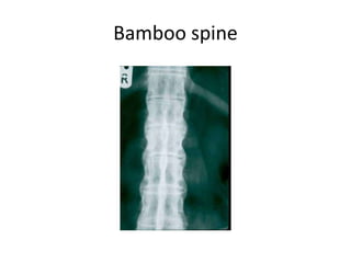
Ankylosing spondylitis
Dr. Sairamakrishnan S
• Bechterew's disease
• Bechterew syndrome
• Marie Strümpell disease
• An systemic chronic autoimmune seronegative
spondyloarthropathy characterized by
– HLA-B27 histocompatability complex positive
(90%)
– primarily affect axial spine.
• Pathoanatomy
– exact mechanism is unknown, but most likely due
to an autoimmune reaction to an environmental
pathogen in a genetically susceptible individual.
• Theories of relation to HLA-B27 include
– HLA-B27 aggregates with peptides in the joint and
leads to a degenerative cascade
– cytotoxic T-cell autoimmune reaction against HLA-
B27
• Human Leukocyte Antigen B*27 is a class I
surface antigen encoded by the B locus in the
major histocompatibility complex (MHC) on
chromosome 6
• Presents microbial antigens to T-cells.
• HLA-B27 is strongly associated with
“seronegative spondyloarthropathies.”
Enthesitis
• Enthesis is defined as the insertion of tendon,
ligaments, or muscle into bone
• Entheses inflammation leads to bony erosion,
surrounding soft-tissue ossification, and
eventually joint ankylosis
• preferentially targets sacroiliac joints, spinal
apophyseal joints, symphysis pubis
• disc space involvement
– inflammation of the annulus lead to bridging
osteophyte formation (syndesmophytes)
• Genetics
– there is a genetic predisposition, but mode of
inheritance is unknown
– HLA-B27 is located on sixth chromosome, B locus
• Epidemiology
– 4:1 male:female
– usually presents in 3rd decade of life
– fewer than 10% of HLA-B27 positive patients have
symptoms of AS
Systemic manifestations
• acute anterior uveitis & iritis
• heart disease (cardiac conduction abnormalities)
• pulmonary fibrosis
• renal amyloidosis
• ascending aortic conditions (aortitis, stenosis,
regurgitation)
• Klebsilella pneumoniae synovitis
– HLA-B27 individuals are more susceptible to Klebsilella
pneumoniae synovitis
Symptoms
• lumbosacral pain and stiffness
– present in most patients
– worse in morning
– Reduces with exercises and not with rest
– insidious onset in 3rd decade of life
• neck and upper thoracic pain
– occurs later in life
– acute neck pain should raise suspicion for fracture
• loss of horizontal gaze
• shortness of breath
– caused by costovertebral joint involvement, leading to
reduced chest expansion
Physical examination
• limitation of chest wall expansion
– < 2cm of expansion is more specific than HLA-B27
for making diagnosis
• decreased spine motion
– Schober test
• Sacroiliac provocative tests
• Hip examination
• kyphotic spine deformity
– chin-on-chest (flexion) deformity of the spine
– caused by multiple microfractures that occur over
time
– chin-brow-to-vertical angle (CBVA)
• measured from standing exam
• useful for preoperative planning
• correction of this angle correlates with improved
surgical outcomes
Radiographs
• spine
• negative in 50% of cases with spine fractures
• squaring of vertebrae with vertical or marginal
syndesmophytes
• late vertebral scalloping (bamboo spine)
• chin-brow to vertical angle
– used to measure chin-on-chest deformity
– useful for preoperative planning for osteotomy
Marginal syndesmophytes
Bamboo spine
• pelvis & lower extremity
• Ferguson pelvic tilt view
• bilateral symmetric sacroiliac erosion
– earliest radiographic sign is erosion of iliac side of sacroiliac
joint
• joint space narrowing
• ankylosis
• CT
– will show bony changes but not active inflammation
– CT is most sensitive test to diagnose cervical fractures
in patients with AS
• MRI
– will detect inflammation, making it the best modality
for early detection of AS in young patients
– obtained with cervical fractures to look for epidural
hemorrhage
Differential Dx - DISH
Treatment
• Physiotherapy
– Main stay of treatment to maintain flexibility
– Never should be done forcefully
• NSAIDS, COX-2 inhibitors
– first line of treatment for pain and stiffness
– oral steroids not recommended
• Spine fractures
– Treated with posterior stabilisation
– high rate of complications
• progressive deformity
• nonunion
• hardware failure
• infection
• Spine deformity
– Lumbar osteotomy
• Thoracolumbar kyphosis
• types
– closing wedge (pedicle subtracting) osteotomy
– vertebral body resection
– single-level opening wedge osteotomy
– multi-segment opening osteotomy
– C7-T1 cervicalthoracic osteotomy
• cervicothoracic kyphotic (chin-on-chest) deformity
• Total hip replacement
– Higher risk of dislocation

Ankylosing spondilitis

  • 1.
  • 2.
    • Bechterew's disease •Bechterew syndrome • Marie Strümpell disease
  • 3.
    • An systemicchronic autoimmune seronegative spondyloarthropathy characterized by – HLA-B27 histocompatability complex positive (90%) – primarily affect axial spine.
  • 4.
    • Pathoanatomy – exactmechanism is unknown, but most likely due to an autoimmune reaction to an environmental pathogen in a genetically susceptible individual. • Theories of relation to HLA-B27 include – HLA-B27 aggregates with peptides in the joint and leads to a degenerative cascade – cytotoxic T-cell autoimmune reaction against HLA- B27
  • 5.
    • Human LeukocyteAntigen B*27 is a class I surface antigen encoded by the B locus in the major histocompatibility complex (MHC) on chromosome 6 • Presents microbial antigens to T-cells. • HLA-B27 is strongly associated with “seronegative spondyloarthropathies.”
  • 6.
    Enthesitis • Enthesis isdefined as the insertion of tendon, ligaments, or muscle into bone • Entheses inflammation leads to bony erosion, surrounding soft-tissue ossification, and eventually joint ankylosis • preferentially targets sacroiliac joints, spinal apophyseal joints, symphysis pubis
  • 7.
    • disc spaceinvolvement – inflammation of the annulus lead to bridging osteophyte formation (syndesmophytes)
  • 8.
    • Genetics – thereis a genetic predisposition, but mode of inheritance is unknown – HLA-B27 is located on sixth chromosome, B locus
  • 9.
    • Epidemiology – 4:1male:female – usually presents in 3rd decade of life – fewer than 10% of HLA-B27 positive patients have symptoms of AS
  • 12.
    Systemic manifestations • acuteanterior uveitis & iritis • heart disease (cardiac conduction abnormalities) • pulmonary fibrosis • renal amyloidosis • ascending aortic conditions (aortitis, stenosis, regurgitation) • Klebsilella pneumoniae synovitis – HLA-B27 individuals are more susceptible to Klebsilella pneumoniae synovitis
  • 13.
    Symptoms • lumbosacral painand stiffness – present in most patients – worse in morning – Reduces with exercises and not with rest – insidious onset in 3rd decade of life • neck and upper thoracic pain – occurs later in life – acute neck pain should raise suspicion for fracture • loss of horizontal gaze • shortness of breath – caused by costovertebral joint involvement, leading to reduced chest expansion
  • 14.
    Physical examination • limitationof chest wall expansion – < 2cm of expansion is more specific than HLA-B27 for making diagnosis • decreased spine motion – Schober test • Sacroiliac provocative tests • Hip examination
  • 15.
    • kyphotic spinedeformity – chin-on-chest (flexion) deformity of the spine – caused by multiple microfractures that occur over time – chin-brow-to-vertical angle (CBVA) • measured from standing exam • useful for preoperative planning • correction of this angle correlates with improved surgical outcomes
  • 18.
    Radiographs • spine • negativein 50% of cases with spine fractures • squaring of vertebrae with vertical or marginal syndesmophytes • late vertebral scalloping (bamboo spine) • chin-brow to vertical angle – used to measure chin-on-chest deformity – useful for preoperative planning for osteotomy
  • 19.
  • 20.
  • 21.
    • pelvis &lower extremity • Ferguson pelvic tilt view • bilateral symmetric sacroiliac erosion – earliest radiographic sign is erosion of iliac side of sacroiliac joint • joint space narrowing • ankylosis
  • 23.
    • CT – willshow bony changes but not active inflammation – CT is most sensitive test to diagnose cervical fractures in patients with AS • MRI – will detect inflammation, making it the best modality for early detection of AS in young patients – obtained with cervical fractures to look for epidural hemorrhage
  • 24.
  • 25.
    Treatment • Physiotherapy – Mainstay of treatment to maintain flexibility – Never should be done forcefully • NSAIDS, COX-2 inhibitors – first line of treatment for pain and stiffness – oral steroids not recommended
  • 26.
    • Spine fractures –Treated with posterior stabilisation – high rate of complications • progressive deformity • nonunion • hardware failure • infection
  • 27.
    • Spine deformity –Lumbar osteotomy • Thoracolumbar kyphosis • types – closing wedge (pedicle subtracting) osteotomy – vertebral body resection – single-level opening wedge osteotomy – multi-segment opening osteotomy – C7-T1 cervicalthoracic osteotomy • cervicothoracic kyphotic (chin-on-chest) deformity
  • 28.
    • Total hipreplacement – Higher risk of dislocation