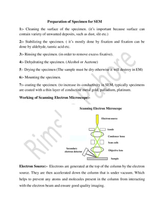
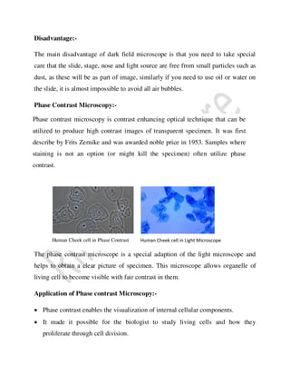
This document discusses different types of microscopes, including their history, parts, uses, and key features. It describes:
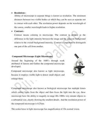
1) The early compound microscope invented by Jansen, capable of 3-9x magnification. Compound microscopes can magnify 40-1000x and have a resolution of 0.25um.
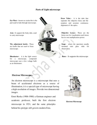
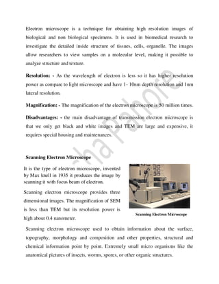
2) Electron microscopes, which use electron beams rather than light and have much higher resolutions of 1-10nm. Scanning electron microscopes provide 3D images while transmission electron microscopes have higher magnification but only show black and white 2D images.
3) Other microscope types like binocular, darkfield, and phase contrast microscopes and their applications in biology research. Pre
This document provides information about bright field microscopes. It describes how bright field microscopes work by using light to illuminate specimens on a slide, which appear dark against a bright background. It outlines the basic components of a microscope like the stage, objectives, and eyepieces. The document discusses using bright field microscopy to view stained specimens and provides tips for microscope care, setup, use, and troubleshooting common problems.
This presentation include information about electron microscope & types of electron microscope i.e. SEM (Scanning electron microscope) & TEM (Transmission electron microscope).
An electron microscope is a microscope that uses a beam of scattered electrons as a source of illumination. It is used to get information about structure, topology, morphology & composition of materials. It has many advantages. Basically there are 4 types of electron microscope but here we will discuss only 2 types.
Transmission electron microscopy is a microscopy technique in which a beam of electrons is transmitted through an ultra-thin specimen, interacting with the specimen as it passes through it. Its resolution & magnification is about 10,000,000x. There are 5 types of transmission electron microscope i.e. BFTEM (Bright field transmision electron microscope), DFTEM (Dark field transmission electron microscope), HRTEM (High resolution transmission electron microscope), EFTEM (Energy filtered transmission electron microscope), ED (Electron diffraction). there are 4 techniques of TEM i.e. negative staining, shadow casting, Freeze fracture replication, freeze etching. It has many applications e.g, for the study of Cancer research, virology, chemical industry, electronic structure etc.
A scanning electron microscope is a type of electron microscope that produces images of a sample by scanning it with a focused beam of electrons. Types of signals produce by SEM include secondary electrons, back scattered electrons, X-rays, light rays. There are many advantages of SEM e.g, Btter resolution, fast imaging easy to operate, work with low voltage etc.
Dark field microscopy produces bright images of unstained samples against a dark background. It works by using a condenser with an opaque disk to block light entering the objective lens directly, allowing only light reflected off the sample to pass through. This causes specimens to appear bright on a dark background. It is useful for viewing transparent or unstained samples like bacteria, cells, and minerals due to the contrast it provides.
this presentation deals with the introduction of some of the commonly used optical microscopes in forensic labs; compound microscope, stereoscopic microscope, comparison microscope, fluorescence microscope and polarized microscope.
Principles, structure and apllications of bright field and dark field microsc...selvaraj227
BRIGHT-FIELD MICROSCOPY. STEPS OF BRIGHT FIELD MICROSCOPY. DARK FIELD MICROSCOPY.USE OF DARK FILED MICROSCOPE.DIFFERENT BETWEEN THE BRIGHT AND DARK FIELD MICROSCOPY
BRIGHT FIELD MICROSCOPY by SIVASANGARI SHANMUGAM
bRIGHT FIELD MICROSCOPY is also called a compound microscope. The name bright - field is derived from the fact that the specimen is dark and contrasted by the surrounding bright viewing field.
This document provides an introduction to phase contrast microscopy and fluorescent microscopy. It discusses that phase contrast microscopy, developed in 1934, uses optical techniques to produce high-contrast images of transparent samples like living cells. It works by converting differences in refractive index to intensity differences visible to the eye. Fluorescent microscopy illuminates samples with high-energy light, causing fluorophores to emit lower-energy light, which can then be filtered and observed. Both techniques allow viewing of unstained, living samples like cells in more detail.
- The document discusses the compound microscope, its history, parts, functions, and use.
- A compound microscope has more than one lens and was invented in the 1590s in the Netherlands. It allows higher magnification than simple microscopes.
- The main parts are the mechanical stage, optical system with objectives and eyepieces, illumination system, and adjustment controls. Objectives magnify the specimen while eyepieces provide a final view.
- Focusing involves using coarse and fine adjustments to bring the specimen into view at different magnifications including with immersion oil.
Phase Contrast Microscopy - Microbiology 1st RAHUL PAL
Phase contrast microscopy uses differences in phase shifts of light waves passing through a specimen to visualize unstained living cells. It allows biologists to study living cells and cell division. Dark field microscopy produces a dark background and bright specimen image using oblique illumination. It is used to view unstained or little absorbed objects like bacteria, algae, and diatoms. Electron microscopy uses a beam of accelerated electrons instead of light for higher resolution imaging of nano-scale structures. Types include analytical electron microscopy, scanning transmission electron microscopy, scanning electron microscopy, and transmission electron microscopy.
Bright field microscopy, Principle and applicationsKAUSHAL SAHU
This document discusses the basics of light microscopy. It begins with a brief history, noting that the first compound microscope was created in 1590, while Antonie van Leeuwenhoek discovered microorganisms and sperm cells using a simple microscope in the 1630s. The basic components of light microscopes are described, including the objective lenses, eyepieces, stage, and condenser. Brightfield microscopy is explained in more detail, noting that it uses transmitted white light and staining to increase contrast. Applications include viewing stained bacteria, tissue sections, and algae. While brightfield microscopy is simple to use, its disadvantages include low contrast and an inability to see transparent, unstained samples clearly.
The document discusses different types of microscopes used to view microscopic specimens. It describes light microscopes, which use lenses and visible light, including brightfield, darkfield, phase contrast, and fluorescence microscopes. It also describes electron microscopes, which use electromagnetic lenses and electrons beams to view specimens, including transmission electron microscopes that pass electrons through thin specimens, and scanning electron microscopes that scan surfaces to produce 3D images. Key aspects and uses of each microscope type are outlined.
INTRODUCTION
HISTORY
TYPES OF MICROSCOPE
Compound Microscope
Dissection Microscope
Scanning Electron Microscope (SEM)
Transmission Electron Microscope (TEM
CARE
PARTS AND FUNCTION
FOCUSING
CONCLUSION
REFERANCE
Microscopy is the technical field of using microscopes to view objects and areas of objects that cannot be seen with the naked eye (objects that are not within the resolution range of the normal eye). There are three well-known branches of microscopy: optical, electron, and scanning probe microscopy, along with the emerging field of X-ray microscopy.
Dark-field microscopy is used to illuminate unstained samples causing them to appear bright against a dark background. This type of microscope contains a special condenser having a central blacked-out area.
Bright-field microscopy, also known as light microscopy, is the simplest optical microscopy technique that uses light to produce a dark image against a bright background. It is the standard microscope used in biology and microbiology laboratories. Bright-field microscopy works by passing light through a specimen, which is differentially absorbed and refracted to produce a contrasting image. The microscope's objective lenses magnify the specimen's image, which is further magnified by the eyepiece for viewing. Magnification ranges from 40-1000x depending on the objective and 10x eyepiece lenses. Bright-field microscopy is used to study animal and plant cells, bacteria, and parasites.
during this ppt of microscopes we will be able to know
INTRODUCTION
DEFINITION
HISTORICAL BACKGROUND
VARIABLES USED IN MICROSCOPY
VARIOUS TYPES OF MICROSCOPES
COMPOUND MICROSCOPE - Structure and Function
USE OF MICROSCOPE
CARE OF MICROSCOPE
defintion
A microscope (Greek: micron = small and scopos = aim)
MICROSCOPE - An instrument for viewing objects that are too small to be seen by the naked or unaided eye
MICROSCOPY - The science of investigating small objects using such an instrument is called microscopy
Types of Microscopes with their applications - Microbiologynote.com
https://microbiologynote.com/types-of-microscopes-with-their-applications/
Youtube Lecture Video:
https://www.youtube.com/watch?v=nuJZtXohFFQ&ab_channel=MicrobiologyNote
This document provides information about different types of microscopes. It discusses simple microscopes, which use a single lens for magnification, and their basic parts and working principle. Compound microscopes are then introduced as using multiple lenses to achieve higher magnification than is possible with a simple lens alone. The key parts of a compound microscope are described, including the eyepiece, objective lenses, body tube, arm, and focus adjustments. It explains that light from the illuminator passes through these parts to produce a magnified image for viewing.
Electron microscope, principle and applicationKAUSHAL SAHU
Introduction
History
Resolution &Magnification of
Electron microscope
Types of electron microscope
1) Transmission electron microscope (TEM)
- Structural parts of TEM
- Principle & Working of TEM
- Sample preparation for TEM
- Advantages & disadvantages of TEM
Scanning electron microscope (SEM)
- Structural parts of SEM
- Principle & Working of SEM
- Sample preparation for SEM
- Advantages & disadvantages of SEM
3) Scanning transmission electron microscope (STEM)
Applications of electron microscope
Conclusion
References
The document discusses phase contrast microscopy, which was developed by Fritz Zernike in the 1930s. It allows living or unstained cells and intracellular components to be visible under a microscope. The phase contrast microscope works by converting small phase changes caused by differences in refractive index of cell structures into visible brightness and darkness differences. This makes organelles and other structures visible without using staining. The phase contrast is achieved using an annular diaphragm and phase rings or filters to shift the phase of light passing through or around the specimen.
The document summarizes the history and components of the confocal microscope. It describes how the confocal microscope was initially conceived in the 1950s but lacked the necessary light sources and computing power. Work in the late 1960s adapted the original concept and allowed for the examination of unstained brain and ganglion cells. Further developments in lasers and computing through the 1980s led to more practical confocal microscopes. Modern confocal microscopes integrate optics, detectors, computers and lasers to produce high-resolution 3D electronic images of samples. Confocal microscopes are now used across various fields including biology and medicine.
Waterborne diseases are caused by microorganisms, toxins, and contaminants spread through contaminated water. They cause illnesses such as cholera, diarrhea, and gastrointestinal problems. Developing countries often lack proper water treatment, especially in rural areas, increasing risk of outbreak. The document then discusses four specific waterborne diseases - diarrhea, cholera, typhoid, and amoebiasis - outlining their causes, symptoms, and treatments.
The document discusses the history and development of electron microscopes. It describes how J.J. Thomson discovered electrons in 1897 and how subsequent scientists like de Broglie, Ruska, and Knoll contributed to the development of the first electron microscope in the 1930s. It then explains the basic workings and components of transmission electron microscopes and scanning electron microscopes, how they produce images, and some examples of their applications in biology and materials science.
This document discusses light microscopes. It begins by defining a microscope as an instrument used to view objects too small to see with the naked eye. It then describes the basic components and workings of light microscopes, including lenses that magnify objects, different types like brightfield and phase contrast, and applications in biology and medicine like pathology. Phase contrast microscopy is explained in more detail, noting how it uses interference of light waves passing through a specimen to visualize differences in brightness of structures. In closing, the document outlines several uses of light microscopes across various fields.
Principles and application of light, phase constrast and fluorescence microscopeMaitriThakor
This document provides an overview of three types of microscopes: light microscopes, phase contrast microscopes, and fluorescence microscopes. It describes the basic principles and components of each microscope type and their applications. Light microscopes use lenses to magnify specimens and are used widely in biology to study cells. Phase contrast microscopes convert phase differences in light passing through specimens into brightness variations, allowing visualization of transparent structures. Fluorescence microscopes use fluorescent dyes and specific wavelengths of light to enhance contrast and study labeled structures within cells.
Viruses are the smallest known infectious agents and lack cellular organization. They contain either DNA or RNA but not both, and are obligate intracellular parasites that depend on host cell machinery for replication. Viruses infect host cells through attachment to receptors, then undergo a replication process involving uncoating, biosynthesis, maturation, and release of new virions. Their structure can be enveloped or nonenveloped, with capsids that have different symmetries and surface proteins important for infection.
LIGHT MICROSCOPY by SIVASANGARI SHANMUGAM
The optical microscope, The functions of a light microscope is based on its ability to focus a beam of light through, which is very small and transparent, to produce an image.
This document provides an overview of microscopy used in diagnostic microbiology. It discusses the history and types of microscopes including bright field, dark field, phase contrast, fluorescence, transmission electron, and scanning electron microscopes. It describes how each microscope works and its applications. Key aspects covered include the use of microscopy to identify microorganisms, detect viruses, and examine cellular structures in detail not visible to the naked eye. Microscopy is an important tool in diagnostic microbiology.
This document discusses various histological tools used to study tissues at the microscopic level. It describes light microscopes, which use visible light and magnification to examine thin tissue slices stained with histological dyes. Electron microscopes are also covered, using electron beams instead of light for higher resolution imaging of cell structures. Specific techniques covered include fluorescence microscopy using fluorescent dyes, polarizing microscopy examining birefringence, and transmission electron microscopy producing 2D images of cell organelles. The history and development of microscopy from early simple microscopes to modern compound and electron microscopes is summarized.
This document discusses various histological tools used to study tissues at the microscopic level. It describes light microscopes, which use visible light and can magnify up to 1000x, and electron microscopes, which use electron beams to achieve much higher magnification up to 1 million times. Key histological techniques mentioned include biopsy, tissue processing, staining, immunohistochemistry, and different types of microscopy like fluorescence and polarizing microscopy. The document provides details on the basic components and functioning of different microscope types used in histology.
This document lists the members of Group One for a microscopy course. It includes 9 students' names and registration numbers. The document then provides an overview of different types of microscopes, including light microscopes, electron microscopes, fluorescence microscopes, and dark field microscopes. It describes the basic setup, working principles, uses and comparisons of these microscopic techniques.
these slides gives information about the types of microscopy. microscopy is divided into two type on the bases of their application which are stereoscopic and biological microscope. another types of microscopy are optical microscope, electron microscope, x-ray microscope and also scanning probe microscopy which perform many function.
Eye microscopy and electron microscopy include differentiation and reflection. Retraction of magnetic fields/electron beams that interact with the image. As well as the scattering of scattered rays or other signals to create the image.
This procedure can be done by inserting a wide-field light sample or by scanning a fine beam over the sample. A microscopy scan probe involves. The interaction of the scanning probe with the surface of the object of interest.
Advances in microscopy transformed living things and exposed the field of histology. And so remain an important strategy for health and natural science.
X-ray microscopy is three-dimensional and unobtrusive. Allowing for repeated photographing of the same sample in situ or 4D subjects. And provides the ability to "see". The sample is readable before devoting it to advanced correction techniques.
The 3D X-ray microscope uses a computed tomography technique, rotating the sample. By 360 degrees and reconstructing images. CT is usually done with a flat panel display. The 3D X-ray microscope uses a series of objectives, e.g., from 4X to 40X, and can include a flat panel.
History of Microscopy
The field of the microscope dates back to at least the 17th century. Early mirrors, single-lens magnifying glasses with limited size. Back to the widespread use of eyeglasses in the 13th century. But the most advanced microscopes first appeared in Europe around 1620 Early.
Microscope doctors included Galileo Galilei, who was discovered in 1610. That he could turn off his telescope to see small objects nearby. And Cornelis Drebbel. Who may have invented the compact microscope in about 1620?
Antonie van Leeuwenhoek developed a simple magnifying microscope. In the 1670s and is often regarded as the first acclaimed microscopist and microbiologist.
Microscope Uses
to view bacteria, parasites, and a variety of human/animal cells
cellular process, cell division
DNA replication
tissue analysis
examining forensic evidence
studying the role of a protein within a cell
studying atomic structures
And in what way are bacteria able to infect human cells, then we use a microscope to study them all. Those studies are done at the micro-level.
We use a microscope to perform the kind of study that we cannot see with the naked eye.
Microscope component
Light
Lence
Optical/Light Microscopy
Bright Field Microscopy
Dark Field Microscopy
Confocal Microscopy
Phase Contrast Microscopy
Fluorescence Microscopy
Electron microscopy
Transmission Electron Microscopy
Scanning Electron Microscopy
Scanning Probe Microscopy
The resolving power of a microscope means
This document provides information about microscopy. It begins by defining microscopy and explaining that microscopes are used to view microorganisms that are too small to see with the naked eye. It then discusses the history and basic principles of microscopy, including magnification, resolution, and numerical aperture. The document outlines the key parts of the microscope and describes different types of microscopes in more detail, including light microscopes (brightfield, darkfield, fluorescence, phase contrast, UV) and electron microscopes (TEM, SEM). It provides examples of how each type of microscope is used.
Microscopy is the technical field of using microscopes to view objects that cannot be seen with the naked eye. There are three main types of microscopy - light microscopy, which uses visible light; electron microscopy, which uses electrons; and scanning probe microscopy, which uses a physical probe. Light microscopes like brightfield, darkfield, phase contrast, and fluorescence microscopes are commonly used to view living and stained specimens. Electron microscopes have much higher resolving power than light microscopes and are able to view much smaller structures. Transmission electron microscopes form images using electrons transmitted through thin specimens while scanning electron microscopes form images from electrons emitted from surfaces.
Introduction to microscopy
Different parts of a microscope & their function
Different types of microscopy
Different types of optical microscopy
Different types of electron microscopy
Different terms used in microscopy
Staining- Simple, Differential, Special
Gram Staining
Microscopes and microscopy are introduced. There are two main types of microscopes - light microscopes, which use optical lenses and light, and electron microscopes, which use a beam of electrons. Light microscopes can use different techniques like brightfield, darkfield, fluorescence, and phase contrast. Electron microscopes have higher resolving power and include transmission electron microscopes and scanning electron microscopes. Sample preparation and staining are important for microscopy as they allow small and transparent specimens to be visualized.
The document summarizes key terms and components of different types of microscopes used in microbiology. It discusses the principles, magnification, resolving power, and limitations of light microscopes, darkfield microscopes, phase contrast microscopes, fluorescence microscopes, and electron microscopes. Examples are provided of specimens visualized under each microscope type, such as paramecium viewed under light and fluorescence microscopes and bacillus subtilis observed with a fluorescence microscope.
The document discusses microscopes and microscopy. It defines a microscope as an optical instrument used to view very small objects magnified hundreds of times. The key parts of a microscope are described including the eyepiece, objective lenses, stage, and illuminator. The history of microscope development from the 16th century to modern electron and scanning probe microscopes is summarized. Common uses of microscopy include tissue analysis, forensic investigation, ecosystem studies, and protein analysis. Advanced microscopy techniques now allow non-invasive in vivo imaging.
Microscope ppt, by jitendra kumar pandey,medical micro,2nd yr, mgm medical co...jitendra Pandey
The document summarizes different types of microscopes used to study microorganisms. It discusses light microscopes like brightfield, darkfield and phase contrast microscopes. It also describes electron microscopes like transmission electron microscopes (TEM) and scanning electron microscopes (SEM) that use electron beams instead of light. TEM images internal structures by transmitting electrons through thin samples while SEM scans sample surfaces using secondary electrons. Sample preparation methods for both light and electron microscopy are also outlined.
Microscopy - Magnification, Resolving power, Principles, Types and ApplicationsNethravathi Siri
Magnification, Resolving power, Principles and Applications of Simple, Compound, Stereozoom, Phase contrast, Fluorescent and Electron microscopes (TEM & SEM).
Microscopy is the technical field that uses microscopes to observe samples which are not in the resolution range of the normal-unaided eye.
The document discusses microscopes, including their history, types, parts, and usage. It notes that the first primitive microscopes date to the late 1300s, but that Galileo Galilei perfected the first known microscope in 1609. The key types discussed are light microscopes, compound microscopes, and electron microscopes like scanning electron microscopes and transmission electron microscopes. The parts of a basic microscope are also outlined, including the objective lens, eyepiece, stage, and controls. Proper microscope care is also briefly covered.
chapter 2- Microscopy.pptx Microscopy related with medicineHarikantSingh4
It is about microscope in details about microscope. It includes simple microscope, fluoresence microscope, electron microscope and dark field microscope. These are described in details in this presentation. This presentation has images also.
The document discusses microscopy and different types of microscopes. It begins by defining microscopy as the technology that makes small objects visible to the human eye using microscopes. It then describes different types of microscopes, focusing on light microscopes and electron microscopes. Electron microscopes use a beam of electrons rather than light, allowing them to achieve much higher magnifications and resolving power than light microscopes. The document discusses the basic components and working principles of transmission electron microscopes and scanning electron microscopes. It also covers sample preparation techniques, applications, limitations, and recent research using electron microscopy.
This document provides information about different types of microscopy. It begins with an introduction to microscopy, then discusses the history and key figures in the development of the microscope. It describes different types of microscopes including light/bright field microscopy, dark field microscopy, phase contrast microscopy, fluorescence microscopy, and electron microscopy. For each type, it provides details on the optical principles, components, and applications. The document aims to inform the reader about the basic concepts and techniques of microscopy.
Similar to Microscope and types of microscope (20)
hematic appreciation test is a psychological assessment tool used to measure an individual's appreciation and understanding of specific themes or topics. This test helps to evaluate an individual's ability to connect different ideas and concepts within a given theme, as well as their overall comprehension and interpretation skills. The results of the test can provide valuable insights into an individual's cognitive abilities, creativity, and critical thinking skills
Nucleophilic Addition of carbonyl compounds.pptxSSR02
Nucleophilic addition is the most important reaction of carbonyls. Not just aldehydes and ketones, but also carboxylic acid derivatives in general.
Carbonyls undergo addition reactions with a large range of nucleophiles.
Comparing the relative basicity of the nucleophile and the product is extremely helpful in determining how reversible the addition reaction is. Reactions with Grignards and hydrides are irreversible. Reactions with weak bases like halides and carboxylates generally don’t happen.
Electronic effects (inductive effects, electron donation) have a large impact on reactivity.
Large groups adjacent to the carbonyl will slow the rate of reaction.
Neutral nucleophiles can also add to carbonyls, although their additions are generally slower and more reversible. Acid catalysis is sometimes employed to increase the rate of addition.
ESPP presentation to EU Waste Water Network, 4th June 2024 “EU policies driving nutrient removal and recycling
and the revised UWWTD (Urban Waste Water Treatment Directive)”
This presentation explores a brief idea about the structural and functional attributes of nucleotides, the structure and function of genetic materials along with the impact of UV rays and pH upon them.
BREEDING METHODS FOR DISEASE RESISTANCE.pptxRASHMI M G
Plant breeding for disease resistance is a strategy to reduce crop losses caused by disease. Plants have an innate immune system that allows them to recognize pathogens and provide resistance. However, breeding for long-lasting resistance often involves combining multiple resistance genes
When I was asked to give a companion lecture in support of ‘The Philosophy of Science’ (https://shorturl.at/4pUXz) I decided not to walk through the detail of the many methodologies in order of use. Instead, I chose to employ a long standing, and ongoing, scientific development as an exemplar. And so, I chose the ever evolving story of Thermodynamics as a scientific investigation at its best.
Conducted over a period of >200 years, Thermodynamics R&D, and application, benefitted from the highest levels of professionalism, collaboration, and technical thoroughness. New layers of application, methodology, and practice were made possible by the progressive advance of technology. In turn, this has seen measurement and modelling accuracy continually improved at a micro and macro level.
Perhaps most importantly, Thermodynamics rapidly became a primary tool in the advance of applied science/engineering/technology, spanning micro-tech, to aerospace and cosmology. I can think of no better a story to illustrate the breadth of scientific methodologies and applications at their best.
ESR spectroscopy in liquid food and beverages.pptxPRIYANKA PATEL
With increasing population, people need to rely on packaged food stuffs. Packaging of food materials requires the preservation of food. There are various methods for the treatment of food to preserve them and irradiation treatment of food is one of them. It is the most common and the most harmless method for the food preservation as it does not alter the necessary micronutrients of food materials. Although irradiated food doesn’t cause any harm to the human health but still the quality assessment of food is required to provide consumers with necessary information about the food. ESR spectroscopy is the most sophisticated way to investigate the quality of the food and the free radicals induced during the processing of the food. ESR spin trapping technique is useful for the detection of highly unstable radicals in the food. The antioxidant capability of liquid food and beverages in mainly performed by spin trapping technique.
EWOCS-I: The catalog of X-ray sources in Westerlund 1 from the Extended Weste...Sérgio Sacani
Context. With a mass exceeding several 104 M⊙ and a rich and dense population of massive stars, supermassive young star clusters
represent the most massive star-forming environment that is dominated by the feedback from massive stars and gravitational interactions
among stars.
Aims. In this paper we present the Extended Westerlund 1 and 2 Open Clusters Survey (EWOCS) project, which aims to investigate
the influence of the starburst environment on the formation of stars and planets, and on the evolution of both low and high mass stars.
The primary targets of this project are Westerlund 1 and 2, the closest supermassive star clusters to the Sun.
Methods. The project is based primarily on recent observations conducted with the Chandra and JWST observatories. Specifically,
the Chandra survey of Westerlund 1 consists of 36 new ACIS-I observations, nearly co-pointed, for a total exposure time of 1 Msec.
Additionally, we included 8 archival Chandra/ACIS-S observations. This paper presents the resulting catalog of X-ray sources within
and around Westerlund 1. Sources were detected by combining various existing methods, and photon extraction and source validation
were carried out using the ACIS-Extract software.
Results. The EWOCS X-ray catalog comprises 5963 validated sources out of the 9420 initially provided to ACIS-Extract, reaching a
photon flux threshold of approximately 2 × 10⁻⁸ photons cm−2
s
−1
. The X-ray sources exhibit a highly concentrated spatial distribution,
with 1075 sources located within the central 1 arcmin. We have successfully detected X-ray emissions from 126 out of the 166 known
massive stars of the cluster, and we have collected over 71 000 photons from the magnetar CXO J164710.20-455217.
The ability to recreate computational results with minimal effort and actionable metrics provides a solid foundation for scientific research and software development. When people can replicate an analysis at the touch of a button using open-source software, open data, and methods to assess and compare proposals, it significantly eases verification of results, engagement with a diverse range of contributors, and progress. However, we have yet to fully achieve this; there are still many sociotechnical frictions.
Inspired by David Donoho's vision, this talk aims to revisit the three crucial pillars of frictionless reproducibility (data sharing, code sharing, and competitive challenges) with the perspective of deep software variability.
Our observation is that multiple layers — hardware, operating systems, third-party libraries, software versions, input data, compile-time options, and parameters — are subject to variability that exacerbates frictions but is also essential for achieving robust, generalizable results and fostering innovation. I will first review the literature, providing evidence of how the complex variability interactions across these layers affect qualitative and quantitative software properties, thereby complicating the reproduction and replication of scientific studies in various fields.
I will then present some software engineering and AI techniques that can support the strategic exploration of variability spaces. These include the use of abstractions and models (e.g., feature models), sampling strategies (e.g., uniform, random), cost-effective measurements (e.g., incremental build of software configurations), and dimensionality reduction methods (e.g., transfer learning, feature selection, software debloating).
I will finally argue that deep variability is both the problem and solution of frictionless reproducibility, calling the software science community to develop new methods and tools to manage variability and foster reproducibility in software systems.
Exposé invité Journées Nationales du GDR GPL 2024
Or: Beyond linear.
Abstract: Equivariant neural networks are neural networks that incorporate symmetries. The nonlinear activation functions in these networks result in interesting nonlinear equivariant maps between simple representations, and motivate the key player of this talk: piecewise linear representation theory.
Disclaimer: No one is perfect, so please mind that there might be mistakes and typos.
dtubbenhauer@gmail.com
Corrected slides: dtubbenhauer.com/talks.html
The debris of the ‘last major merger’ is dynamically youngSérgio Sacani
The Milky Way’s (MW) inner stellar halo contains an [Fe/H]-rich component with highly eccentric orbits, often referred to as the
‘last major merger.’ Hypotheses for the origin of this component include Gaia-Sausage/Enceladus (GSE), where the progenitor
collided with the MW proto-disc 8–11 Gyr ago, and the Virgo Radial Merger (VRM), where the progenitor collided with the
MW disc within the last 3 Gyr. These two scenarios make different predictions about observable structure in local phase space,
because the morphology of debris depends on how long it has had to phase mix. The recently identified phase-space folds in Gaia
DR3 have positive caustic velocities, making them fundamentally different than the phase-mixed chevrons found in simulations
at late times. Roughly 20 per cent of the stars in the prograde local stellar halo are associated with the observed caustics. Based
on a simple phase-mixing model, the observed number of caustics are consistent with a merger that occurred 1–2 Gyr ago.
We also compare the observed phase-space distribution to FIRE-2 Latte simulations of GSE-like mergers, using a quantitative
measurement of phase mixing (2D causticality). The observed local phase-space distribution best matches the simulated data
1–2 Gyr after collision, and certainly not later than 3 Gyr. This is further evidence that the progenitor of the ‘last major merger’
did not collide with the MW proto-disc at early times, as is thought for the GSE, but instead collided with the MW disc within
the last few Gyr, consistent with the body of work surrounding the VRM.
The debris of the ‘last major merger’ is dynamically young
Microscope and types of microscope
1. University of Sindh Jamshoro
Name: Rimsha Pahore
Group: Group B
Subject: Biological Techniques
Assignment topic: Microscope and its Types
2. Microscope and its Types
The word microscope taken from Ancient Greek Language means small and scope
means to see, it is an instrument used to see small objects that are too small to be
seen by the naked eye.
Simply a microscope is an instrument that produces an accurately enlarged image
of small objects.
Early Microscope
The first microscope is invented by Jansen by mounting two lenses in a tube and
the objected lens is positioned close to the object and produces an image that is
picked up and magnified further by the second lens, called eye piece.
Variables used in Microscopy:-
Magnification: Degree of enlargement. No of the times the length or diameter
of an object is multiplied.
It depends upon optical tube length, focal length of objective, magnifying
power of eye piece,
So to find total magnification the magnification of the eyepiece is multiple with
the magnification of the objective lense.
This Early Microscope invented
by Jansen having no tripod and
was capable of magnifying three
to nine times the true size.
3. Resolution:-
Ability of microscope to separate things is known as resolution. The minimum
distances between two visible bodies at which they can be seen as separate not
in contact with each other. The resolution power depends on the wavelength of
the source, smaller wavelength leads to higher resolution.
Contrast:-
Contrast means coloring in microscope. The contrast is defined as the
difference in the light intensity between the image and the adjacent background
relative to the overall background intensity. Contrast is important to distinguish
one part of the cell from another.
Compound Microscope (Light Microscope)
Compound microscope also known as biological microscope has multiple lenses
which collect lights from the object and then focus the light into the eye, these
microscope have the ability to magnify in range 40x-1000x very minute objects to
a substantial size, clearly showing the smallest details. And the resolution power of
the compound microscope is 0.25um.
The ocular lense in light microscope has magnification of 10x normal vision.
Around the beginning of the 1600’s through work
attributed of Jansen and Galileo the compound microscope
developed.
Compound microscope also known as light microscope,
because it employs visible light to detect small objects and
enlarge them.
4. Electron Microscope:-
Parts of Light microscope
Eye Piece:- known as ocular this is the
part used to look through microscope.
Base:- It supports the microscope
Objective Lenses:- These are the
lenses use for visualization each lenses
has its own multiplication power.
Arm:- It support the body tube, used
to carry microscope
The adjustment knobs:- These
are knobs that are used to focus
microscope.
Stage- where the specimen usually
mounted onto glass slide for
observation.
Illuminator:- it is the light source
for a microscope, compound
microscope uses a low voltage bulb
as an illuminator.
An electron microscope is a microscope that uses a
beam of accelerated electrons as a source of
illumination, it is a special type of microscope having
a high resolution of images. Provide two dimensional
images.
Electron Microscope
Ernst Ruska (1906-1988) a German engineer and
academic professor, built the first electron
microscope in 1931, and the same principles
behind his protype still govern modern Ems.
Draw Tube:- it is the tube that
separates the objective lense and the
eyepiece and assumes continuous
alignment of the optics.
5. Electron microscope is a technique for obtaining high resolution images of
biological and non biological specimens. It is used in biomedical research to
investigate the detailed inside structure of tissues, cells, organelle. The images
allow researchers to view samples on a molecular level, making it possible to
analyze structure and texture.
Resolution: - As the wavelength of electron is less so it has higher resolution
power as compare to light microscope and have 1- 10nm depth resolution and 1nm
lateral resolution.
Magnification: - The magnification of the electron microscope is 50 million times.
Disadvantages: - the main disadvantage of transmission electron microscope is
that we only get black and white images and TEM are large and expensive, it
requires special housing and maintenances.
Scanning Electron Microscope
Scanning electron microscope used to obtain information about the surface,
topography, morphology and composition and other properties, structural and
chemical information point by point. Extremely small micro organisms like the
anatomical pictures of insects, worms, spores, or other organic structures.
It is the type of electron microscope, invented
by Max knell in 1935 it produces the image by
scanning it with focus beam of electron.
Scanning electron microscope provides three
dimensional images. The magnification of SEM
is less than TEM but its resolution power is
high about 0.4 nanometer.
Scanning Electron Microscope
6. Preparation of Specimen for SEM
1:- Cleaning the surface of the specimen. (it’s important because surface can
contain variety of unwanted deposits, such as dust, slit etc.)
2:- Stabilizing the specimen. ( it’s mostly done by fixation and fixation can be
done by aldehyde, tannic acid etc.
3:- Rinsing the specimen. (in order to remove excess fixative).
4:- Dehydrating the specimen. (Alcohol or Acetone)
5:- Drying the specimen (The sample must be dry otherwise it will destroy in EM)
6:- Mounting the specimen.
7:- coating the specimen. (to increase its conductivity in SEM, typically specimens
are coated with a thin layer of conductive metal gold, palladium, platinum.
Working of Scanning Electron Microscope:-
Electron Source:- Electrons are generated at the top of the column by the electron
source. They are then accelerated down the column that is under vacuum. Which
helps to prevent any atoms and molecules present in the column from interacting
with the electron beam and ensure good quality imaging.
7. Condenser and objective lens: - The electromagnetic lenses are used to control
the path of the electrons. The condenser lens defines the size of the electron beam
which defines resolution, while the objective lens main role is the focusing of the
beam onto the sample.
Scanning Coils: - After beam is focused, scanning coils are used to deflect the
beam in the X and Y axes so that it scans in the raster fashion over the surface of
the sample.
Different types of the electrons are emitted from the samples upon interacting with
the electron beam. A back scattered electron detector is placed above the sample to
help detect backscattered electron.
Secondary Electron Detector: - it is placed at the side of the electron chamber at
an angle, in order to increase the efficiency of detecting secondary electrons which
can provide more detailed surface information.
Some Pictures under Scanning Electron Microscope
However there are so many uses of SEM but these are expensive and large and
special training is required to operate them and they are only limited to solid
samples. It also carries a small risk of radiation exposure associated with the
electrons that scatter from beneath the sample surface.
Polyester Thread
Red Blood Cells
8. Binocular Microscope:-
A binocular microscope head works by taking one light beam and splitting it into
two parts that then go to the eyes, the prism that does is known as splitter prism.
The main advantage of binocular microscope over monocular is that it has high
magnification power, trinocular microscopes are also available they contain third
eye piece. The third eyepiece of this type of microscope allows you to mount a
camera onto the eyepiece.
Applications of Binocular Microscope:-
The binocular microscope widely use in scientific laborites to perform
researches.
The binocular microscope used in application into cell biology, research, blood
microscopy, urine analysis, biotechnology, pathology etc.
Disadvantages of Binocular Microscope:-
The binocular microscope are much heavier and expensive than simple
microscopes, they require a regular maintenance check for smooth functioning.
Binocular Microscope
Binocular microscope is any optical microscope with
two eyepieces to significantly ease viewing and
observe slide samples. Most of the microscope sold
today is binocular microscope
9. Dark Field Microscopy:-
Dark field microscopy is a technique used to observe unstained samples causing
them to appear brightly against a dark, almost purely black background. So there is
no need of staining.
A dark microscopy is used to examine live microorganisms that either invisible in
the ordinary light microscope or cannot stained by the standard methods so there
we can use dark microscopy. Recently dark field microscopy has regained some of
its popularity when combined with other illumination techniques such as fluoresce.
The dark field microscopy can be perform both in light or electron microscope by
blocking the light source causing light to scatter as it hits the specimen.
Applications of Dark Microscopy:-
Dark field microscopy is used to study marine organisms such as algae,
plankton, diatoms, insects yeast etc.
To study live bacterium.
More detailed view of external features, outline boundaries, edges.
It is used for viewing objects that are unstained, transparent, and absorb little or
no light.
It’s useful for viewing diamonds and precious stones and minerals.
Bacteria Colony Snow Flake
10. Disadvantage:-
The main disadvantage of dark field microscope is that you need to take special
care that the slide, stage, nose and light source are free from small particles such as
dust, as these will be as part of image, similarly if you need to use oil or water on
the slide, it is almost impossible to avoid all air bubbles.
Phase Contrast Microscopy:-
The phase contrast microscope is a special adaption of the light microscope and
helps to obtain a clear picture of specimen. This microscope allows organelle of
living cell to become visible with fair contrast in them.
Application of Phase contrast Microscopy:-
Phase contrast enables the visualization of internal cellular components.
It made it possible for the biologist to study living cells and how they
proliferate through cell division.
Human Cheek cell in Phase Contrast Human Cheek cell in Light Microscope
Phase contrast microscopy is contrast enhancing optical technique that can be
utilized to produce high contrast images of transparent specimen. It was first
describe by Frits Zernike and was awarded noble price in 1953. Samples where
staining is not an option (or might kill the specimen) often utilize phase
contrast.
11. Disadvantage of Phase Contrast Microscopy
The phase contrast microscope is not ideal for thick organisms or particles, thick
specimen can appear distorted. Annuli or rings in contrast microscope limit the
aperture to some extent which decrease resolution.
Camera Lucida:-
It works on simple optical principle reflecting beam of light through a prism and a
plane mirror. The microscope image of the object is reflected by the prism on the
plane mirror and there from the image is reflected on to the plane paper. The
observer moves the pencil on the lines of the image and draws a correct and
faithful figure of the object on the paper.
Before the applications of the photography, the camera Lucida was of considerable
importance to draughtsman. The advantage claimed for it was its cheapness,
smallness, and portability, but nowadays has no more importance.
The word has been taken from Latin which means light
chamber. Camera Lucida is a optical device when attached
with a compound microscope helps drawing microscope
images of objects on paper.
That’s how it works. And was first
invented by William hyde
Wollaston in 1806