LEUKEMIA
JAHNAVI YADAV
Dept of Oral Pathology
CONTENTS
Introduction And Definition
Etiology and Risk Factors
Classification
Treatment
Reference
INTRODUCTION
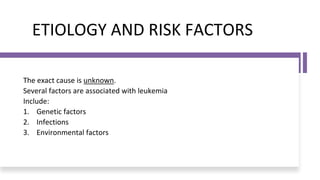
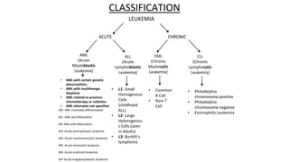
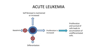
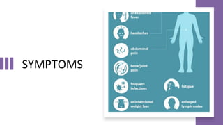
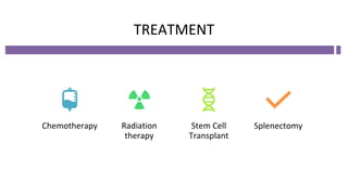
• Leukemia is a type of blood cancer
that begins in the bone marrow.
Leukemia is a malignant disorders
of hematopoietic stem cells
characteristically associated with increase
number of white cells in bone
marrow/peripheral blood.
{Davidson’s Principles and
Practice of Medicine, 21st edition}
ETIOLOGY AND RISK FACTORS
The exact cause is unknown.
Several factors are associated with leukemia
Include:
1. Genetic factors
2. Infections
3. Environmental factors
CLASSIFICATION
ACUTE CHRONIC
AML
(Acute
Myeloblastic
Leukemia)
ALL
(Acute
Lymphoblastic
Leukemia)
CML
(Chronic
Myelocytic
Leukemia)
CLL
(Chronic
Lymphocytic
Leukemia)
LEUKEMIA
• AML with certain genetic
abnormalities.
• AML with multilineage
dysplasia
• AML related to previous
chemotherapy or radiation
• AML otherwise not specified
M0- AML minimally differentiated
M1- AML w/o Maturation
M2-AML with Maturation
M3- Acute promyelocytic leukemia
M4- Acute myelomonocytic leukemia
M5- Acute monocytic leukemia
M6- Acute erythroid leukemia
M7-Acute megakaryoblastic leukemia
• L1- Small
Homogenous
Cells
(childhood
ALL)
• L2- Large
Heterogenou
s Cells (seen
in Adults)
• L3- Burkitt’s
lymphoma
• Common
B Cell
• Rare T
Cell
• Philadelphia
chromosome positive
• Philadelphia
chromosome negative
• Eosinophilic Leukemia
ACUTE LEUKEMIA
Differentiation
Self Renewal is maintained
or increased
Apoptosis Proliferation is
increased
Proliferation
and survival of
cells leads to
accumulation of
undifferentiated
cells.
ACUTE MYELOID LEUKEMIA
• Most common in adults.
• Peak incidence after 60 years.
• Minor fraction of leukemia in childhood(10-15%)
• Blast cells and Auer Rods seen in peripheral
blood smear.
• Periodic Acid Schiff Stain (PAS) – positive
• Peroxidase and Sudan Black B - negative
ACUTE LYMPHOID LEUKEMIA
• Acute Lymphoblastic Leukemia are neoplasm composed of immature B or T
cells.
• Occurs primarily in children between age group of 2-10 years.
• Blasts cells on peripheral blood smear.
• Auer rods absent.
• PAS -negative.
• Peroxidase and Sudan black B –positive.
CHRONIC MYELOCYTIC LEUKEMIA
• By WHO definition, CML is established by identification of the clone of
hematopoietic stem cell that possesses the balanced reciprocal translocation
between chromosomes 9 and 22, forming Philadelphia chromosomes.
Ref. Harsh Mohan Textbook Of Pathology, 7th Edition
• Commonly seen in Elderly Population and has male predominance of 2:1
• Etiology is unknown.
CHRONIC LYMPHOCYTIC LEUKEMIA
• Neoplastic disorder characterized by monoclonal proliferation of
immunologically incompetent, slowly dividing, mature B-lymphocytes.
Ref. Harsh Mohan Textbook Of Pathology, 7th Edition
• CLL is the most common form of leukemia of adults in Europe and the USA.
• Male: Female= 2:1
SYMPTOMS
ORAL MANIFESTATIONS
• Gingival Bleeding
• Gingival Hypertrophy
• Jaw Pain
• Exudation from gingivae
• Spontaneous hemorrhage
• Nonspecific ulcerations
• Loose teeth
• Halitosis
• Lymphadenopathy
LAB DIAGNOSIS
• Haemoglobin– Low (Anemia-Normocytic)
• Platelet- Low (Thrombocytopenia-below 50,000/ul)
• WBC- Markedly HIGH (50,000-1,00,000/cmm)
• Blood film shows large no of blast cells
• Bone Marrow- Reduced erythropoiesis & hypercellularity
• Periodic acid-Schiff stain is positive in AML.
• Sudan Black B and Myeloperoxidase is positive in ALL.
TREATMENT
Chemotherapy Radiation
therapy
Stem Cell
Transplant
Splenectomy
REFERENCE
• Harsh Mohan Textbook Of Pathology, 7th Edition
• Shafer's Textbook of Oral Pathology, 8th Edition
• Textbook of Clinical Medicine, 4th Edition
• Davidson’s Principles and Practice of Medicine, 21st edition

Leukemia presentation

  • 1.
  • 2.
    CONTENTS Introduction And Definition Etiologyand Risk Factors Classification Treatment Reference
  • 3.
    INTRODUCTION • Leukemia isa type of blood cancer that begins in the bone marrow. Leukemia is a malignant disorders of hematopoietic stem cells characteristically associated with increase number of white cells in bone marrow/peripheral blood. {Davidson’s Principles and Practice of Medicine, 21st edition}
  • 4.
    ETIOLOGY AND RISKFACTORS The exact cause is unknown. Several factors are associated with leukemia Include: 1. Genetic factors 2. Infections 3. Environmental factors
  • 5.
    CLASSIFICATION ACUTE CHRONIC AML (Acute Myeloblastic Leukemia) ALL (Acute Lymphoblastic Leukemia) CML (Chronic Myelocytic Leukemia) CLL (Chronic Lymphocytic Leukemia) LEUKEMIA • AMLwith certain genetic abnormalities. • AML with multilineage dysplasia • AML related to previous chemotherapy or radiation • AML otherwise not specified M0- AML minimally differentiated M1- AML w/o Maturation M2-AML with Maturation M3- Acute promyelocytic leukemia M4- Acute myelomonocytic leukemia M5- Acute monocytic leukemia M6- Acute erythroid leukemia M7-Acute megakaryoblastic leukemia • L1- Small Homogenous Cells (childhood ALL) • L2- Large Heterogenou s Cells (seen in Adults) • L3- Burkitt’s lymphoma • Common B Cell • Rare T Cell • Philadelphia chromosome positive • Philadelphia chromosome negative • Eosinophilic Leukemia
  • 6.
    ACUTE LEUKEMIA Differentiation Self Renewalis maintained or increased Apoptosis Proliferation is increased Proliferation and survival of cells leads to accumulation of undifferentiated cells.
  • 7.
    ACUTE MYELOID LEUKEMIA •Most common in adults. • Peak incidence after 60 years. • Minor fraction of leukemia in childhood(10-15%) • Blast cells and Auer Rods seen in peripheral blood smear. • Periodic Acid Schiff Stain (PAS) – positive • Peroxidase and Sudan Black B - negative
  • 8.
    ACUTE LYMPHOID LEUKEMIA •Acute Lymphoblastic Leukemia are neoplasm composed of immature B or T cells. • Occurs primarily in children between age group of 2-10 years. • Blasts cells on peripheral blood smear. • Auer rods absent. • PAS -negative. • Peroxidase and Sudan black B –positive.
  • 9.
    CHRONIC MYELOCYTIC LEUKEMIA •By WHO definition, CML is established by identification of the clone of hematopoietic stem cell that possesses the balanced reciprocal translocation between chromosomes 9 and 22, forming Philadelphia chromosomes. Ref. Harsh Mohan Textbook Of Pathology, 7th Edition • Commonly seen in Elderly Population and has male predominance of 2:1 • Etiology is unknown.
  • 10.
    CHRONIC LYMPHOCYTIC LEUKEMIA •Neoplastic disorder characterized by monoclonal proliferation of immunologically incompetent, slowly dividing, mature B-lymphocytes. Ref. Harsh Mohan Textbook Of Pathology, 7th Edition • CLL is the most common form of leukemia of adults in Europe and the USA. • Male: Female= 2:1
  • 11.
  • 12.
    ORAL MANIFESTATIONS • GingivalBleeding • Gingival Hypertrophy • Jaw Pain • Exudation from gingivae • Spontaneous hemorrhage • Nonspecific ulcerations • Loose teeth • Halitosis • Lymphadenopathy
  • 13.
    LAB DIAGNOSIS • Haemoglobin–Low (Anemia-Normocytic) • Platelet- Low (Thrombocytopenia-below 50,000/ul) • WBC- Markedly HIGH (50,000-1,00,000/cmm) • Blood film shows large no of blast cells • Bone Marrow- Reduced erythropoiesis & hypercellularity • Periodic acid-Schiff stain is positive in AML. • Sudan Black B and Myeloperoxidase is positive in ALL.
  • 14.
  • 15.
    REFERENCE • Harsh MohanTextbook Of Pathology, 7th Edition • Shafer's Textbook of Oral Pathology, 8th Edition • Textbook of Clinical Medicine, 4th Edition • Davidson’s Principles and Practice of Medicine, 21st edition