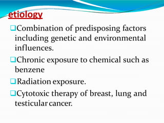
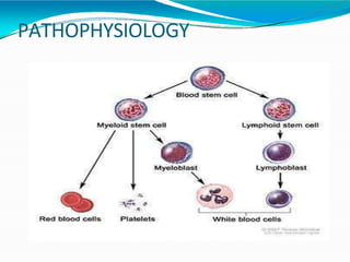
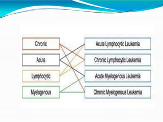
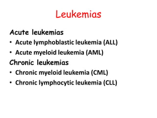
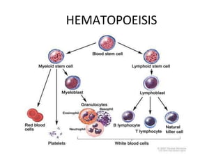
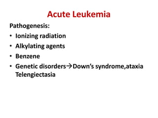
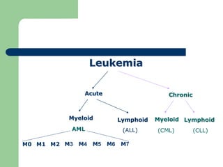
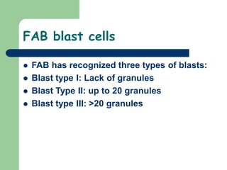
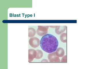
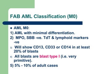
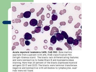
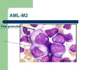
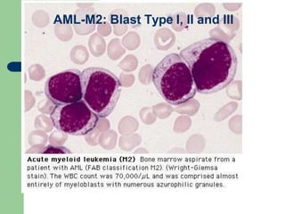
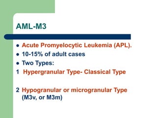
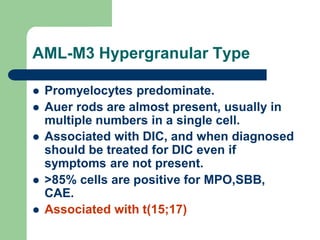
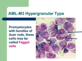
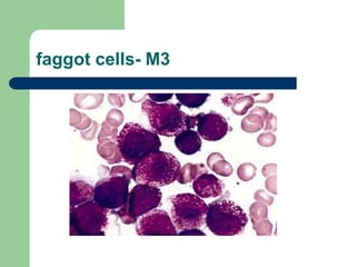
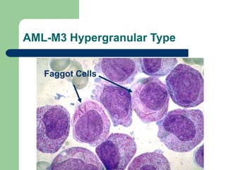
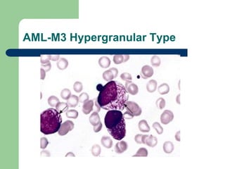
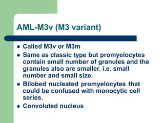
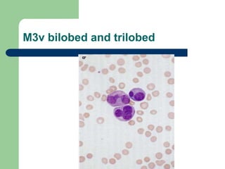
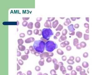
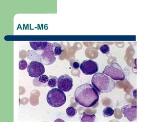
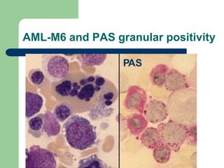
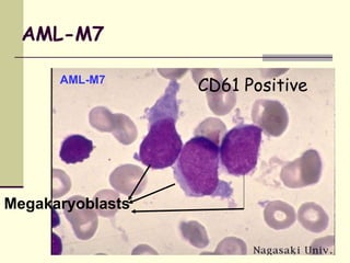
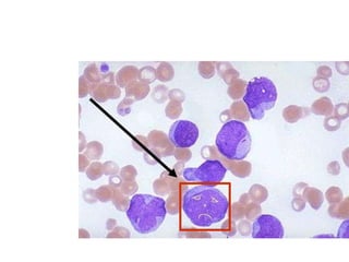
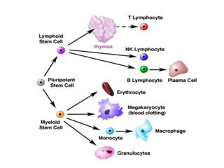
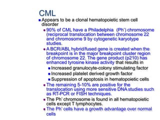
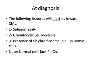
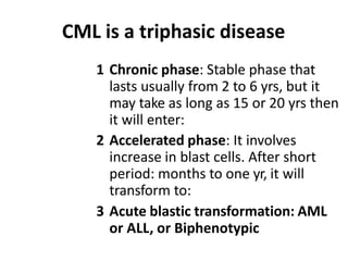
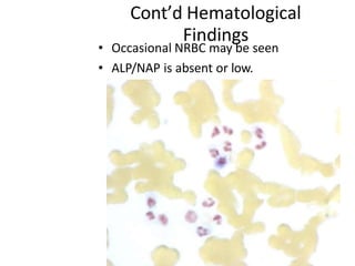
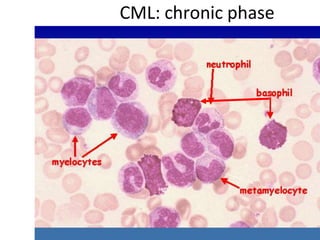
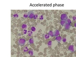
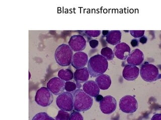
Dr Saket Kumar
Definition
It is a group of malignant
disorder, affecting the blood and
blood –forming tissue of the bone
marrow lymph system and spleen.
⚫Theword Leukemia comes from
the Greek leukos which means
"white" and aima which means
"blood".
⚫The stem cells are committed to
produce specific types of blood
cells. Lymphoid stem cells produce
eitherT or B lymphocytes.
⚫Myeloid stem cellsdifferentiate
into three broad cell types:
RBCs, WBCs, and platelets.
Function of the bone marrow
The bone marrow is found in the inside of
bones. The marrow in the large bones of
adults produces blood cells. Approximately
4% of our total bodyweight consists of bone
marrow.
Thereare two typesof bone marrow:
⚫ 1. Red marrow, made up mainly of myeloid
tissue.
⚫2. Yellow marrow, made up mostly of fat
cells.
⚫Red marrowcan be found in the flat
bones, such as the breast bone, skull,
vertebrae, shoulder blades, hip bone
and ribs. Red marrow can also be
found at the ends of long bones,
such as the humerus and femur.
⚫White blood cells (lymphocytes), red blood
cells and platelets are produced in the red
marrow. Red blood cells carryoxygen, white
blood cells fight diseases. Platelets are
essential for blood clotting.
⚫Yellow marrowcan be found in the insideof
the middle section of long bones.
etiology
Combination of predisposing factors
including genetic and environmental
influences.
Chronic exposure to chemical such as
benzene
Radiation exposure.
Cytotoxic therapy of breast, lung and
testicularcancer.
PATHOPHYSIOLOGY
Leukemias
Acute leukemias
• Acute lymphoblastic leukemia (ALL)
• Acute myeloid leukemia (AML)
Chronic leukemias
• Chronic myeloid leukemia (CML)
• Chronic lymphocytic leukemia (CLL)
Acute leukemias
• Uncontrolled proliferation of blast
(lymphoblast or myeloblast) cells in
bone marrow.
• Normally in BM blast cells are < 5%
of total cells.
• > 30% blasts (FAB classification) /
>20% blasts (WHO classification –
recent)
Acute lymphoblastic leukemia
• Clonal proliferation and accumulation
of lymphoblasts in blood, bone
marrow and other organs
originates in single B or T
lymphocyte progenitor
• Disorder
• Heterogenous disease with different
biological subtypes
HEMATOPOEISIS
Acute lymphoblastic leukemia
• ~ 30% of all childhood
malignancies. Most common
malignancy.
• Incidence in adults : 20% of
acute leukemias
• ALL five times more common than AML.
• ~ peak incidence age 2-5 years
• Boys > girls
Acute Leukemia
Pathogenesis:
• Ionizing radiation
• Alkylating agents
• Benzene
• Genetic disordersDown’s syndrome,ataxia
Telengiectasia
FAB classification
• French-American-British CFN – 1976
• Based on cell morphology & cell
cytochemistry .
Cytochemistry
• Myeloperoxidase stain - Myeloid cells
• Sudan Black B- Myeloid cells
• Periodic acid schiff – Lymhoblasts (L1 type ALL)
Acute lymphoblastic leukemia
(FAB classification)
• L1 : small lymphoblasts with
coarse,condensed chromatin &
inconsistent 1-2 punched out nucleoli.
75% cases
• L2 : large lymphoblasts of variable size
with 1-2 nucleoli. 20% cases
• L3 : large cells, fine chromatin,
vacuolated cytoplasm. 5% cases
L1 type acute lymphoblastic leukemia
ALL L2 Type - PBS
ALL L3 Type – cytoplasmic vacuolation
BasW
ed o
H
n O Classification-2001
Cell Morphology in PBS & BM.
Cytochemistry
• Myeloperoxidase stain - Myeloid cells
• Sudan Black B- Myeloid cells
• Periodic acid schiff – Lymhoblasts (L1 type ALL)
Immunophenotyping – McAntibodies against cell
surface antigen / nuclear antigens (CD
molecules)
Molecular genetics – PCR, RT-PCR –fusion genes &
products.
Cytogenetics
ALL-WHO Classification-2001
• Precursor B ALL / Acute
lymphoblastic lymphoma - 80% of
cases
• Precursor T ALL / Acute
lymphoblastic lymphoma - 15-20%
of cases
Acute Myeloid Leukemia
(AML)
HEMATOPOEISIS
Leukemia
Acute Chronic
Lymphoid
(CLL)
Myeloid
(CML)
Lymphoid
(ALL)
Myeloid
AML
M0 M1 M2 M3 M4 M5 M6 M7
AML
 Clonal malignancies  increased numbers of
immature myeloid cells in the marrow and
blood.
 If left untreated is fatal within a few weeks or
months
 It can present as an acute, illness in a patient
who is otherwise healthy, or as a final
outcome of other Myeloproliferative diseases
(CMPD) or Myelodysplastic syndromes
(MDS).
Age Onset
 AML is the most common form of acute
leukemia during the first few months of
life.
 AML is rare during childhood, while ALL
is the most common during childhood.
 AML is the most frequent during the
middle and late years of life.
AML General Clinical Features
 Often critically ill
 Fever, fatigue, anemia symptoms
 Infections (usually respiratory)
 Bleeding, purpura, spontaneous bruising,
epistaxis
 Leukostatic signs
 Infiltration of tissues (e.g. gum hypertrophy)
 Splenomegaly,
Hepatomegaly,Lymphoadenopathy
LAB studies
1- CBC
 Anemia, neutropenia, thrombocytopenia
 High blast counts in most cases.
 Total White Blood Count exceeds 50
x109/L.
 Blast counts >20% (20-80%)
 May be presented as Aleukemic leukemia:
Pancytopenia with no blasts in PB
 Or presented as subleukemic: TWBC
<15,ooo but with blast cells in Peripheral
Blood.
 NRBC may be seen
 Usually Differential count shows: Blast
cells more than other cells in Blood film.
Bone Marrow
 Hypercellular
 Packed with blast cells of the same
morphology found in clusters.
 Confirms diagnosis (>20% blast Cells)
Cytochemistry
 MPO or SBB is positive in at least 3% of
the cells.
 Esterase +vity :
– NSE (ANBE): Monocytic cells positive
 PAS: M6 and M7 show granular positivity
 LAP or NAP score Low
AML Classification
 The French-American-British (FAB)
cooperative group classification, depends on:
 Morphology
 Cytochemical staining
 Immunophenotyping in selected cases.
AML - Morphological Classification
(FAB)
 M0 – Undifferentiated AML
 M1 - AML without maturation
 M2 - AML with maturation
 M3 - Promyelocytic
 M4 - Myelomonocytic
 M5 - Monoblastic
 M6 - Erythroleukaemic
 M7 - Megakaryoblastic
FAB blast cells
 FAB has recognized three types of blasts:
 Blast type I: Lack of granules
 Blast Type II: up to 20 granules
 Blast type III: >20 granules
Blast Type I
FAB AML Classification (M0)
 AML M0:
1) AML with minimal differentiation.
2) MPO, SBB -ve. TdT & lymphoid markers
-ve
3) Will show CD13, CD33 or CD14 in at least
20% of blasts
4) All blasts are blast type I (i.e. very
primitive).
5) 5% - 10% of adult cases
AML-M1
AML without maturation
2 Myeloblasts type I and type II
3> 90% of NECs in marrow are blasts
4- At least 3% of blasts MPO +ve or
SBB +ve.
5- 10 - 20% of adult cases
AML-M2
AML with maturation
>10% of NECs are promyelocytes to
neutrophils (full range of myelocytic
differentiation)
Type I, II, III blasts are present.
blasts are MPO,SBB positive.
AML-M2
 <20% stain positive with NSE.
 t(8;21) is associated with this type
of leukemia, causing fusion of
AML1/ETO genes.
 8- 30 - 40% of adult cases.
 Good prognosis.
AML-M2
Few granules
AML-M2: Blast Type I, II and III
AML-M3
 Acute Promyelocytic Leukemia (APL).
 10-15% of adult cases
 Two Types:
1 Hypergranular Type- Classical Type
2 Hypogranular or microgranular Type
(M3v, or M3m)
AML-M3 Hypergranular Type
 Promyelocytes predominate.
 Auer rods are almost present, usually in
multiple numbers in a single cell.
 Associated with DIC, and when diagnosed
should be treated for DIC even if
symptoms are not present.
 >85% cells are positive for MPO,SBB,
CAE.
 Associated with t(15;17)
T(15:17) in AML – M3
 Associated with RARα-PML fusion
geneblocks maturation.
 Responds to ATRA (All trans-
retinoic acid) which binds to RARα-
PML protein antagonize its action.
 Example of targeted therapy
 Arsenic trioxide also leads to
differentiation by degrading RARα-
PML protein
AML-M3 Hypergranular Type
Promyelocytes
with bundles of
Auer rods, these
cells may be
called Faggot
cells
faggot cells- M3
AML-M3 Hypergranular Type
Faggot Cells
AML-M3 Hypergranular Type
AML-M3v (M3 variant)
 Called M3v or M3m
 Same as classic type but promyelocytes
contain small number of granules and the
granules also are smaller. i.e. small
number and small size.
 Bilobed nucleated promyelocytes that
could be confused with monocytic cell
series.
 Convoluted nucleus
M3v bilobed and trilobed
AML M3v
AML-M4
 Acute Myelomonocytic Leukemia
 Monocytic series: >20% of marrow
cells are monoblasts, promonocytes,
and mononcytes;
 Myeloid series: Myeloblasts,
promyelocytes, myelocytes and other
mature cells represent 30-80% of
NECs.
AML-M4
 Positive SE and NSE (inhibited by NaF).
 Variable positivity for MPO, and SBB.
 10-15% of adult cases
Monocytic
Myeloid
AML-M4Eo
 Eo stands for Eosinophils.
 So it is acute myelomonocytic leukemia with
eosinophilia.
 Eosinophils are abnormal in appearance
since they contain basophilic granules
admixed with normal eosinophilic granules.
 Associated with Inv(16) that confers good
prognosis
AML-M4Eo
Gingival Infiltration in Monocytic
(AML M4 eos/M5) Variant of AML
AML-M5: Acute Monocytic Leukemia
 10-15% of adult cases
 High rate of 11q23 abnormalities.
 Two subtypes:
– AML-M5a: Acute Monocytic
Leukemia without differentiation.
– AML-M5b: Acute monocytic
Leukemia with differentiation.
AML-M5 Diagnosis
 AML M5a:
 >80% of NECs are monoblasts
 NSE +ve (but NaF sensitive)
AML-M5b
 >80% of NECs are monocytic series:
monoblasts, promonocytes, and monocytes.
 Promonocytes have less chromatin
condensation than monocytes and have
convoluted nuclei.
 NSE Positive (NaF sensitive)
AML-M5a-MONOBLASTS
AML-M5b (Promoncytes, monocytes)
Extramedullary disease (ie, myeloid /granulocytic
sarcoma/myeloblastoma)
Can also have involvement of lymph nodes,
intestine, mediastinum, ovaries, uterus
AML-M6
 Also called Di Guglielmo syndrome or
erythroleukemia
 <5% of adult cases
 >50% of marrow nucleated cells are
erythroblasts, and
 Of the remaining 20% are myeloblasts, if less
then MDS should be considered.
 Dyserythropoiesis in the form of:
Megaloblastoid, bizarre nuclear shape, and
multinucleated giant forms.
AML-M6
 Usually terminate as M1,M2, or M4
 Strong granular PAS positive
erythroblasts.
 Immunophenotyping: The surface marker
Glycophorin is positive.
AML-M6
AML-M6 and PAS granular positivity
PAS
AML-M7
 Acute Megakaryoblastic Leukemia
 <5% of adult cases
 >20% of marrow are megakaryoblasts.
 Usually diagnosed by
immunophenotyping: CD61 is positive.
 Megakaryoblasts are PAS positive.
AML-M7
AML-M7
Megakaryoblasts
CD61 Positive
PAS diffuse granular positive: 3 Megakaryoblast
(AML-M7) with one strong positive neutrophil
AML Cytogenetic Abnormalities
 50-70% AML shows cytogenetic
abnormalities.
 10% of AML is associated with trisomy 8,
and is associated with bad prognosis.
 t(8;21) in AML M2 somewhat better
prognosis.
 t(15;17) in AML M3 good prognosis.
 Abnormalities in chromosome 11q23 in
AML M4 and M5.—bad prognosis.
 Inv(16) in AML M4Eo- good prognosis.
WHO CLASSIFICATION OF
AML
AML –WHO Classification
1.AML with recurrent genetic abnormalities.
T(8:21), t(16), inv(16), t(15:17), 11q23.
2. AML with MDS like features :bad
prognosis.
With prior MDS
With multilineage dysplasia
3. AML therapy related (chemotherapy/
radiotherapy)—bad prognosis
4. AML not otherwise specified (FAB type
M0-M7 except for M3) /Biphenotypic
leukemia
CHRONIC MYELOID LEUKEMIA
CML
Chronic Myeloid Leukemia
• Abbreviated as CML
• Synonyms:
1- Chronic myelogenous leukemia
2- Chronic granulocytic leukemia
CML
 Is characterized by an unregulated
proliferation of myeloid elements in the
bone marrow, liver and spleen, leading to
marked leukocytosis and organomegaly.
 Incidence
20% of all leukemias
Primarily affects adults 25-60 years old,
with a peak incidence at 40-59.
Etiology
Anything that can induce
chromosomal aberrations such as
ionizing radiation, alkylating agents,
and exposure to other biologically
active chemicals.
CML
 Appears to be a clonal hematopoietic stem cell
disorder
 90% of CML have a Philadelphia (Ph’) chromosome
(reciprocal translocation between chromosome 22
and chromosome 9 by cytogenetic karyotype
studies.
 A BCR/ABL hybrid/fused gene is created when the
breakpoint is in the major breakpoint cluster region
of chromosome 22. The gene product (p210) has
enhanced tyrosine kinase activity that results in
 Increased granulocyte-colony stimulating factor
 Increased platelet derived growth factor
 Suppression of apoptosis in hematopoietic cells
 The remaining 5-10% are positive for the
translocation using more sensitive DNA studies such
as RT-PCR or FISH techniques.
 The Ph’ chromosome is found in all hematopoietic
cells except T lymphocytes.
 The Ph’ cells have a growth advantage over normal
cells
At diagnosis
• The following features will alert us toward
CML:
• 1- Splenomegaly
• 2- Granulocytic Leukocytosis
• 3- Presence of Ph chromosome in all leukemic
cells.
• Note: Normal cells lack Ph Ch.
CML is a triphasic disease
1 Chronic phase: Stable phase that
lasts usually from 2 to 6 yrs, but it
may take as long as 15 or 20 yrs then
it will enter:
2 Accelerated phase: It involves
increase in blast cells. After short
period: months to one yr, it will
transform to:
3 Acute blastic transformation: AML
or ALL, or Biphenotypic
Clinical Course: Phases of CML
Chronic phase
Median 4–6 years
stabilization
Accelerated phase
Median duration
up to 1 year
Blastic phase (blast crisis)
Median survival
3–6 months
Terminal phase
Advanced phases
In CML usually
• Myelopoiesis that involve primarily
the granulocytic series is increased.
• Also, common, megakaryopoiesis is
increased and manifested as high PLT
count.
• But erythroid hyperplasia and
polycythemia occur only rarely
Cytogenetics
• In more than 90% of CML cases Philadelphia
chromosome is present in the leukemic cells.
This leads to the formation of BCR-ABL gene
that would be translated to a novel chimeric
protein with oncogenic activity.
BCR-ABL
• ABL gene is normally found in Ch. 9.
• This gene normally encodes for a
tyrosine kinase.
• But after fusion with BCR gene on
ch.22, the chimeric protein (P210 kda)
oncoprotein has very far greater TK
activity than the normal ABL gene
product.
Clinical Findings
• Symptoms are related to:
– Splenomegaly
– Hemorrhage from different sites
– Anemia
50% of CML cases
• 50% of CML cases are diagnosed
accidentally:
– During pregnancy
– Before blood donation
– After RTA
– Other causes that let the patient to visit
physicians.
Symptoms
• Loss of energy-Easy Fatiguability
• Shortness of breath on exertion.
• Weight loss
• Night sweat
• Fever
• Hemorrhage from various sites
• Left Upper quadrant discomfort
• Splenomegaly
• Hepatomegaly
• Lymphadenopathy
• Bone tenderness
Hematological Findings
• Anemia but not severe
• TWBC range: 20-200x109/L, but counts as
high as 1000x109/L have been reported.
– ¾ have WBC counts > 100 x 109/L
• BF shows left shift- full spectrum of
normally appeared cells in the granulocytic
series (but they are not functionally normal,
however), ranging from blast forms to
mature neutrophil.
• But myelocytes and polymorphs
predominate- Myelocyte peak, and PMN
peak
Cont’d Hematological Findings
• Blasts are usually less than 10%.
• Basophilia
• For your knowledge: Basophilia may
alert us for a malignancy.
• Eosinophilia
• PLT are usually increased (300-600
x109/L), but may be normal or
decreased.
Cont’d Hematological
Findings
• Occasional NRBC may be seen
• ALP/NAP is absent or low.
CML: chronic phase
Accelerated phase
Blast Transformation
CHRONIC LYMPHOCYTIC LEUKEMIA
CLL
 This is predominantly a disease of the elderly;
> 90% are over 50 and 2/3 are over 60;
male:female ratio is 2:1
 Is characterized by peripheral and bone
marrow lymphocytosis and a survival of a few
years to > 10 years
 This is a B cell abnormality
 The lymphocytes appear normal, but are
immunologically incompetent. However,
some functionally normal B cells remain and
there is a normal T cell pool
 Etiology
Genetic factors are important since it runs in
families
 Clinical course
The pace of the disease varies and is
dependent on the rate of accumulation of
abnormal lymphocytes
Median survival is 3-4 years, but 10-15%
survive > 10years
There is no tendency for blast
transformation, but complications of
advanced disease result from progressive
accumulation of long-lived, poorly functional
lymphocytes.
 Signs and symptoms
Organomegaly and lymphadenopathy
Often discovered accidentally
Fatigue
 Lab features
Absolute lymphocytosis of 10-150 x 109/L
Lymphocytes usually appear normal, but they
are markedly fragile and smudge cells are
seen on the peripheral smear
It is not necessary to do a bone marrow biopsy
for diagnosis.
Anemia occurs late in the disease and may be
due to decreased production secondary to
marrow infiltration, hypersplenism, or
autoimmune hemolytic anemia: the same
things may cause neutropenia or
thrombocytopenia
Hypogammaglobulinemia as the disease
progresses
CLL WITH SMUDGE CELLS
Smudge cell
THANKYOU

Leukaemia.pptx

  • 1.
  • 2.
    Definition It is agroup of malignant disorder, affecting the blood and blood –forming tissue of the bone marrow lymph system and spleen.
  • 3.
    ⚫Theword Leukemia comesfrom the Greek leukos which means "white" and aima which means "blood".
  • 4.
    ⚫The stem cellsare committed to produce specific types of blood cells. Lymphoid stem cells produce eitherT or B lymphocytes. ⚫Myeloid stem cellsdifferentiate into three broad cell types: RBCs, WBCs, and platelets.
  • 5.
    Function of thebone marrow The bone marrow is found in the inside of bones. The marrow in the large bones of adults produces blood cells. Approximately 4% of our total bodyweight consists of bone marrow. Thereare two typesof bone marrow: ⚫ 1. Red marrow, made up mainly of myeloid tissue. ⚫2. Yellow marrow, made up mostly of fat cells.
  • 6.
    ⚫Red marrowcan befound in the flat bones, such as the breast bone, skull, vertebrae, shoulder blades, hip bone and ribs. Red marrow can also be found at the ends of long bones, such as the humerus and femur.
  • 7.
    ⚫White blood cells(lymphocytes), red blood cells and platelets are produced in the red marrow. Red blood cells carryoxygen, white blood cells fight diseases. Platelets are essential for blood clotting. ⚫Yellow marrowcan be found in the insideof the middle section of long bones.
  • 8.
    etiology Combination of predisposingfactors including genetic and environmental influences. Chronic exposure to chemical such as benzene Radiation exposure. Cytotoxic therapy of breast, lung and testicularcancer.
  • 9.
  • 11.
    Leukemias Acute leukemias • Acutelymphoblastic leukemia (ALL) • Acute myeloid leukemia (AML) Chronic leukemias • Chronic myeloid leukemia (CML) • Chronic lymphocytic leukemia (CLL)
  • 12.
    Acute leukemias • Uncontrolledproliferation of blast (lymphoblast or myeloblast) cells in bone marrow. • Normally in BM blast cells are < 5% of total cells. • > 30% blasts (FAB classification) / >20% blasts (WHO classification – recent)
  • 13.
    Acute lymphoblastic leukemia •Clonal proliferation and accumulation of lymphoblasts in blood, bone marrow and other organs originates in single B or T lymphocyte progenitor • Disorder • Heterogenous disease with different biological subtypes
  • 14.
  • 15.
    Acute lymphoblastic leukemia •~ 30% of all childhood malignancies. Most common malignancy. • Incidence in adults : 20% of acute leukemias • ALL five times more common than AML. • ~ peak incidence age 2-5 years • Boys > girls
  • 16.
    Acute Leukemia Pathogenesis: • Ionizingradiation • Alkylating agents • Benzene • Genetic disordersDown’s syndrome,ataxia Telengiectasia
  • 17.
    FAB classification • French-American-BritishCFN – 1976 • Based on cell morphology & cell cytochemistry . Cytochemistry • Myeloperoxidase stain - Myeloid cells • Sudan Black B- Myeloid cells • Periodic acid schiff – Lymhoblasts (L1 type ALL)
  • 18.
    Acute lymphoblastic leukemia (FABclassification) • L1 : small lymphoblasts with coarse,condensed chromatin & inconsistent 1-2 punched out nucleoli. 75% cases • L2 : large lymphoblasts of variable size with 1-2 nucleoli. 20% cases • L3 : large cells, fine chromatin, vacuolated cytoplasm. 5% cases
  • 19.
    L1 type acutelymphoblastic leukemia
  • 20.
  • 21.
    ALL L3 Type– cytoplasmic vacuolation
  • 22.
    BasW ed o H n OClassification-2001 Cell Morphology in PBS & BM. Cytochemistry • Myeloperoxidase stain - Myeloid cells • Sudan Black B- Myeloid cells • Periodic acid schiff – Lymhoblasts (L1 type ALL) Immunophenotyping – McAntibodies against cell surface antigen / nuclear antigens (CD molecules) Molecular genetics – PCR, RT-PCR –fusion genes & products. Cytogenetics
  • 23.
    ALL-WHO Classification-2001 • PrecursorB ALL / Acute lymphoblastic lymphoma - 80% of cases • Precursor T ALL / Acute lymphoblastic lymphoma - 15-20% of cases
  • 24.
  • 25.
  • 26.
  • 27.
    AML  Clonal malignancies increased numbers of immature myeloid cells in the marrow and blood.  If left untreated is fatal within a few weeks or months  It can present as an acute, illness in a patient who is otherwise healthy, or as a final outcome of other Myeloproliferative diseases (CMPD) or Myelodysplastic syndromes (MDS).
  • 28.
    Age Onset  AMLis the most common form of acute leukemia during the first few months of life.  AML is rare during childhood, while ALL is the most common during childhood.  AML is the most frequent during the middle and late years of life.
  • 29.
    AML General ClinicalFeatures  Often critically ill  Fever, fatigue, anemia symptoms  Infections (usually respiratory)  Bleeding, purpura, spontaneous bruising, epistaxis  Leukostatic signs  Infiltration of tissues (e.g. gum hypertrophy)  Splenomegaly, Hepatomegaly,Lymphoadenopathy
  • 30.
    LAB studies 1- CBC Anemia, neutropenia, thrombocytopenia  High blast counts in most cases.  Total White Blood Count exceeds 50 x109/L.  Blast counts >20% (20-80%)
  • 31.
     May bepresented as Aleukemic leukemia: Pancytopenia with no blasts in PB  Or presented as subleukemic: TWBC <15,ooo but with blast cells in Peripheral Blood.  NRBC may be seen  Usually Differential count shows: Blast cells more than other cells in Blood film.
  • 32.
    Bone Marrow  Hypercellular Packed with blast cells of the same morphology found in clusters.  Confirms diagnosis (>20% blast Cells)
  • 33.
    Cytochemistry  MPO orSBB is positive in at least 3% of the cells.  Esterase +vity : – NSE (ANBE): Monocytic cells positive  PAS: M6 and M7 show granular positivity  LAP or NAP score Low
  • 34.
    AML Classification  TheFrench-American-British (FAB) cooperative group classification, depends on:  Morphology  Cytochemical staining  Immunophenotyping in selected cases.
  • 35.
    AML - MorphologicalClassification (FAB)  M0 – Undifferentiated AML  M1 - AML without maturation  M2 - AML with maturation  M3 - Promyelocytic  M4 - Myelomonocytic  M5 - Monoblastic  M6 - Erythroleukaemic  M7 - Megakaryoblastic
  • 36.
    FAB blast cells FAB has recognized three types of blasts:  Blast type I: Lack of granules  Blast Type II: up to 20 granules  Blast type III: >20 granules
  • 37.
  • 38.
    FAB AML Classification(M0)  AML M0: 1) AML with minimal differentiation. 2) MPO, SBB -ve. TdT & lymphoid markers -ve 3) Will show CD13, CD33 or CD14 in at least 20% of blasts 4) All blasts are blast type I (i.e. very primitive). 5) 5% - 10% of adult cases
  • 40.
    AML-M1 AML without maturation 2Myeloblasts type I and type II 3> 90% of NECs in marrow are blasts 4- At least 3% of blasts MPO +ve or SBB +ve. 5- 10 - 20% of adult cases
  • 42.
    AML-M2 AML with maturation >10%of NECs are promyelocytes to neutrophils (full range of myelocytic differentiation) Type I, II, III blasts are present. blasts are MPO,SBB positive.
  • 43.
    AML-M2  <20% stainpositive with NSE.  t(8;21) is associated with this type of leukemia, causing fusion of AML1/ETO genes.  8- 30 - 40% of adult cases.  Good prognosis.
  • 44.
  • 45.
    AML-M2: Blast TypeI, II and III
  • 46.
    AML-M3  Acute PromyelocyticLeukemia (APL).  10-15% of adult cases  Two Types: 1 Hypergranular Type- Classical Type 2 Hypogranular or microgranular Type (M3v, or M3m)
  • 47.
    AML-M3 Hypergranular Type Promyelocytes predominate.  Auer rods are almost present, usually in multiple numbers in a single cell.  Associated with DIC, and when diagnosed should be treated for DIC even if symptoms are not present.  >85% cells are positive for MPO,SBB, CAE.  Associated with t(15;17)
  • 48.
    T(15:17) in AML– M3  Associated with RARα-PML fusion geneblocks maturation.  Responds to ATRA (All trans- retinoic acid) which binds to RARα- PML protein antagonize its action.  Example of targeted therapy  Arsenic trioxide also leads to differentiation by degrading RARα- PML protein
  • 49.
    AML-M3 Hypergranular Type Promyelocytes withbundles of Auer rods, these cells may be called Faggot cells
  • 50.
  • 51.
  • 52.
  • 53.
    AML-M3v (M3 variant) Called M3v or M3m  Same as classic type but promyelocytes contain small number of granules and the granules also are smaller. i.e. small number and small size.  Bilobed nucleated promyelocytes that could be confused with monocytic cell series.  Convoluted nucleus
  • 54.
  • 55.
  • 56.
    AML-M4  Acute MyelomonocyticLeukemia  Monocytic series: >20% of marrow cells are monoblasts, promonocytes, and mononcytes;  Myeloid series: Myeloblasts, promyelocytes, myelocytes and other mature cells represent 30-80% of NECs.
  • 57.
    AML-M4  Positive SEand NSE (inhibited by NaF).  Variable positivity for MPO, and SBB.  10-15% of adult cases
  • 58.
  • 59.
    AML-M4Eo  Eo standsfor Eosinophils.  So it is acute myelomonocytic leukemia with eosinophilia.  Eosinophils are abnormal in appearance since they contain basophilic granules admixed with normal eosinophilic granules.  Associated with Inv(16) that confers good prognosis
  • 61.
  • 62.
    Gingival Infiltration inMonocytic (AML M4 eos/M5) Variant of AML
  • 63.
    AML-M5: Acute MonocyticLeukemia  10-15% of adult cases  High rate of 11q23 abnormalities.  Two subtypes: – AML-M5a: Acute Monocytic Leukemia without differentiation. – AML-M5b: Acute monocytic Leukemia with differentiation.
  • 64.
    AML-M5 Diagnosis  AMLM5a:  >80% of NECs are monoblasts  NSE +ve (but NaF sensitive)
  • 65.
    AML-M5b  >80% ofNECs are monocytic series: monoblasts, promonocytes, and monocytes.  Promonocytes have less chromatin condensation than monocytes and have convoluted nuclei.  NSE Positive (NaF sensitive)
  • 66.
  • 67.
  • 68.
    Extramedullary disease (ie,myeloid /granulocytic sarcoma/myeloblastoma) Can also have involvement of lymph nodes, intestine, mediastinum, ovaries, uterus
  • 69.
    AML-M6  Also calledDi Guglielmo syndrome or erythroleukemia  <5% of adult cases  >50% of marrow nucleated cells are erythroblasts, and  Of the remaining 20% are myeloblasts, if less then MDS should be considered.  Dyserythropoiesis in the form of: Megaloblastoid, bizarre nuclear shape, and multinucleated giant forms.
  • 70.
    AML-M6  Usually terminateas M1,M2, or M4  Strong granular PAS positive erythroblasts.  Immunophenotyping: The surface marker Glycophorin is positive.
  • 71.
  • 72.
    AML-M6 and PASgranular positivity PAS
  • 73.
    AML-M7  Acute MegakaryoblasticLeukemia  <5% of adult cases  >20% of marrow are megakaryoblasts.  Usually diagnosed by immunophenotyping: CD61 is positive.  Megakaryoblasts are PAS positive.
  • 74.
  • 75.
    PAS diffuse granularpositive: 3 Megakaryoblast (AML-M7) with one strong positive neutrophil
  • 76.
    AML Cytogenetic Abnormalities 50-70% AML shows cytogenetic abnormalities.  10% of AML is associated with trisomy 8, and is associated with bad prognosis.  t(8;21) in AML M2 somewhat better prognosis.  t(15;17) in AML M3 good prognosis.  Abnormalities in chromosome 11q23 in AML M4 and M5.—bad prognosis.  Inv(16) in AML M4Eo- good prognosis.
  • 77.
  • 78.
    AML –WHO Classification 1.AMLwith recurrent genetic abnormalities. T(8:21), t(16), inv(16), t(15:17), 11q23. 2. AML with MDS like features :bad prognosis. With prior MDS With multilineage dysplasia 3. AML therapy related (chemotherapy/ radiotherapy)—bad prognosis 4. AML not otherwise specified (FAB type M0-M7 except for M3) /Biphenotypic leukemia
  • 79.
  • 82.
    Chronic Myeloid Leukemia •Abbreviated as CML • Synonyms: 1- Chronic myelogenous leukemia 2- Chronic granulocytic leukemia
  • 83.
    CML  Is characterizedby an unregulated proliferation of myeloid elements in the bone marrow, liver and spleen, leading to marked leukocytosis and organomegaly.  Incidence 20% of all leukemias Primarily affects adults 25-60 years old, with a peak incidence at 40-59.
  • 84.
    Etiology Anything that caninduce chromosomal aberrations such as ionizing radiation, alkylating agents, and exposure to other biologically active chemicals.
  • 85.
    CML  Appears tobe a clonal hematopoietic stem cell disorder  90% of CML have a Philadelphia (Ph’) chromosome (reciprocal translocation between chromosome 22 and chromosome 9 by cytogenetic karyotype studies.  A BCR/ABL hybrid/fused gene is created when the breakpoint is in the major breakpoint cluster region of chromosome 22. The gene product (p210) has enhanced tyrosine kinase activity that results in  Increased granulocyte-colony stimulating factor  Increased platelet derived growth factor  Suppression of apoptosis in hematopoietic cells  The remaining 5-10% are positive for the translocation using more sensitive DNA studies such as RT-PCR or FISH techniques.  The Ph’ chromosome is found in all hematopoietic cells except T lymphocytes.  The Ph’ cells have a growth advantage over normal cells
  • 86.
    At diagnosis • Thefollowing features will alert us toward CML: • 1- Splenomegaly • 2- Granulocytic Leukocytosis • 3- Presence of Ph chromosome in all leukemic cells. • Note: Normal cells lack Ph Ch.
  • 87.
    CML is atriphasic disease 1 Chronic phase: Stable phase that lasts usually from 2 to 6 yrs, but it may take as long as 15 or 20 yrs then it will enter: 2 Accelerated phase: It involves increase in blast cells. After short period: months to one yr, it will transform to: 3 Acute blastic transformation: AML or ALL, or Biphenotypic
  • 88.
    Clinical Course: Phasesof CML Chronic phase Median 4–6 years stabilization Accelerated phase Median duration up to 1 year Blastic phase (blast crisis) Median survival 3–6 months Terminal phase Advanced phases
  • 89.
    In CML usually •Myelopoiesis that involve primarily the granulocytic series is increased. • Also, common, megakaryopoiesis is increased and manifested as high PLT count. • But erythroid hyperplasia and polycythemia occur only rarely
  • 90.
    Cytogenetics • In morethan 90% of CML cases Philadelphia chromosome is present in the leukemic cells. This leads to the formation of BCR-ABL gene that would be translated to a novel chimeric protein with oncogenic activity.
  • 91.
    BCR-ABL • ABL geneis normally found in Ch. 9. • This gene normally encodes for a tyrosine kinase. • But after fusion with BCR gene on ch.22, the chimeric protein (P210 kda) oncoprotein has very far greater TK activity than the normal ABL gene product.
  • 92.
    Clinical Findings • Symptomsare related to: – Splenomegaly – Hemorrhage from different sites – Anemia
  • 93.
    50% of CMLcases • 50% of CML cases are diagnosed accidentally: – During pregnancy – Before blood donation – After RTA – Other causes that let the patient to visit physicians.
  • 94.
    Symptoms • Loss ofenergy-Easy Fatiguability • Shortness of breath on exertion. • Weight loss • Night sweat • Fever • Hemorrhage from various sites • Left Upper quadrant discomfort • Splenomegaly • Hepatomegaly • Lymphadenopathy • Bone tenderness
  • 95.
    Hematological Findings • Anemiabut not severe • TWBC range: 20-200x109/L, but counts as high as 1000x109/L have been reported. – ¾ have WBC counts > 100 x 109/L • BF shows left shift- full spectrum of normally appeared cells in the granulocytic series (but they are not functionally normal, however), ranging from blast forms to mature neutrophil. • But myelocytes and polymorphs predominate- Myelocyte peak, and PMN peak
  • 96.
    Cont’d Hematological Findings •Blasts are usually less than 10%. • Basophilia • For your knowledge: Basophilia may alert us for a malignancy. • Eosinophilia • PLT are usually increased (300-600 x109/L), but may be normal or decreased.
  • 97.
    Cont’d Hematological Findings • OccasionalNRBC may be seen • ALP/NAP is absent or low.
  • 98.
  • 99.
  • 100.
  • 101.
    CHRONIC LYMPHOCYTIC LEUKEMIA CLL This is predominantly a disease of the elderly; > 90% are over 50 and 2/3 are over 60; male:female ratio is 2:1  Is characterized by peripheral and bone marrow lymphocytosis and a survival of a few years to > 10 years  This is a B cell abnormality  The lymphocytes appear normal, but are immunologically incompetent. However, some functionally normal B cells remain and there is a normal T cell pool
  • 102.
     Etiology Genetic factorsare important since it runs in families  Clinical course The pace of the disease varies and is dependent on the rate of accumulation of abnormal lymphocytes Median survival is 3-4 years, but 10-15% survive > 10years There is no tendency for blast transformation, but complications of advanced disease result from progressive accumulation of long-lived, poorly functional lymphocytes.  Signs and symptoms Organomegaly and lymphadenopathy Often discovered accidentally Fatigue
  • 103.
     Lab features Absolutelymphocytosis of 10-150 x 109/L Lymphocytes usually appear normal, but they are markedly fragile and smudge cells are seen on the peripheral smear It is not necessary to do a bone marrow biopsy for diagnosis. Anemia occurs late in the disease and may be due to decreased production secondary to marrow infiltration, hypersplenism, or autoimmune hemolytic anemia: the same things may cause neutropenia or thrombocytopenia Hypogammaglobulinemia as the disease progresses
  • 104.
    CLL WITH SMUDGECELLS Smudge cell
  • 105.