The document describes the LENSTAR LS 900 optical biometry device. It provides 8 measurements in a single procedure, including corneal thickness, anterior chamber depth, lens thickness, axial length, keratometry, white-to-white distance, pupilometry, and eccentricity of the optical axis. The measurements are precise, taken on the true optical axis using optical coherence technology. The device allows for fast, patient-friendly measurements and integrated IOL power calculation.
The document discusses patterns of strabismus, specifically the A pattern and V pattern. The A pattern involves relative convergence on upgaze and divergence on downgaze, while the V pattern is the opposite with relative divergence on upgaze and convergence on downgaze. Variants include the X, Y, lambda, and diamond patterns. The etiology of these patterns involves dysfunction of the horizontal, vertical, or oblique eye muscles. Clinical features may include anomalous head posture, amblyopia, and abnormal retinal correspondence. Diagnosis involves measuring alignment in upgaze and downgaze while preventing accommodation.
The document discusses various formulas used for calculating intraocular lens (IOL) power, including SRK, SRK2, Holladay, Haigis, and Holladay 2. It explains the factors these formulas account for such as axial length, corneal power, anterior chamber depth, and how they have evolved over generations to improve accuracy. Special considerations for calculating IOL power in cases involving prior refractive surgery, silicone oil filling, posterior staphyloma, and using optical biometry devices are also summarized.
Optical and Non-optical Methods of Measuring Axial Length of EyeRabindraAdhikary
This document discusses various optical and non-optical methods of measuring axial length of the eye. It begins by defining axial length and noting its importance in intraocular lens power calculations. It then describes ultrasonic (A-scan) biometry, the historical standard, and optical biometry techniques like partial coherence interferometry used in devices like the IOLMaster 500. Key advantages of optical techniques are discussed as well as limitations of ultrasound. Details are provided on performing both immersion and non-immersion ultrasound techniques and interpreting the results.
The document discusses Pentacam, a diagnostic tool that uses a rotating Scheimpflug camera to capture 50 images in 2 seconds and create a 3D model of the anterior eye segment. It has applications in assessing corneal ectasia, refractive surgery, corrected intraocular pressure, corneal aberrations, IOL power calculation, and densitometry. The Pentacam provides curvature, pachymetry, and elevation maps. It can detect ectasia by identifying if the highest curvature, thinnest thickness, and steepest elevation points coincide. The Pentacam is also used to measure corneal aberrations via Zernike analysis and calculate accurate IOL power for patients with previous refractive surgery or cataracts.
This document discusses biometry, which involves measuring the eye to determine the ideal intraocular lens power for cataract surgery. It notes that biometry errors are the second most common cause of claims in cataract malpractice cases. It describes various techniques for measuring the corneal curvature and axial length of the eye, including manual and automated keratometry, ultrasound A-scan, and optical biometers. It also discusses considerations for biometry in special cases and different intraocular lens calculation formulas.
This document provides guidelines for prescribing glasses in children. It discusses that the pediatric eye is different from the adult eye in terms of axial length, corneal curvature, and lens power. The goals of prescribing glasses in children are to provide a focused retinal image and achieve optimal balance between accommodation and convergence. It is more difficult to prescribe glasses for children due to lack of subjective response and poor attention. American guidelines provide recommendations on refractive errors that warrant correction at different ages. Factors like emmetropization, amblyopia risk, and presence of strabismus are considered. Frame selection depends on the child's condition and age, aiming for correct fit, comfort, safety, and not hindering nasal development.
Heidelberg Retinal Tomography II (HRT II) is a diagnostic imaging technique that uses confocal laser scanning to generate 3D topographic images of the optic disc and retinal nerve fiber layer. It provides quantitative measurements of parameters like cup-to-disc ratio, rim area, and cup shape that are useful for diagnosing and monitoring glaucoma. The HRT II obtains multiple optical sections to build a 3D image with a resolution of 10 micrometers per pixel. It has good test-retest reproducibility and can detect glaucomatous nerve damage earlier than conventional techniques. However, small optic discs continue to present challenges for accurate classification of glaucoma status.
The presentation presents some treatment modalities as regards AI.This is to keep you thinking more on how to approach a case of AI in terms of management.
Ultrasonography uses ultrasound to image tissues within the body. A-scan ultrasonography provides a one-dimensional view of the eye by measuring the echoes of ultrasound waves. It can be used to detect and measure tumors, assess eye structures for IOL calculation, and interpret pathology. The ultrasound is reflected at interfaces between tissues, appearing as spikes on the display. Immersion techniques provide more accurate measurements than contact techniques by avoiding compression artifacts. Limitations include artifacts, small lesions, missed foreign bodies, and misalignment issues.
This document provides information about optical biometry and the IOL Master device. It discusses the principles and history of optical interferometry, intended uses of the IOL Master including axial length measurement, corneal curvature measurement, and IOL power calculation. Screen layouts and measurements taken by the IOL Master are described. Advantages include highly accurate and non-contact measurements, while limitations include inability to measure in cases of severe media opacities or poor patient cooperation.
This document provides information on contact lens options for managing keratoconus. It discusses various soft lens designs, scleral lenses, corneal lenses, and hybrid lens systems. For corneal lenses, it describes two fitting philosophies - apical bearing and apical clearance. Specific lens designs are also outlined, including Soper, McGuire, NiCone, and ROSE K lenses, which are designed to closely fit the irregular shape of the cornea in keratoconus. The document provides details on parameters like total diameter, base curve radius, optic zone diameter, and materials for fitting these specialized lenses.
The document summarizes the Amsler grid, a diagnostic tool used since 1945 to screen for and monitor macular diseases. It consists of a grid with a central dot that patients look at to detect any distortions, gaps, or blurred areas in their central vision. Various versions are available, including ones with different colors, patterns of lines, or dot sizes to test specific parts of the visual field and detect different types of visual abnormalities that could indicate conditions like macular degeneration or glaucoma. The procedure involves having patients view the grid with each eye separately at 16 inches and report any anomalies in the lines of the grid.
The optical center of a lens is the point where light rays pass through without deviation. It is important for the optical center to be directly in front of the pupil for optimum vision. Decentering a lens, or moving it so the optical center is no longer in front of the pupil, introduces a prismatic effect. The amount of prismatic effect, measured in prism diopters, is calculated by multiplying the distance the lens is decentered in centimeters by the lens power in diopters. Decentering a lens with a spherical prescription or cylinder introduces different prismatic effects depending on the orientation of the cylinder axis relative to the direction of decentration.
Pediatric Ophthalmic dispensing in different visual problemsRaju Kaiti
Pediatric dispensing, introduction, different from adult dispensing, frame selection, lens selection, special case fitting, Do's and Dont's, Measurements, Down's syndrome, albinism, aphakia, strabismus, syndromes
The document discusses lensometry, which is the process of using a lensometer or lensmeter to measure the optical properties of lenses. A lensometer projects lines that allow optometrists to determine information like the sphere, cylinder, and axis measurements specified in a prescription. It can also verify the accuracy of lenses and detect their type (spherical, astigmatic, prismatic). Lensometers are used to properly fit lenses into frames and ensure prescriptions are correct. The document outlines the history of the lensometer's invention and provides details on its use, parts, manual operation, and the measurements it can obtain for different lens types like bifocals.
This document provides guidance on selecting eyeglass frames. It discusses that the primary purposes of frames are to hold lenses to provide optimal vision and be physically comfortable. Additional considerations for frame selection include appearance, fashion, prescription needs, face shape, coloring, nose shape, hairline, image, work environment, habits, budget, and fashion trends. General guidelines are to ensure proper positioning of lenses within the frames based on factors like face shape, width, brow position, and proportion. Color and frame style should complement the face. Frame material and design should account for prescription, activities, and durability needs. Fashion has also become an important factor in frame selection.
Contact lens options in keratoconus hiraHira Dahal
Rigid gas permeable lenses are commonly used to correct vision in keratoconus, though some patients cannot tolerate them. Piggyback or hybrid lens systems can improve comfort by combining a rigid lens with a soft lens. Newer options like mini-scleral lenses and Rose K lenses are designed specifically for keratoconus, vaulting the irregular cornea to improve vision. The Boston PROSE treatment also creates a new smooth optical surface over the cornea using customized prosthetic lenses. While fitting lenses for keratoconus can be challenging, contact lenses are often able to restore vision without surgery.
Amblyopia is a condition of reduced vision in one or both eyes that is not caused by structural eye problems. It occurs during early childhood development when there is inadequate visual stimulation to one or both eyes. Common causes include strabismus, significant refractive error differences between the eyes, form deprivation, and abnormal binocular interaction. Treatment involves correcting any refractive errors and using occlusion therapy or drugs to blur vision in the non-amblyopic eye, forcing use of the amblyopic eye. Occlusion therapy is the most common treatment but requires compliance to achieve results. Other options include penalization, visual stimulation, and drugs, but occlusion remains the standard first approach. Success depends on early diagnosis and treatment before age 7.
This document discusses the verification process for contact lenses. It has two main stages - laboratory and clinical. In the laboratory, lenses are checked to ensure their parameters match what was ordered. Clinically, lenses should be verified upon receipt to ensure the correct lens was dispensed. Parameters like radius of curvature, diameters, thickness and power must be measured for both rigid and soft contact lenses using various techniques and instruments. On-eye verification is also important to assess fit and comfort. The goal of verification is to ensure patients receive high quality lenses that meet specifications and provide good vision.
This document discusses different types of tinted lenses, including their purposes and materials. It covers integral tints produced during manufacturing by adding metals or metal oxides to glass. Surface coatings deposit metallic oxides onto glass through evaporation. Plastic lenses are dyed by immersing them in organic dyes. Various tint colors like yellow, red, purple, and brown are explained in terms of the materials used and their applications. Integral tints provide consistent tinting while surface coatings and dyes allow tinting of any prescription.
Biometry is used to measure the eye to determine the correct intraocular lens power for cataract surgery. It involves measuring the corneal power with keratometry and the eye length with axial length measurement. The optimal method is optical biometry which measures both simultaneously while allowing the patient to fixate, improving accuracy. Special cases like high myopia, prior refractive surgery, or pathology require adjusted measurement techniques or formulas to calculate the lens power accurately.
The document provides information on axial length measurement techniques using ultrasound (A-scan) biometry. It discusses average axial lengths, accuracy of measurements, examination procedure, potential sources of error for different techniques, instrument settings, and special measurement considerations. Key points include:
- The average axial length of a normal eye is 23.06mm, ranging mostly from 22-24.5mm.
- Accuracy of A-scan ultrasound is ±0.1mm. Differences between eyes should be ≤0.3mm.
- Potential sources of error include corneal compression, fluid excess, misalignment, inappropriate eye type settings.
- Gates, gain, and eye type settings impact accuracy and must be optimized.
- Special
Glare testing and dark adaptation assessment are important for evaluating ocular conditions that impact vision in low light or with glare. Glare refers to discomfort or reduced vision caused by excessive brightness in the visual field. There are several types of glare and various instruments to test for its effects. Dark adaptation measures the eye's recovery of sensitivity in low light over time and provides information about rod and cone function. Factors like pre-adaptation light levels and stimulus properties influence the dark adaptation curve. Abnormal curves may indicate conditions affecting the outer retina or retinal pigment epithelium. Management can include absorptive glasses worn before bright light exposure.
This document discusses retinal correspondence and abnormal retinal correspondence. It defines retinal correspondence as the relationship between paired retinal visual cells in the two eyes that allows for single binocular vision. Abnormal retinal correspondence occurs when the fovea of one eye corresponds to an extrafoveal area in the other eye, resulting in eccentric fixation but maintained binocular vision. The document describes tests to assess normal versus abnormal retinal correspondence, including the Bagolini striated glasses test, red filter test, and Hering-Bielschowsky after-image test.
The cover test is used to qualitatively measure strabismus. It involves covering each eye separately while having the patient fixate on a target. This allows the examiner to observe any movement in the uncovered eye, indicating the presence or absence of a manifest deviation. There are three main types of cover tests: direct cover test to detect manifest squint, cover-uncover test to detect heterophoria, and alternate cover test to differentiate between unilateral and alternating squint and determine if the deviation is concomitant or paralytic. The results of the cover test help diagnose the type of strabismus present.
Contact lens fitting in keratoconus copykamal thakur
This document discusses keratoconus and contact lens fitting options for keratoconus patients. It begins by describing the different types and stages of keratoconus cones. It then discusses the various contact lens options including soft lenses, rigid gas permeable lenses, and scleral lenses. For rigid gas permeable lenses, it explains the different fitting philosophies of apical bearing, apical clearance, and three point touch. Specific lens designs like Rose K2 and scleral lenses are also summarized. Key factors for determining the appropriate contact lens are also listed.
The document describes the Maddox Wing, which is used to measure heterophoria and small heterotropia at near. It works on the principle of dissociating fusion by having the right eye see a vertical white arrow and horizontal red arrow, while the left eye sees vertical and horizontal lines of numbers. The examiner asks the patient to report the number each arrow is pointing at to determine the amount of deviation. It allows simultaneous measurement of horizontal, vertical, and cyclophoric deviations in a handheld instrument that can be used for children. However, it cannot be used for patients without accommodation or at a distance, and is not suitable if abnormal retinal correspondence or suppression are present.
-IOL formula
1st generation formula : SRK, Binkhost
2nd generation formula : SRK II
3rd generation formula: Hoffer Q, Holladay 1, SRK/T
4th generation formula: Haigis, Holladay 2, Olsen
-The Hoffer Q, Holladay I, and SRK/T formula are all commonly used.
IOL power calculation is challenging in eyes with prior refractive surgery or other special situations. In eyes with prior radial keratotomy, standard keratometry overestimates corneal power due to flattening outside the central optical zone. Multiple methods of IOL power calculation should be used, including topography to measure the flattest central corneal power. A study comparing methods in eyes with prior RK found IOL power calculation using topographic keratometry was least accurate compared to formulas from the ESCRS calculator. No single method provided reliable results, highlighting the difficulty in IOL power calculation for eyes with prior refractive surgery.
The document provides information about preparing for and taking the SAT and SAT Subject Tests. It includes:
1) An overview of the SAT sections (Critical Reading, Mathematics, and Writing) and SAT Subject Tests, explaining their formats and content.
2) Tips for preparing, such as taking challenging classes, practicing with free online tools from the College Board website, and reviewing sample questions in the booklet.
3) Strategies for taking the tests, like answering easier questions first, educated guessing, managing time, and returning to skipped questions if possible.
How a professional should go about building their brand, at various points in their career. Also why they need to do so. Presentation by Jessie Paul, CEO, Paul Writer made to the team at Ontrac on April 17, 2011
The document discusses the importance of social media in sales and how buyers are increasingly conducting initial research online before engaging with sales representatives. It outlines the various social media platforms and the types of content that can be shared on each platform at different stages of the sales process to generate awareness, lead generation, nurturing prospects, and engaging with customers. The document also cautions about appropriate and safe use of social media by employees to build personal brands that enhance the corporate brand.
Photo Inventory Project. This is a catholic church in my neighborhood. It was recently remodeled and it steps away from a tradition catholic church look by have modernized versions of very traditional symbols.
Thought Leadership – How it can be Used to Drive BusinessPaul Writer
Presented by Pam Stanford - VP - Global Marketing Center, IBM at Paul Writer's Great Indian Marketing Summit - IT Edition, 1 Feb, 2013, Bangalore
webcast of the session available at http://www.24framesdigital.com/paulwriter/webcast/010213/in.asp
Four student veterans spoke to faculty about their transition from military to student life. They found it difficult to adjust to the lack of structure and camaraderie of the military. Student veterans miss having clear tasks and appreciate accommodations from faculty for their unique needs, such as from training conflicts or PTSD triggers like alarms. The presentation provided information on PTSD, its symptoms, and supports available through the Vet Center.
Laboratorio di marketing digitale Smau 2013 - Andrea BoarettoPersonalive srl
Slide dell'evento svoltosi il 24 ottobre 2013 presso l'edizione milanese di Smau del Laboratorio di marketing digitale Smau 2013 powered by Marketing Reloaded e condotto da Andrea Boaretto con le testimonianze di Marzia Moretti di Beenz e Guido Arata di Done Group
In this document, the author celebrates the one year anniversary of the Library Buzz newsletter by thanking colleagues for their contributions which have made the newsletter a success. It summarizes that over the past year, colleagues shared professional development opportunities and participated in or created programs, displays, and interviews that were featured in the newsletter. The author hopes for continued support of the newsletter and welcomes more librarians to have their own columns.
This document provides guidance for a consulting project assignment on a top U.S. retailer. It outlines the 5 steps of the project, including researching the selected retailer, conducting a SWOT analysis, analyzing omni-channel strategies, choosing a key competitor, and making recommendations. The document then describes library resources for conducting the required research, emphasizing databases for company information, SWOT analysis, and relevant articles. It demonstrates how to search specific databases like Mergent Online, Business Insights, and ProQuest Central Business to find financial data, history, and articles about the selected retailer and its competitors.
El documento habla sobre varios temas como los medios de transporte, el semáforo y sus colores, un objeto con alas que no es un ave, la perseverancia y su importancia para alcanzar el éxito, y un video sobre la perseverancia. También menciona cohetes que van al espacio y la realización de un dibujo ciego con crayones y una punta.
CRO (conversion rate optimization) is a process of increasing sales by improving the user experience on a website. It involves observing user behavior data from tools like Google Analytics to find areas for improvement ("leaks"), gathering additional data through user testing and clickstream analytics, then testing variations through A/B or multivariate testing. An example is described of a football pitch provider that saw a 73.1% uplift in conversions and a six figure increase in sales by streamlining their checkout process based on insights from these CRO techniques.
Mobile Advertising: è solo una questione di nuovi schermi? Personalive srl
Intervento di Andrea Boaretto, Head of Marketing Projects della School of Management del Politecnico di Milano, nell'ambito della Conversazione IAA Italian Chapter dal titolo “I-phone, I-pad, nuovi schermi per mobile advertising” svoltasi a Milano il 22 novembre 2001 presso la sede di Sipra
Mvo workshop presentatie sheets slimmer werken - UWV 10-10-2013Glenn van der Burg
Workshop gegeven voor bedrijven in Nijmegen tijdens UWV bijeenkomst. Doel van workshop is ondernemers het proces en tools aanbieden om MVO samen met hun werknemers vorm te geven.
The document describes the transformation of a vacant house in the Fox Valley from an unoccupied home that sat on the market for 231 days with over 60 showings but no second visits, to a valued home after undergoing renovations and staging. The staging involved taking before and after photos throughout the home that highlighted the improvements, and it resulted in a second showing and an offer being made only two days after it became a show home.
This document provides information about FFSJ, a file splitting and joining program. FFSJ consists of File Splitter and File Joiner programs that allow users to split large files into smaller chunks for storage and transmission, and then join the split files back together. The document outlines FFSJ's main features, how to download and use FFSJ, command line interface details, and provides examples of commands for splitting, joining, and generating MD5 checksums of files.
This presentation was created by Berkeley College librarians for Prof. Kaplan's GEC 112 course running on the Woodbridge campus in Fall 2014. This will guide students in finding the resources they need for the invention assignment.
SUN Telecom’s SUN-OT5000-plus Series handheld OTDR, featuring compact design, excellent stability,
user-friendliness and cost-effectiveness, is the newest instrument for testing FTTx network. The hotkeys
enable convenient events review and analysis. With TraceManager software, you can save and transfer
test data from OTDR to PC for further analysis, reporting and printing.
The document discusses improving non-invasive blood analysis by expanding the medical spectral window. It proposes measuring optical properties of blood through the thumb webbing rather than the finger to increase light absorption. An experiment shows transmission increasing 13-fold when measuring the thumb webbing versus finger at 569nm. Absorption is amplified by taking the ratio between the peak counts and integration times for each. This method enhances signal and reduces noise compared to existing techniques targeting the finger.
The SUN-OT7000 PON OTDR is a portable, lightweight instrument used to test fiber optic networks. It can measure fiber characteristics like length, loss, and splices. It identifies faults or breaks in fiber. It has a high resolution and short dead zone, making it suitable for testing short fibers. Auto measurement and analysis functions simplify testing and improve efficiency. It can test through splitters for PON networks.
The document discusses improving non-invasive blood analysis by expanding the medical spectral window. It summarizes prior work using optical properties like absorption and scattering of blood to detect medical problems. The author's research targets the thumb webbing instead of finger for measurements, finding absorption is amplified over 13 times compared to the finger. This allows using wavelengths below 600nm normally avoided due to high tissue absorption. The technique improved signal quality and detected additional error sources compared to prior methods.
App090 en non-contacting-online-layer-thickness-measurement-foilsISATECK
The document describes a combination sensor that can non-contactingly and non-destructively measure the layer thickness of foils in production. It uses both a capacitive and eddy current sensor to compensate for mechanical changes and ensure accuracy of 1 micrometer. The sensor can take cross-profiles by traversing and its integrated electronics provide limit detection and data collection functionality for quality control.
Our research aims to improve non-invasive blood analysis by targeting the thumb webbing instead of the finger. This extends the medical spectral window by decreasing sample thickness to allow better absorption measurements at wavelengths below 600nm. Experimental results show light transmission increases significantly when measuring the webbing versus the finger. Further analysis of absorption differentials could help correlate more medical problems. Additional data from varied subjects is needed to validate this approach.
The document discusses improving non-invasive blood analysis by measuring optical properties of blood outside the typical medical spectral window of 600-1000nm. It summarizes using different wavelengths to detect various medical problems and describes how the Beer-Lambert law models light absorption. The authors hypothesize that measuring the thumb webbing rather than the index finger can improve accuracy by decreasing sample thickness and signal noise. Two experiments show transmission increases over 10x with the thumb webbing and amplification of the 569nm absorption peak outside the medical window decreases artifacts and noise.
The document discusses improving non-invasive blood analysis by measuring optical properties of blood outside the typical medical spectral window of 600-1000nm. It summarizes using different wavelengths to detect various medical problems and explains how the Beer-Lambert law models light absorption. The authors hypothesize that measuring the thumb webbing rather than the index finger can improve accuracy by decreasing sample thickness and signal noise. Two experiments show the thumb webbing provides a stronger optical signal with less noise and artifacts compared to the index finger. This amplification could enable more accurate non-invasive blood analysis outside the medical spectral window.
The SUN-OT4900 handheld OTDR is a lightweight, easy-to-use optical network tester for fiber installation, troubleshooting, and maintenance. It features a 3.5-inch touchscreen, hotkeys for automatic testing, and up to 8 hours of battery life. The SUN-OT4900 provides dual-wavelength testing at 1310/1550nm with high dynamic range and accuracy for characterizing fiber optic networks.
App036 en thickness-monitoring-straightening-plantsISATECK
Optical laser displacement sensors are used on straightening plants to measure material thickness and prevent damage. Two sensors are mounted above and below moving panels to detect double sheets and measure upright edges accurately by coupling distance measurements. The sensors are protected from harsh ambient conditions like dirt and scale in enclosed housings flushed with compressed air to prevent deposits.
App093 en deformation-measurement-lightweight-structuresISATECK
Two laser-based optical displacement measurement systems are used to record surface contours of a lightweight structure under load without affecting it. A draw-wire displacement sensor also measures the movement of a movable subframe with 19 reference points over a meter. This allows deformation to be recorded at all loading stages as high precision data from the sensors is acquired by a PC for analysis. The optoNCDT and WDS systems were selected for their high accuracy, ruggedness, ease of use, and price/performance ratio, with the optoNCDT also enabling non-contact measurement with a visible laser and high spatial resolution.
The document describes the MProbeVisVHR thin film measurement system. It can measure film thicknesses with a precision of 0.01nm or 0.01% and accuracy of 0.2% or 1 nm. It can measure a wide range of thin films and thicknesses from 15nm to 350um. Measurements include thickness, optical constants, and surface roughness. The system provides real-time analysis and has an extensive materials library. It is a flexible desktop system that can integrate with external systems.
App048 en measurement-system-cut-packaging-piecesISATECK
The document describes a laser-optical displacement sensor system used to measure cut packaging pieces. The system scans the surface of cardboard pieces to measure topography and determine groove, edge, and length measurements with high accuracy. It provides measurement results to correct production processes and ensure quality. Key features of the optoNCDT sensor include a 1mm measuring range, 0.3% accuracy, 1um resolution, and 10KHz bandwidth.
The SUN-OPM-PON200 is a handheld optical power meter designed for Passive Optical Network (PON) field testing. It simultaneously measures power levels on all PON wavelengths. Features include a visual fault locator, memory for 1000 test records, 3 color LED indicators for pass/warning/fail readings, and a USB interface for data transfer. It is useful for installing and maintaining Fiber to the X networks.
This document describes a measurement system used to digitally display the size of a roll slit on a coating machine. Two inductive precision gauging sensors are mounted on the roll frame and contact a measurement surface attached to the roll guide to measure small displacements indicating the slit size. The sensors and compact measuring amplifiers send data to display units showing the slit size at both ends of the rolls separately since the height can differ. The system was selected for its easy fitting and use, high accuracy and resolution, rugged design, and economical implementation.
The document describes a laser-optical displacement sensor system for profile measurements in quality assurance applications such as the tire industry. The optoNCDT system uses non-contact laser sensors that can measure profiles with high accuracy of 0.03% over a 30mm range and 0.005% resolution at 10,000 measurements per second. The system setup includes laser sensors, connecting cables, a power supply, interface cards, clamps, a measurement bench and software to meet requirements of a 30mm thickness measurement range with 0.1mm accuracy and 500mm width range with 0.1mm accuracy at adjustable traversing speeds up to 10m/min.
The SUN-OT4900 handheld OTDR from SUN Telecom is a lightweight, easy-to-use optical network tester for fiber installation, troubleshooting, transformation, and maintenance. It has a 3.5-inch LCD screen, simple graphical interface, touchscreen and keyboard controls, and auto-sleep and hotkey functions to simplify testing. The SUN-OT4900 provides dual-wavelength testing at 1310nm and 1550nm with up to 8 hours of battery life and can test fiber links up to 120km in length.
SUN Telecom’s SUN-OT4900 series of handheld Optical Time Domain Reflector (OTDR) is a high
performance and cost-effective optical network analysis tester, with the features of lightweight, easy to
use, highly intelligent and having a hotkey for automatic rapid testing.
The SUN-OT4900 handheld OTDR from SUN Telecom is a lightweight, easy-to-use optical network tester for fiber installation, troubleshooting, transformation, and maintenance. It has a 3.5-inch LCD screen, simple graphical interface, touchscreen and keyboard controls, and hotkeys for automatic rapid testing. The OTDR can test at 1310/1550nm wavelengths, has a battery life of up to 8 hours, and includes PC software for generating test reports. It is suitable for metropolitan, WAN, and CATV network applications.
The SUN-OT4900 handheld OTDR from SUN Telecom is a lightweight, easy-to-use optical network tester for fiber installation, troubleshooting, transformation, and maintenance. It has a 3.5-inch LCD screen, simple graphical interface, touchscreen and keyboard controls, and auto-sleep and hotkey functions to simplify testing. The SUN-OT4900 provides dual-wavelength testing at 1310nm and 1550nm with up to 8 hours of battery life and can test fiber links up to 120km in length.
Este documento presenta el volumen 4 número 18 de la Lista de Estrabismo y Oftalmología Pediátrica (LEOP). Incluye casos nuevos para discusión, casos presentados anteriormente para comentarios, y un informe sobre implantes de lentes intraoculares en menores de 2 años. El documento también anuncia la reunión anual de la Sociedad Argentina de Oftalmología Infantil.
Este documento presenta dos casos clínicos de estrabismo para su discusión en la próxima edición de LEOP. El primer caso es de una paciente con alta miopía unilateral tratada con éxito mediante el cálculo de la posición del ecuador ocular y un gran retroceso del músculo recto medio. El segundo caso es de una paciente con exotropía magna del ojo derecho con movilidad nula, enviado por la Dra. Marcela Bordaberry para su discusión.
Este documento presenta un resumen de 4 casos clínicos enviados por oftalmólogos pediátricos de Argentina, México y España para ser discutidos en la próxima edición de LEOP (Lista de Estrabismo y Oftalmología Pediátrica). Incluye comentarios de varios expertos sobre un caso anterior enviado por una oftalmóloga de Buenos Aires. Además, contiene información sobre el directorio y consejo de redacción de LEOP.
Este documento presenta información sobre la Lista de Estrabismo y Oftalmología Pediátrica (LEOP), incluyendo los directores, editores y consejo de redacción. Además, presenta tres casos clínicos nuevos enviados por especialistas para ser discutidos, y un caso clínico presentado anteriormente para ser comentado. Finalmente, incluye discusiones de casos anteriores por varios expertos.
Este documento presenta dos casos clínicos de estrabismo para ser discutidos. El primer caso involucra a un paciente con exotropía que fue operado previamente, mientras que el segundo caso describe a una paciente con estrabismo congénito que desarrolló una exotropía consecutiva y ahora presenta descompensación de la DVD. Los expertos comparten sus opiniones sobre los posibles protocolos quirúrgicos y conductas a seguir en cada caso.
Este resumen describe un caso clínico de un paciente con diplopía vertical y tortícolis. Se presentan los detalles de la exploración oftalmológica del paciente, incluyendo los resultados del cover test que muestran desviaciones verticales positivas mayores en visión lejana. Se pide opinión sobre el caso para su evaluación.
Este resumen describe un caso clínico enviado por el Dr. Arturo Castellanos Bracamontes sobre un paciente masculino de 10 años que mostraba exotropía desde los 3 años. A los 5 años le aplicaron botox en ambos músculos rectos laterales mejorando la desviación horizontal pero luego apareció hipertropía bilateral alternante. Actualmente muestra hiperfunción del ojo izquierdo con incomitancia y nistagmo de oclusión bilateral con diplopía vertical en el ojo izquierdo. Varios oftalmólogos comentan este caso discuti
Este documento presenta tres casos clínicos de estrabismo y oftalmología pediátrica para ser discutidos. El primer caso presentado por el Dr. Arturo Castellanos involucra a un paciente con antecedente de exotropía que desarrolló endotropía e hipertropía después de aplicar botox. El segundo caso presentado por la Dra. Gloria Isaza describe a un paciente con paresia congénita del oblicuo superior izquierdo que no respondió a dos miectomías del oblicuo inferior izquierdo hiperfuncionante. El tercer
Este documento presenta 4 casos clínicos de estrabismo y oftalmología pediátrica enviados por diferentes especialistas para ser discutidos. Incluye detalles sobre los antecedentes, exámenes y hallazgos de cada paciente, así como imágenes ilustrativas. Los especialistas comparten sus opiniones y propuestas de manejo para cada caso.
Este resumen proporciona una descripción concisa del documento en 3 oraciones:
El documento presenta 4 casos clínicos enviados por diferentes médicos para ser discutidos. Incluye información sobre los pacientes, sus síntomas y exámenes realizados. Los casos son comentados por otros expertos quienes comparten sus diagnósticos, tratamientos recomendados y preguntas sobre los casos.
Este documento presenta 4 casos clínicos de estrabismo para ser discutidos por expertos. Incluye información sobre pacientes con desviaciones oculares verticales y horizontales, así como resultados quirúrgicos. Los especialistas comparten detalles clínicos y piden opiniones sobre diagnósticos y tratamientos propuestos.
Este documento presenta un resumen de la Leop Vol. III # VII de julio de 2012. Incluye la dirección, el directorio y la información de contacto. También presenta dos casos clínicos enviados por colegas para ser comentados, junto con comentarios sobre casos presentados en Leops anteriores. Finalmente, invita a los miembros a enviar nuevos casos para ser discutidos en la próxima edición.
Este resumen describe 4 casos clínicos enviados a la Lista de Estrabismo y Oftalmología Pediátrica (LEOP) para su discusión. El primer caso presenta una paciente con esotropía variable y limitación de la abducción. Los otros 3 casos describen pacientes pediátricos con diferentes tipos de desviaciones oculares que fueron sometidos a cirugía, y se solicitan comentarios sobre el manejo quirúrgico. La LEOP provee un foro para la discusión de casos clínicos complejos entre especialistas en estrabismo.
Este documento presenta un directorio y resumenes de 5 casos clínicos enviados por diferentes médicos para su discusión como parte de la Lista de Estrabismo y Oftalmología Pediátrica (LEOP). Incluye también un informe sobre las características anatómicas y funcionales del músculo recto superior escrito por el Dr. Julio Prieto-Díaz.
Este documento presenta el caso de un niño vietnamita de 5 años con estrabismo convergente y anisometropía que fue tratado irregularmente con anteojos y parche. Tras una cirugía de recesión de los músculos rectos medios y recesión del oblicuo inferior derecho, el paciente desarrolló una limitación de la elevación del ojo derecho, posiblemente debido a un síndrome de antielevación. Una segunda cirugía no resolvió completamente el problema. Los expertos discuten posibles causas y tratamientos
Este documento presenta dos casos clínicos enviados para discusión. El primer caso involucra a un niño de 5 años con estrabismo, hipertropía y discos ópticos asimétricos que fue tratado con anteojos y parche pero mostró adherencia irregular. Se sometió a cirugía de rectos medios y oblicuo inferior derecho, luego una revisión por limitación de supraducción del OD. El segundo caso es de un hombre de 62 años con desviación y diplopía desde los 25 años, con hallazgos de TC. Se discuten posibles diagnóstic
Este documento presenta 4 casos clínicos de estrabismo enviados por diferentes médicos para su discusión. Incluye detalles sobre los pacientes como edades, antecedentes, exámenes y hallazgos. Se solicita opinión sobre diagnósticos y tratamientos propuestos. También incluye noticias sobre investigaciones recientes sobre quistes pineales en niños y adolescentes.
How to Get CNIC Information System with Paksim Ga.pptxdanishmna97
Pakdata Cf is a groundbreaking system designed to streamline and facilitate access to CNIC information. This innovative platform leverages advanced technology to provide users with efficient and secure access to their CNIC details.
HCL Notes and Domino License Cost Reduction in the World of DLAUpanagenda
Webinar Recording: https://www.panagenda.com/webinars/hcl-notes-and-domino-license-cost-reduction-in-the-world-of-dlau/
The introduction of DLAU and the CCB & CCX licensing model caused quite a stir in the HCL community. As a Notes and Domino customer, you may have faced challenges with unexpected user counts and license costs. You probably have questions on how this new licensing approach works and how to benefit from it. Most importantly, you likely have budget constraints and want to save money where possible. Don’t worry, we can help with all of this!
We’ll show you how to fix common misconfigurations that cause higher-than-expected user counts, and how to identify accounts which you can deactivate to save money. There are also frequent patterns that can cause unnecessary cost, like using a person document instead of a mail-in for shared mailboxes. We’ll provide examples and solutions for those as well. And naturally we’ll explain the new licensing model.
Join HCL Ambassador Marc Thomas in this webinar with a special guest appearance from Franz Walder. It will give you the tools and know-how to stay on top of what is going on with Domino licensing. You will be able lower your cost through an optimized configuration and keep it low going forward.
These topics will be covered
- Reducing license cost by finding and fixing misconfigurations and superfluous accounts
- How do CCB and CCX licenses really work?
- Understanding the DLAU tool and how to best utilize it
- Tips for common problem areas, like team mailboxes, functional/test users, etc
- Practical examples and best practices to implement right away
Cosa hanno in comune un mattoncino Lego e la backdoor XZ?Speck&Tech
ABSTRACT: A prima vista, un mattoncino Lego e la backdoor XZ potrebbero avere in comune il fatto di essere entrambi blocchi di costruzione, o dipendenze di progetti creativi e software. La realtà è che un mattoncino Lego e il caso della backdoor XZ hanno molto di più di tutto ciò in comune.
Partecipate alla presentazione per immergervi in una storia di interoperabilità, standard e formati aperti, per poi discutere del ruolo importante che i contributori hanno in una comunità open source sostenibile.
BIO: Sostenitrice del software libero e dei formati standard e aperti. È stata un membro attivo dei progetti Fedora e openSUSE e ha co-fondato l'Associazione LibreItalia dove è stata coinvolta in diversi eventi, migrazioni e formazione relativi a LibreOffice. In precedenza ha lavorato a migrazioni e corsi di formazione su LibreOffice per diverse amministrazioni pubbliche e privati. Da gennaio 2020 lavora in SUSE come Software Release Engineer per Uyuni e SUSE Manager e quando non segue la sua passione per i computer e per Geeko coltiva la sua curiosità per l'astronomia (da cui deriva il suo nickname deneb_alpha).
GraphSummit Singapore | The Art of the Possible with Graph - Q2 2024Neo4j
Neha Bajwa, Vice President of Product Marketing, Neo4j
Join us as we explore breakthrough innovations enabled by interconnected data and AI. Discover firsthand how organizations use relationships in data to uncover contextual insights and solve our most pressing challenges – from optimizing supply chains, detecting fraud, and improving customer experiences to accelerating drug discoveries.
Goodbye Windows 11: Make Way for Nitrux Linux 3.5.0!SOFTTECHHUB
As the digital landscape continually evolves, operating systems play a critical role in shaping user experiences and productivity. The launch of Nitrux Linux 3.5.0 marks a significant milestone, offering a robust alternative to traditional systems such as Windows 11. This article delves into the essence of Nitrux Linux 3.5.0, exploring its unique features, advantages, and how it stands as a compelling choice for both casual users and tech enthusiasts.
Driving Business Innovation: Latest Generative AI Advancements & Success StorySafe Software
Are you ready to revolutionize how you handle data? Join us for a webinar where we’ll bring you up to speed with the latest advancements in Generative AI technology and discover how leveraging FME with tools from giants like Google Gemini, Amazon, and Microsoft OpenAI can supercharge your workflow efficiency.
During the hour, we’ll take you through:
Guest Speaker Segment with Hannah Barrington: Dive into the world of dynamic real estate marketing with Hannah, the Marketing Manager at Workspace Group. Hear firsthand how their team generates engaging descriptions for thousands of office units by integrating diverse data sources—from PDF floorplans to web pages—using FME transformers, like OpenAIVisionConnector and AnthropicVisionConnector. This use case will show you how GenAI can streamline content creation for marketing across the board.
Ollama Use Case: Learn how Scenario Specialist Dmitri Bagh has utilized Ollama within FME to input data, create custom models, and enhance security protocols. This segment will include demos to illustrate the full capabilities of FME in AI-driven processes.
Custom AI Models: Discover how to leverage FME to build personalized AI models using your data. Whether it’s populating a model with local data for added security or integrating public AI tools, find out how FME facilitates a versatile and secure approach to AI.
We’ll wrap up with a live Q&A session where you can engage with our experts on your specific use cases, and learn more about optimizing your data workflows with AI.
This webinar is ideal for professionals seeking to harness the power of AI within their data management systems while ensuring high levels of customization and security. Whether you're a novice or an expert, gain actionable insights and strategies to elevate your data processes. Join us to see how FME and AI can revolutionize how you work with data!
Sudheer Mechineni, Head of Application Frameworks, Standard Chartered Bank
Discover how Standard Chartered Bank harnessed the power of Neo4j to transform complex data access challenges into a dynamic, scalable graph database solution. This keynote will cover their journey from initial adoption to deploying a fully automated, enterprise-grade causal cluster, highlighting key strategies for modelling organisational changes and ensuring robust disaster recovery. Learn how these innovations have not only enhanced Standard Chartered Bank’s data infrastructure but also positioned them as pioneers in the banking sector’s adoption of graph technology.
Pushing the limits of ePRTC: 100ns holdover for 100 daysAdtran
At WSTS 2024, Alon Stern explored the topic of parametric holdover and explained how recent research findings can be implemented in real-world PNT networks to achieve 100 nanoseconds of accuracy for up to 100 days.
Unlock the Future of Search with MongoDB Atlas_ Vector Search Unleashed.pdfMalak Abu Hammad
Discover how MongoDB Atlas and vector search technology can revolutionize your application's search capabilities. This comprehensive presentation covers:
* What is Vector Search?
* Importance and benefits of vector search
* Practical use cases across various industries
* Step-by-step implementation guide
* Live demos with code snippets
* Enhancing LLM capabilities with vector search
* Best practices and optimization strategies
Perfect for developers, AI enthusiasts, and tech leaders. Learn how to leverage MongoDB Atlas to deliver highly relevant, context-aware search results, transforming your data retrieval process. Stay ahead in tech innovation and maximize the potential of your applications.
#MongoDB #VectorSearch #AI #SemanticSearch #TechInnovation #DataScience #LLM #MachineLearning #SearchTechnology
Maruthi Prithivirajan, Head of ASEAN & IN Solution Architecture, Neo4j
Get an inside look at the latest Neo4j innovations that enable relationship-driven intelligence at scale. Learn more about the newest cloud integrations and product enhancements that make Neo4j an essential choice for developers building apps with interconnected data and generative AI.
Dr. Sean Tan, Head of Data Science, Changi Airport Group
Discover how Changi Airport Group (CAG) leverages graph technologies and generative AI to revolutionize their search capabilities. This session delves into the unique search needs of CAG’s diverse passengers and customers, showcasing how graph data structures enhance the accuracy and relevance of AI-generated search results, mitigating the risk of “hallucinations” and improving the overall customer journey.
For the full video of this presentation, please visit: https://www.edge-ai-vision.com/2024/06/building-and-scaling-ai-applications-with-the-nx-ai-manager-a-presentation-from-network-optix/
Robin van Emden, Senior Director of Data Science at Network Optix, presents the “Building and Scaling AI Applications with the Nx AI Manager,” tutorial at the May 2024 Embedded Vision Summit.
In this presentation, van Emden covers the basics of scaling edge AI solutions using the Nx tool kit. He emphasizes the process of developing AI models and deploying them globally. He also showcases the conversion of AI models and the creation of effective edge AI pipelines, with a focus on pre-processing, model conversion, selecting the appropriate inference engine for the target hardware and post-processing.
van Emden shows how Nx can simplify the developer’s life and facilitate a rapid transition from concept to production-ready applications.He provides valuable insights into developing scalable and efficient edge AI solutions, with a strong focus on practical implementation.
Infrastructure Challenges in Scaling RAG with Custom AI modelsZilliz
Building Retrieval-Augmented Generation (RAG) systems with open-source and custom AI models is a complex task. This talk explores the challenges in productionizing RAG systems, including retrieval performance, response synthesis, and evaluation. We’ll discuss how to leverage open-source models like text embeddings, language models, and custom fine-tuned models to enhance RAG performance. Additionally, we’ll cover how BentoML can help orchestrate and scale these AI components efficiently, ensuring seamless deployment and management of RAG systems in the cloud.
In his public lecture, Christian Timmerer provides insights into the fascinating history of video streaming, starting from its humble beginnings before YouTube to the groundbreaking technologies that now dominate platforms like Netflix and ORF ON. Timmerer also presents provocative contributions of his own that have significantly influenced the industry. He concludes by looking at future challenges and invites the audience to join in a discussion.
GraphRAG for Life Science to increase LLM accuracyTomaz Bratanic
GraphRAG for life science domain, where you retriever information from biomedical knowledge graphs using LLMs to increase the accuracy and performance of generated answers
GraphRAG for Life Science to increase LLM accuracy
Lenstar ls 900[1]
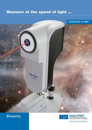
1. 1511.7220032.02010 - 04.08 - 3
Comprehensive measurements Pick your star ... Measure at the speed of light ...
for optimal IOC calculation ...
LENSTAR LS 900®
Intuitive and open
Combined with the IOL power calculator, LENSTAR features a
sophisticated data base to handle the user’s preferred IOL collec-
tion. All standard parameters, including three power ranges
with independent step sizes for the available IOL power as well
as complementary data fields allow to store complete informati-
on on the IOL used.
Advance to the future ...
HAAG-STREIT
Gartenstadtstrasse 10 Biometry
CH-3098 Koeniz/Switzerland
Phone +41 31 978 01 11
Fax +41 31 978 02 82
info@haag-streit.ch
www.haag-streit.com
7
2. Explore new dimensions ... Ergonomic solutions LENSTAR LS 900® technical
for you and your patients ... specifications ...
Measured variables Optical radiation
Complete optical biometry – Corneal thickness (CT) Eye length measurement (A-Scan) and central fixation
Measurement range 300 – 800 μm Light source Superluminescence diode
including CCT and lens thick- Display resolution 0.1 μm Wave length 820 nm
In-vivo repeatability (1.s) +/- 2 μm Power on patient’s eye < 0.6 mW
ness Accuracy of glass plate thickness +/- 2 μm max. load per patient/day 30 000 s
LS 900 on HMS 901 IT height adjus-
Anterior chamber depth (AC) Keratometry
table instrument table combined with Measurement range 1.5 - 5.5 mm Light source LED
a small form factor bare bone PC. Display resolution 0.01 mm Wave length 950 nm
In-vivo repeatability (1.s) +/- 50 μm Power on patient’s eye < 0.2 mW
Align once, get all Accuracy of glass plate thickness +/- 30 μm max. load per patient/day 30 000 s
results – Lens thickness (LT)
Measurement range 0.5 – 6.5 mm
Peripheral fixation
Light source LED
fastest biometrical Display resolution
In-vivo repeatability
0.01 mm
(1.s) +/- 50 μm
Wave length
Power on patient’s eye
570 nm
< 0.02 mW
assessment Accuracy of glass plate thickness +/- 30 μm max. load per patient/day 30 000 s
Eye length (AL) Green illumination
Measurement range 14 – 32 mm Light source LED
Display resolution 0.01 mm Wave length 525 nm
In-vivo repeatability (1.s) +/- 30 μm Power on patient’s eye < 0.2 mW
Noncontact, highest Accuracy of glass plate thickness (+/-) +/- 30 μm max. load per patient/day 600 s
precision – Space saving solution; LS 900 on HMS
Keratometry
Measurement range for radius 5 – 10.5 mm
The combination of radiation generated corresponds to laser class I,
according to IEC 60825-1.
901 instrument table in combination
all measures on with a laptop.
Display resolution
In-vivo repeatability
0.01 mm
(1.s) +/- 20 μm
the true Accuracy of measuring sphere
Measurement range for axis angle
+/- 25 μm
0-180°
optical axis Display resolution
In-vivo repeatability
1°
(1.s) +/- 5°
Accuracy of test object (R7.85/R8.35) +/- 1°
White-to-white distance
Measurement range 7 - 16 mm
Display resolution 0.01 mm
In-vivo repeatability (1.s) +/- 0.3 mm
Accuracy of test object (+/-) +/- 0.1 mm
Pupillometry
Measurement range 2 – 13 mm
Display resolution 0.01 m
In-vivo repeatability (1.s) +/- 0.1 mm
Accuracy of test object (+/-) +/- 0.05 mm
The above mentioned measurement ranges base on the standard set-
ting of the device for automatic measurement and analysis.
The user may change these setting manually to facilitate measure-
ments outside the standard measurement range.
Using LS 900 on a Unit is easy becau-
se of the separation of the examinati-
on unit from the PC used, example LS
8 9
3. The first optical biometry
of the entire eye ...
Eight Measurements in one shot
Precise measurement of eye parameters is critical
in modern cataract treatment.
The LENSTAR LS 900 provides the surgeon with
all necessary parameters needed to calculate the
optimal IOL using latest multivariable formulas in
one singe measurement. The measurement
includes corneal thickness, anterior chamber
depth, lens thickness, axial length, keratometry,
white to white distance, pupilometry and eccentri-
city of the optical axis.
Fast and patient friendly
measurements
The measurement process of the LENSTAR LS 900
is optimised to ensure maximum patient comfort
and minimum process time. The device has to be
aligned only once to get all measurements in a
single shot. Blinking of the patient is detected and
only good measurements are taken for the analy-
sis.
Precision on the true visual axis
The patient fixates directly on the measurement
beam. This ensures that all readings are taken on
the true optical axis. Furthermore all length
measurement are assessed with optical coherence
biometry, leading to unmatched precision and
accuracy. Multiple markers ensure a stable and reli-
able measurement of the corneal curvature.
3
4. The future begins now ...
The all in one optical biometer
Optical coherence biometry revolutionised cataract surgery, the
LENSTAR LS 900 is about to revolutionise optical biometry.
State of the arte, multivariable IOL calculation formulas
demand more than just the axial length and keratometry values
of the eye. LENSTAR LS 900 provides the user with a complete
biometrical assessment of the patient’s eye in a single measure-
ment procedure, including lens thickness, anterior chamber
depth (lens position) and central corneal thickness. Ey
Central corneal thickness (CCT)
CCT is measured using optical coherence technology, leading Lens
to unmatched accuracy and precision. Reproducibility of this thickness
measurement is as good as ± 2μm, providing one of the base
parameters for possible laser correction of the cataract suregery. ACD
Corneal thickness
Keratometry
A 32 marker pattern ensures precise assessment of the corneal
curvature of the patient. The distribution of the marker on two
concentric circles allows stable measurements even with non-
compliant patients.
Pupillometry
The software allows the measurement of the patient’s pupil
diameter in ambient light condition. Adjusting respective light
levels allows to asses the patients adaption capabilities.
White to white
For sulcus fixated IOLs and for the calculation of the IOL power
using Holliday II formula, LENSTAR SL 900 measures white to
white distance (horizontal Iris width).
4
5. Unlimited optical biometry ...
Lens thickness
Modern multivariable IOL calculation formulas use the patients lens
thickness as an input parameter. LENSTAR LS 900 provides the user
with the measurement of true lens thickness on the optical axis of the
patient using optical coherence technology. No more estimation or
additional ultrasound based measurement, to get this important para-
meter.
Anterior chamber depth (ACD)
Just like all other length measures ACD is assessed with laser optical
coherence technology. Combined with the CCT measurement, LEN-
ye length STAR provides the user with the anatomical as well as with the ACD
as measured by ultrasound biometer.
Axial length (AL)
Optical coherence technology using a super luminescence diode as
light source allows the measurement of the axial length of the
patient’s eye on the true optical axis in unmatched precision and
through dens cataracts.
Eccentricity of the optical axis
The eccentricity of the optical axis, an important parameter for laser
refractive procedures, is measured with respect to the centre of the
white to white circle but also with respect to the pupil centre, the refe-
rence to the refractive laser systems.
Special eye conditions
All of the described measurements are available for the measurement of
the “normal” eye cataract patient as well as for the aphakic, pseudophakic
and silicone oil filled eye conditions. In case of an error, you may even
change the selected eye condition after competition of the measurement
procedure.
5
6. Reach for the stars ...
Best measurements for Ready for the future in
optimum IOL prediction IOL power calculation
LENSTAR provides the user with a complete assessment of the The integrated IOL power calculator incorporates all state of the
human eye in highest precision using optical coherence techno- art IOL prediction formulas such as Haigis, Holladay, Hoffer Q
logy. The patient fixates the measurement beam, ensuring that SRK II and SRK/T. Measuring more than just all parameters for
all length measurements are taken on the true optical axis. currently standard formulas, LENSTAR is ready of the next
generation of IOL power estimation formulas.
6
7. 1511.7220032.02010 - 04.08 - 3
Comprehensive measurements Pick your star ... Measure at the speed of light ...
for optimal IOC calculation ...
LENSTAR LS 900®
Intuitive and open
Combined with the IOL power calculator, LENSTAR features a
sophisticated data base to handle the user’s preferred IOL collec-
tion. All standard parameters, including three power ranges
with independent step sizes for the available IOL power as well
as complementary data fields allow to store complete informati-
on on the IOL used.
Advance to the future ...
HAAG-STREIT
Gartenstadtstrasse 10 Biometry
CH-3098 Koeniz/Switzerland
Phone +41 31 978 01 11
Fax +41 31 978 02 82
info@haag-streit.ch
www.haag-streit.com
7
8. Explore new dimensions ... Ergonomic solutions LENSTAR LS 900® technical
for you and your patients ... specifications ...
Measured variables Optical radiation
Complete optical biometry – Corneal thickness (CT) Eye length measurement (A-Scan) and central fixation
Measurement range 300 – 800 μm Light source Superluminescence diode
including CCT and lens thick- Display resolution 0.1 μm Wave length 820 nm
In-vivo repeatability (1.s) +/- 2 μm Power on patient’s eye 0.6 mW
ness Accuracy of glass plate thickness +/- 2 μm max. load per patient/day 30 000 s
LS 900 on HMS 901 IT height adjus-
Anterior chamber depth (AC) Keratometry
table instrument table combined with Measurement range 1.5 - 5.5 mm Light source LED
a small form factor bare bone PC. Display resolution 0.01 mm Wave length 950 nm
In-vivo repeatability (1.s) +/- 50 μm Power on patient’s eye 0.2 mW
Align once, get all Accuracy of glass plate thickness +/- 30 μm max. load per patient/day 30 000 s
results – Lens thickness (LT)
Measurement range 0.5 – 6.5 mm
Peripheral fixation
Light source LED
fastest biometrical Display resolution
In-vivo repeatability
0.01 mm
(1.s) +/- 50 μm
Wave length
Power on patient’s eye
570 nm
0.02 mW
assessment Accuracy of glass plate thickness +/- 30 μm max. load per patient/day 30 000 s
Eye length (AL) Green illumination
Measurement range 14 – 32 mm Light source LED
Display resolution 0.01 mm Wave length 525 nm
In-vivo repeatability (1.s) +/- 30 μm Power on patient’s eye 0.2 mW
Noncontact, highest Accuracy of glass plate thickness (+/-) +/- 30 μm max. load per patient/day 600 s
precision – Space saving solution; LS 900 on HMS
Keratometry
Measurement range for radius 5 – 10.5 mm
The combination of radiation generated corresponds to laser class I,
according to IEC 60825-1.
901 instrument table in combination
all measures on with a laptop.
Display resolution
In-vivo repeatability
0.01 mm
(1.s) +/- 20 μm
the true Accuracy of measuring sphere
Measurement range for axis angle
+/- 25 μm
0-180°
optical axis Display resolution
In-vivo repeatability
1°
(1.s) +/- 5°
Accuracy of test object (R7.85/R8.35) +/- 1°
White-to-white distance
Measurement range 7 - 16 mm
Display resolution 0.01 mm
In-vivo repeatability (1.s) +/- 0.3 mm
Accuracy of test object (+/-) +/- 0.1 mm
Pupillometry
Measurement range 2 – 13 mm
Display resolution 0.01 m
In-vivo repeatability (1.s) +/- 0.1 mm
Accuracy of test object (+/-) +/- 0.05 mm
The above mentioned measurement ranges base on the standard set-
ting of the device for automatic measurement and analysis.
The user may change these setting manually to facilitate measure-
ments outside the standard measurement range.
Using LS 900 on a Unit is easy becau-
se of the separation of the examinati-
on unit from the PC used, example LS
900 on a HS 2010 unit.
8 9
9. Explore new dimensions ... Ergonomic solutions LENSTAR LS 900® technical
for you and your patients ... specifications ...
Measured variables Optical radiation
Complete optical biometry – Corneal thickness (CT) Eye length measurement (A-Scan) and central fixation
Measurement range 300 – 800 μm Light source Superluminescence diode
including CCT and lens thick- Display resolution 0.1 μm Wave length 820 nm
In-vivo repeatability (1.s) +/- 2 μm Power on patient’s eye 0.6 mW
ness Accuracy of glass plate thickness +/- 2 μm max. load per patient/day 30 000 s
LS 900 on HMS 901 IT height adjus-
Anterior chamber depth (AC) Keratometry
table instrument table combined with Measurement range 1.5 - 5.5 mm Light source LED
a small form factor bare bone PC. Display resolution 0.01 mm Wave length 950 nm
In-vivo repeatability (1.s) +/- 50 μm Power on patient’s eye 0.2 mW
Align once, get all Accuracy of glass plate thickness +/- 30 μm max. load per patient/day 30 000 s
results – Lens thickness (LT)
Measurement range 0.5 – 6.5 mm
Peripheral fixation
Light source LED
fastest biometrical Display resolution
In-vivo repeatability
0.01 mm
(1.s) +/- 50 μm
Wave length
Power on patient’s eye
570 nm
0.02 mW
assessment Accuracy of glass plate thickness +/- 30 μm max. load per patient/day 30 000 s
Eye length (AL) Green illumination
Measurement range 14 – 32 mm Light source LED
Display resolution 0.01 mm Wave length 525 nm
In-vivo repeatability (1.s) +/- 30 μm Power on patient’s eye 0.2 mW
Noncontact, highest Accuracy of glass plate thickness (+/-) +/- 30 μm max. load per patient/day 600 s
precision – Space saving solution; LS 900 on HMS
Keratometry
Measurement range for radius 5 – 10.5 mm
The combination of radiation generated corresponds to laser class I,
according to IEC 60825-1.
901 instrument table in combination
all measures on with a laptop.
Display resolution
In-vivo repeatability
0.01 mm
(1.s) +/- 20 μm
the true Accuracy of measuring sphere
Measurement range for axis angle
+/- 25 μm
0-180°
optical axis Display resolution
In-vivo repeatability
1°
(1.s) +/- 5°
Accuracy of test object (R7.85/R8.35) +/- 1°
White-to-white distance
Measurement range 7 - 16 mm
Display resolution 0.01 mm
In-vivo repeatability (1.s) +/- 0.3 mm
Accuracy of test object (+/-) +/- 0.1 mm
Pupillometry
Measurement range 2 – 13 mm
Display resolution 0.01 m
In-vivo repeatability (1.s) +/- 0.1 mm
Accuracy of test object (+/-) +/- 0.05 mm
The above mentioned measurement ranges base on the standard set-
ting of the device for automatic measurement and analysis.
The user may change these setting manually to facilitate measure-
ments outside the standard measurement range.
Using LS 900 on a Unit is easy becau-
se of the separation of the examinati-
on unit from the PC used, example LS
8 9
10. 1511.7220032.02010 - 04.08 - 3
Comprehensive measurements Pick your star ... Measure at the speed of light ...
for optimal IOC calculation ...
LENSTAR LS 900®
Intuitive and open
Combined with the IOL power calculator, LENSTAR features a
sophisticated data base to handle the user’s preferred IOL collec-
tion. All standard parameters, including three power ranges
with independent step sizes for the available IOL power as well
as complementary data fields allow to store complete informati-
on on the IOL used.
Advance to the future ...
HAAG-STREIT
Gartenstadtstrasse 10 Biometry
CH-3098 Koeniz/Switzerland
Phone +41 31 978 01 11
Fax +41 31 978 02 82
info@haag-streit.ch
www.haag-streit.com
7