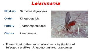
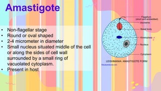
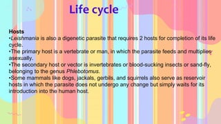
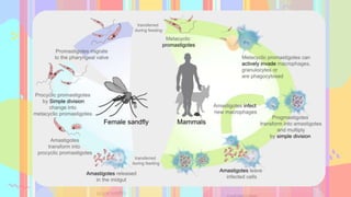
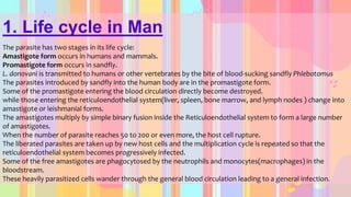
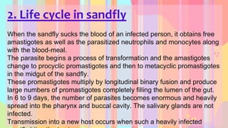
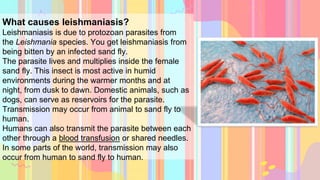
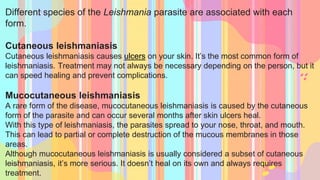
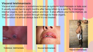
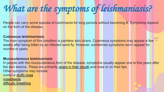
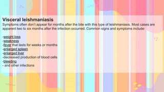
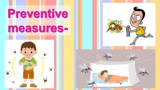
By the end of this presentation we’ll be able to learn about- -Geographical distribution of leishmania parasites- Know the different stages of leishmania parasites and their morphology.-Describe the lifecycle of leishmania.-Causes and pathogenesis of leishmania -Preventive measures of leishmaniasis
1. LIFE CYCLE, PATHOGENICITY,
CAUSES, SYMPTOMS AND
CONTROL OF LEISHMANIA
Supervisor- Dr. Archana Kumar
Institute- Amity institute of biotechnology
Made by- Aditi Tanwar bsz-107
2. By the end of this presentation we’ll be able to learn
about-
-Geographical distribution of leishmania parasites
- Know the different stages of leishmania parasites
and their morphology.
-Describe the lifecycle of leishmania.
-Causes and pathogenesis of leishmania
-Preventive measures of leishmaniasis
3.
4.
5.
6. Introduction
Leishmania
• It is genus of trypanosomatid protozoa, which causes a fatal vector-
borne parasitic disease called Leishmaniasis.
• It is spread by the bite of sandflies of the genus Phlebotomus in the
old world, and of the genus Lutzomyia in the new world.
• Leishmaniasis is caused by a protozoa parasite from over 20
Leishmania species. Over 90 sandfly species are known to transmit
Leishmania parasites.
• Leishmaniasis is the second largest parasitic killer in the world (after
malaria) and is endemic in many parts of America, Asia and South
America.
7. Important species
L. donovani
L. tropica
L. mexicana
L. braziliensis
• L. major
L. lainsoni
L. guanensis,
etc.
8. Morphology
Hosts
• Definitive host- man
• Intermediate host- female sand fly
• Reservoir of infection- dog(China and Brazil), man (India), rodents
9Africa) and jackals (Russia).
Habitat – In reticulo-endothelial system of vertebrate hosts.
Stages
• Promastigote
• Amastigote
9. Leptomonad or promastigote stage
• Thin elongate cells with an anterior
kinetoplast.
• Flagellar stage
• Spindle shaped
• 15-20 micrometer in length
• 1-2 micrometer in width
• Large nucleus centrally situated
• It is found in the midgut of the invertebrates
host or sandfly.
10. Amastigote
• Non-flagellar stage
• Round or oval shaped
• 2-4 micrometer in diameter
• Small nucleus situated middle of the cell
or along the sides of cell wall
surrounded by a small ring of
vacuolated cytoplasm.
• Present in host
11. Life cycle
Hosts
•Leishmania is also a digenetic parasite that requires 2 hosts for completion of its life
cycle.
•The primary host is a vertebrate or man, in which the parasite feeds and multiplies
asexually.
•The secondary host or vector is invertebrates or blood-sucking insects or sand-fly,
belonging to the genus Phlebotomus.
•Some mammals like dogs, jackals, gerbils, and squirrels also serve as reservoir
hosts in which the parasite does not undergo any change but simply waits for its
introduction into the human host.
12.
13. 1. Life cycle in Man
The parasite has two stages in its life cycle:
Amastigote form occurs in humans and mammals.
Promastigote form occurs in sandfly.
L. donovani is transmitted to humans or other vertebrates by the bite of blood-sucking sandfly Phlebotomus
The parasites introduced by sandfly into the human body are in the promastigote form.
Some of the promastigote entering the blood circulation directly become destroyed.
while those entering the reticuloendothelial system(liver, spleen, bone marrow, and lymph nodes ) change into
amastigote or leishmanial forms.
The amastigotes multiply by simple binary fusion inside the Reticuloendothelial system to form a large number
of amastigotes.
When the number of parasite reaches 50 to 200 or even more, the host cell rupture.
The liberated parasites are taken up by new host cells and the multiplication cycle is repeated so that the
reticuloendothelial system becomes progressively infected.
Some of the free amastigotes are phagocytosed by the neutrophils and monocytes(macrophages) in the
bloodstream.
These heavily parasitized cells wander through the general blood circulation leading to a general infection.
14. 2. Life cycle in sandfly
When the sandfly sucks the blood of an infected person, it obtains free
amastigotes as well as the parasitized neutrophils and monocytes along
with the blood-meal.
The parasite begins a process of transformation and the amastigotes
change to procyclic promastigotes and then to metacyclic promastigotes
in the midgut of the sandfly.
These promastigotes multiply by longitudinal binary fusion and produce
large numbers of promastigotes completely filling the lumen of the gut.
In 6 to 9 days, the number of parasites becomes enormous and heavily
spread into the pharynx and buccal cavity. The salivary glands are not
infected.
Transmission into a new host occurs when such a heavily infected
15. Pathogenesis
• Infections range from asymptomatic to
progressive fully developed kala-azar.
• Incubation period is usually 2-4 months
• Symptoms begins with low grade fever
followed by progressive anemia and
protrusion of the abdomen from enlarged
liver and spleen.
• In acute with chills, fever up to 104 degree
Fahrenheit and vomiting, death may occur
within 6-12 months
• Immediate cases of death is usually an
invasion of a secondary pathogen than the
body is unable to combat.
16. What causes leishmaniasis?
Leishmaniasis is due to protozoan parasites from
the Leishmania species. You get leishmaniasis from
being bitten by an infected sand fly.
The parasite lives and multiplies inside the female
sand fly. This insect is most active in humid
environments during the warmer months and at
night, from dusk to dawn. Domestic animals, such as
dogs, can serve as reservoirs for the parasite.
Transmission may occur from animal to sand fly to
human.
Humans can also transmit the parasite between each
other through a blood transfusion or shared needles.
In some parts of the world, transmission may also
occur from human to sand fly to human.
17. Different species of the Leishmania parasite are associated with each
form.
Cutaneous leishmaniasis
Cutaneous leishmaniasis causes ulcers on your skin. It’s the most common form of
leishmaniasis. Treatment may not always be necessary depending on the person, but it
can speed healing and prevent complications.
Mucocutaneous leishmaniasis
A rare form of the disease, mucocutaneous leishmaniasis is caused by the cutaneous
form of the parasite and can occur several months after skin ulcers heal.
With this type of leishmaniasis, the parasites spread to your nose, throat, and mouth.
This can lead to partial or complete destruction of the mucous membranes in those
areas.
Although mucocutaneous leishmaniasis is usually considered a subset of cutaneous
leishmaniasis, it’s more serious. It doesn’t heal on its own and always requires
treatment.
18. Visceral leishmaniasis
Visceral leishmaniasis is sometimes known as systemic leishmaniasis or kala azar.
It usually occurs two to eight months after being bitten by a sand fly. It damages
internal organs, such as your spleen and liver. It also affects your bone marrow, as
well as your immune system through damage to these organs.
The condition is almost always fatal if it’s not treated.
Visceral leishmaniasis
Mucosal leishmaniasis
Cutaneous leishmaniasis
19. What are the symptoms of leishmaniasis?
People can carry some species of Leishmania for long periods without becoming ill. Symptoms depend
on the form of the disease.
Cutaneous leishmaniasis
The main symptom of this condition is painless skin ulcers. Cutaneous symptoms may appear a few
weeks after being bitten by an infected sand fly. However, sometimes symptoms won’t appear for
months or years.
Mucocutaneous leishmaniasis
In people with the mucocutaneous form of the disease, symptoms usually appear one to five years after
the skin lesions. These are primarily ulcers in their mouth and nose or on their lips.
Other symptoms may include:
runny or stuffy nose
nosebleeds
difficulty breathing
20. Visceral leishmaniasis
Symptoms often don’t appear for months after the bite with this type of leishmaniasis. Most cases are
apparent two to six months after the infection occurred. Common signs and symptoms include:
-weight loss
-weakness
-fever that lasts for weeks or months
-enlarged spleen
-enlarged liver
-decreased production of blood cells
-bleeding
- and other infections
21. There’s no vaccine or prophylactic medication available. The only way to prevent leishmaniasis is to
avoid getting bitten by a sand fly.
Follow these steps to help prevent being bitten by a sand fly:
-Wear clothing that covers as much skin as possible. Long pants, long-sleeved shirts tucked into pants,
and high socks are recommended.
-Use insect repellent on any exposed skin and on the ends of your pants and sleeves. The most effective
insect repellants contain DEET.
-Spray indoor sleeping areas with insecticide.
-Sleep on the higher floors of a building. The insects are poor fliers.
-Avoid the outdoors between dusk and dawn. This is when sand flies are most active.
-Use screens and air conditioning indoors when possible. Using fans might make it more difficult for the
insects to
fly.
-Use a bed net tucked into your mattress. Sand flies are much smaller than mosquitos, so you need a
tightly woven net. Spray the net with insecticide containing pyrethroid if possible.
Buy bed nets, insecticides, and repellents before traveling to high-risk areas.
22.
23. ^ Schnur, L. F (1989). "On the Clinical Manifestations and Parasites of Old World Leishmaniases and Leishmania Tropica
Causing Visceral Leishmaniasis". In Hart, D. T. (ed.). Leishmaniasis. pp. 939–43. doi:10.1007/978-1-4613-1575-9_119. ISBN
978-1-4612-8862-6.
^ Ryan KJ; Ray CG (editors) (2004). Sherris Medical Microbiology (4th ed.). McGraw Hill. pp. 749–54. ISBN 0-8385-8529-9.
^ Myler P; Fasel N (editors) (2008). Leishmania: After The Genome. Caister Academic Press. ISBN 978-1-904455-28-8.
^ Ansari MY, Equbal A, Dikhit MR, Mansuri R, Rana S, Ali V, Sahoo GC, Das P (Nov 2015). "Establishment of Correlation
between In-Silico &In-Vitro Test Analysis against Leishmania HGPRT to inhibitors". International Journal of Biological
Macromolecules. 83: 78–96. doi:10.1016/j.ijbiomac.2015.11.051. PMID 26616453.
^ WHO (2010) Annual report. Geneva
^ Cox, F. E. G (2002). "History of Human Parasitology". Clinical Microbiology Reviews. 15 (4): 595–612.
doi:10.1128/CMR.15.4.595-612.2002. PMC 126866. PMID 12364371.
^ Yanik, M.; et al. (2004). "The psychological impact of cutaneous leishmaniasis". Clinical and Experimental Dermatology. 29
(5): 464–467. doi:10.1111/j.1365-2230.2004.01605.x. PMID 15347324. S2CID 11543741.